As a graduate student in Canada in the late sixties and early seventies, one of my duties was to act as a demonstrator in freshman undergraduate chemistry laboratories. During one session, in which the class experiment involved observing colour changes during a reaction in solution, a student passed the casual remark “I wish I could actually see what was happening in there…” I cannot remember my reply, but I imagine that we both laughed it off as wishful thinking. However, this year's Lonsdale Lecture, given by Arwen Pearson of the Hamburg Centre for Ultrafast Imaging, Germany, entitled “Visualising molecules in motion: crystallography as a tool to probe structure and dynamics” was perhaps an indication that after 46 years, that student's dream is on the verge of becoming reality.
As is now customary, the Young Crystallographers’ Group Satellite Meeting began on Monday and continued on Tuesday morning, consisting of invited plenary talks and short oral presentations. Two plenary talks were given by Sally Price of University College London and James Errey of Heptares Therapeutics, and the Parkin Lecture was given by Jonathan Brooks-Bartlett of Oxford University. A Teaching Session for the Young Crystallographers followed, entitled “Forgotten methods in Crystallography”. Bob Eady of Liverpool University, and Paul Raithby of Bath University provided the first two talks. Mike Glazer of the Universities of Oxford and Warwick spoke on “Plotting three-dimensional information in two dimensions”, after which earnest-looking students could be seen clutching facsimiles of Wulff Nets. The main meeting programme commenced at noon on Tuesday, and continued with three parallel sessions on each of Tuesday afternoon, Wednesday (all day), and Thursday morning.
There was no specific theme to this year's Spring Meeting, but a pleasingly diverse range of topics was assembled by the Biological, Chemical, Physical and Industrial Crystallography Groups, and a more detailed account of the meeting is contained in the supplemental material.
Returning to Arwen Pearson's Lonsdale Lecture, the aim of structural studies is to understand how structure leads to function, but macromolecules are dynamic, flexible objects, and the average ensemble structure determined in a crystallographic experiment sees all conformations at once (an average), and dynamic information is lost. In order to understand the dynamics of a system, a number of methodologies have been developed to enable time-resolved structural measurements. Trapping methodologies may be used to determine the structures of metastable intermediates, comprising mechanistic trapping, “on-the-fly” cryo-trapping of longer-lived intermediates and serendipitous intermediate trapping. Mechanistic trapping may include altering reaction conditions, using mutants, using altered substrates, and driving systems into steady states.
Pump probe time-resolved studies are used but their limitations are low signal/noise (S/N) ratios due to very short pulses, and generation of only one data point per cycle. The experiments need to be repeated many times in order to raise the S/N ratio, and also need to be repeated with different values of Δt (the time lapse between excitation and probe pulses) in order to amass a series of time-resolved data sets. As a consequence many crystals are needed. X-ray crystallographic experiments based on the Hadamard Transform (HATRX), in which time resolution is defined by the underlying periodicity of the probe pulse sequence, have resulted in greatly-improved S/N ratios when compared with those for the fastest pump-probe experiments that depend upon a single pulse. The availability of methods such as HATRX, Laue crystallography, and serial femtosecond crystallography (SFX) mean that time-resolved experiments are becoming increasingly accessible.
However, for time-resolved experiments, the reaction must be rapidly and uniformly initiated throughout the crystal. The mode of initiation depends upon the process that is to be observed. For a slow (>ms) reaction, rapid mixing will suffice but for fast (<ms) reactions the most effective way to do this is to use intense light pulses (a laser pulse or a T-jump IR pulse) to drive photochemical reactions. Unfortunately, most biomacromolecules are not activated by light. To address this challenge, attempts have been made to synthesize and characterize a variety of photocages (synthetic molecules whose biological activity is controlled by light) in order to develop a library of light-activated compounds that will be of general use to the structural biology community.
Mike Zaworotko of the University of Limerick, Eire, began his Chemical Crystallography Group plenary talk with a quote from John Maddox in 1988, the then editor of Nature. “One of the continuing scandals in the physical sciences is that it remains in general impossible to predict the structure of even the simplest crystalline solid from a knowledge of its chemical composition.” That composition and structure profoundly impact the properties of crystalline solids, has provided impetus for exponential growth in the field of crystal engineering over the past 25 years. In that time crystal engineering has evolved from structure design (form) to control over bulk properties (function). Two classes of functional materials: multicomponent pharmaceutical materials (MPMs) and hybrid ultra-microporous materials (HUMs) are examples of important materials, which scientists are attempting to improve by design.
MPMs, such as co-crystals, have emerged at the pre-formulation stage of drug development. This results from their modular and designable nature, which facilitates the discovery of new crystal forms of active pharmaceutical ingredients (APIs). Co-crystals can be classified into molecular co-crystals (MCCs) that contain only neutral components (coformers) and ionic co-crystals (ICCs), which comprise at least one ionic coformer that is a salt, e.g. glucose/sodium chloride. ICCs offer much greater diversity in terms of composition and properties than single component crystal forms and are amenable to design. Co-crystals of lithium chloride and leucine, for example, have been produced and may be used in the treatment of mental disorders that require lithium to penetrate the blood brain barrier and exert therapeutic effects in the central nervous system. These novel co-crystal forms may be used to lower the oral dose required to achieve therapeutic concentrations of lithium in the brain, thus reducing peripheral toxicity.
HUMs are built from metal or metal cluster “nodes” and combinations of organic and inorganic “linkers”. There is a need for cheap, robust porous materials for carbon capture at pre- and post-combustion stages, and also for direct air capture of CO2. Selectivity is important, with CO2/N2 and CO2/water vapour uptake ratios of 200:1 and 100:1 respectively, considered essential. A hybrid ultra-microporous material with ~7 Å pore has been made with organic linkers (pyrazine) and inorganic pillars (hexafluorosilicate). The Zn analogue in the SIFSIX series (SIFSIX-3-Zn) has set a benchmark selectivity for CO2/N2 (>3000) and CO2/CH4 (>1000). Ethylene is the most produced commodity at 150 m tonnes per annum but it is rarely pure, containing 1–2% acetylene as a contaminant. These gases are difficult to separate and the best HUM in terms of selectivity is SIFSIX-2-Cu-i, which also happens to have the second highest uptake.
In an entertaining session entitled “Tips, Tricks & Trials”, Horst Puschmann, of Durham University, Durham, UK described “The pesky CIF – and how to tame it.” When it comes to routine, small molecule structures, the hard part is often not the structure determination itself, but the correct and consistent reporting of these structures (or sets of structures). As a scientific field, crystallography is extremely lucky to have a tried, tested, and checkable data exchange format – the much loved (and loathed) CIF file. Generating these files in a consistent, complete, and true-to-fact way is harder than it may at first seem.
A CIF file generated through structure determination contains details of data collection and reduction, refinement, atomic coordinates, thermal parameters, molecular geometry, and auxiliary information. CheckCIF is a service operated by the IUCr in which a user submits a CIF for it to be checked prior to publication or archiving. CheckCIF will cause alerts if there are inconsistencies or omissions in the original CIF, and the submitter is expected to correct the errors before resubmission.
According to Puschmann, CIF is great! It has rigorous syntax, clear definition, and it allows for structure data interchange regardless of structure origination. As an archiving tool it enables verification of every step in the structure determination, and best of all, it is human-readable. However, if a user wishes to modify a CIF file, it should not be done by hand, but should be run through structure solution software such as OLEX2, which is free to download. Recent updates to the world's “favourite” crystallographic software package comprise the inclusion of reflection (hkl) data, models (.res), and the recommended way to run and report SQUEEZE (a routine for refining disordered solvent molecules).
The late afternoon session of the Physical Crystallography Group entitled “Modelling crystals and crystallographic data” was opened by Carole Morrison, of the University of Edinburgh, UK with a talk entitled “Frustrated MOFs: insight from modelling when crystallography is stumped.”
Metal-organic frameworks (MOFs) continue to attract high interest from the scientific community due to their promise in fields of guest-specific gas sorption, separation, drug delivery, and catalysis. However, these structurally versatile materials often have soft mechanical properties that distort or even collapse upon application of temperature, shear stress or hydrostatic pressure. This poses problems for the sintering and pelletizing steps required to shape MOF powders into industrially useful morphologies. Young's Modulus and hardness measurements were made on evacuated crystals of both Zr–UiO–67 and Zr–UiO–abdc MOFs, confirming that the latter structure is more flexible and the former more rigid. It is clear that the bowed ligand (UiO-abdc) offers greater resilience to external pressures, and that to make and characterize frustrated MOFs requires combined studies of structure, dynamics, and mechanical properties.
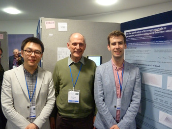
Matt Cliffe, of the University of Cambridge, UK (one of two ICDD Bursary recipients (Figure 1) continued MOF chemistry with his talk “Correlated defects in Hafnium and Zirconium MOFs”.
ICDD bursary recipients Yue Wu of Oxford University (left) and Matt Cliffe of Cambridge University (right) with David Rendle.

Defects are crucial to the chemistry of MOFs with recent studies demonstrating the prevalence of defects, especially ligand vacancies (missing linkers), in MOF chemistry. These defects may improve sorption properties, catalytic activity, and ionic conductivity. In many functional materials, it is not just the presence of defects but their interactions and correlations that determine their properties. Correlated defects can be accommodated in a MOF, by including “modulators” (ligands such as trifluoroacetic acid (TFA) able to bind to clusters but not form part of the network structure). Using a combination of anomalous Hf-K edge X-ray diffraction, total scattering and electron diffraction measurements, it is clear that these defects are not just of ligand vacancies, but also include Hf cluster absences.
“The Future of Structural Science” provided four interesting talks from quite different viewpoints. John Spence, of Arizona State University, USA spoke on “Time resolved molecular imaging using XFELs”, David Keen (ISIS Facility, Rutherford Appleton Laboratory, UK) asked the question “Does neutron diffraction have a role?”, Derek Wann of the University of York, UK described electron diffraction as a tool for determining molecular structures and investigating dynamics in the gas phase, and Peter Wood from the Cambridge Crystallographic Data Centre (CCDC) looked at the future of structural databases.
SFX using ultrashort pulses from X-ray free electron lasers (XFELs) has enabled studies of light-triggered dynamics of biomolecules. The use of femtosecond X-ray pulses, instead of freezing, to avoid radiation damage to the crystals has opened the way to the study of protein dynamics at room temperature at atomic resolution without damage. A continuously refreshed supply of hydrated protein nano crystals must be supplied. This “diffract before destroy” mode then also allows study of irreversible processes in proteins for which crystals large enough for macromolecular crystallography (MX) cannot be grown. Microcrystals of photoactive yellow protein (a bacterial blue light photoreceptor) were used as a model system and high resolution, time-resolved difference electron density maps were obtained, allowing the determination of structures of reaction intermediates to a resolution of 1.4 Å.
Some membrane protein nanocrystals have been grown in lipid cubic phase (LCP) and the resulting nanocrystals have been injected into the pulsed X-ray beam in a viscous “toothpaste” via a specially designed grease gun. The LCP jet delivers the crystals at the rate of the X-ray pulses so a high hit rate is obtained, and the low flow rate (1–300 nl min−1) avoids wastage of precious protein. XFEL is important because (a) radiation damage is avoided, (b) room temperature structures are possible, thus avoiding the need to freeze the crystals, (c) there is better time resolution (picoseconds), (d) irreversible reactions may be studied, (e) there is no need for large crystals – nanocrystals will suffice, (f) the optical pump laser absorption length is comparable with the nanocrystal size, (g) diffusion times are short for the nanocrystals in the mixing jet, and (h) higher resolution (~1.8 Å) is possible for some proteins.
According to Keen, neutron scattering has played a vital role in our understanding of structural science over many years. Neutrons are highly penetrating, sensitive to magnetic structures, very useful in the location of H atoms in the presence of heavier atoms, and have no intensity fall-off at high Q values. Two distinct disadvantages, however, are low flux and limited access to a source of neutrons, although regarding the latter, the Spallation Neutron Source at ISIS in the UK is considered to be one of the best instrumental facilities in the world. Early successes with neutron diffraction include the structures of Vitamin B12 (Hodgkin, 1967) and Myoglobin (Schoenborn, 1969), with the structure of liquid lead interpreted (Chamberlain, 1950) in terms of the distribution in separation distance of neighbouring pairs of atoms (Pair Distribution Function, PDF). Neutron diffraction most certainly does have a future role in structural science – in new areas of magnetism, PDF analysis, in protein crystallography, and in non-ambient studies. Sample size (how small can be tolerated?), how big a structure can be solved, and how accurate will the result be, are under question. However, neutron facilities are “big science” and as such, unfortunately, come under political scrutiny.
Electrons have a larger scattering cross section than X-rays, they are less damaging and they are easier to create and manipulate. Electron diffraction has been a staple technique in determining the structures of gaseous molecules for nearly a century. However, until recently most electron diffraction experiments used a continuous electron beam, which has restricted study to that of static structures of molecules. As molecules are constantly vibrating, this time-averaged information is essentially a “blurred” image, like a photograph of a fast moving object taken with a long exposure camera. With the availability of Ti: sapphire lasers, it is now possible to capture sharp, near instantaneous diffraction images from molecular species using a pulsed electron beam. By combining the laser pump (700 fs) and electron probe (70 keV) techniques, one can watch molecular structures as they evolve over a period of time – the so-called “molecular movie”.
In contrast to the previous talks, it was instructive to hear how crystallographic data should be stored. In 1948 a curated database of crystallographic structures was first envisaged, and then in 1965 the Cambridge Structural Database (CSD) began as a fledgling project, and the Protein Database (PDB) followed in 1971. Now, in 2016, there are some 827 982 structures in the CSD and partnerships have been developed with some journal publishers such that crystallographic data are passed automatically to the CSD. Links to ChemSpider and to DataCite have also been established. One challenge in database curation is in coping with the trends in structure determination. As well as single crystal X-ray data, X-ray powder data, neutron single crystal and powder data, and electron diffraction data are generating structural coordinates. In addition, when powder diffraction fails to solve a crystal structure, crystal structure prediction methods may be used. How should a predicted structure be classified? Wood contended that in the future – say 50 years’ time – all journal articles should be linked to the database, which will contain millions of structural datasets, perhaps including those of inorganic structures. Databases should be integrated into scientific workflows. With such an increase in amount of data available, more effective searching methods will be needed, and of course adequate funding to ensure sustainability.
Powder diffraction means real crystallography on real materials under real conditions! Examples of real materials include Li batteries, fuel cells, C60, nanomaterials, paracetamol, pencil “lead”, proteins and turbine blades. With this dramatic statement began the Physical Crystallography Group plenary lecture, this year given by Bill David FRS of the ISIS Facility, Rutherford Appleton Laboratory, UK. His talk, entitled “120 years of Powder Diffraction 1916–2036” looked at how powder diffraction has progressed from its beginnings at the start of the last century to the present date.
The persons originally responsible for developing powder diffraction as an analytical tool were Max von Laue, Peter Debye, Paul Scherrer, Albert Hull, Irving Langmuir and William Coolidge. Hull was something of a polymath, studying first Greek at Yale, and then physics. Among the powder patterns he produced were those of iron, silicon, aluminium, magnesium, sodium, lithium, nickel, and graphite. Notable advances were made in powder diffraction by Bertram Warren (1934) when he studied the structure of glass, and investigated nonperiodic and nearly periodic structures through quantitative measurement of X-ray intensities. Hugo Rietveld in 1966, with his method of least-squares refinement of a theoretical powder pattern, calculated from a known crystal structure, with its measured experimental powder pattern, paved the way for Robert von Dreele's ubiquitous software package (GSAS – General Structure Analysis System), which enables structure refinement from single crystal and/or powder data collected with either X-rays or neutrons. Rietveld was awarded the Aminoff Prize in 1995 for this contribution to crystallography. Bill David himself made a significant contribution by developing software (DASH) for structure determination from powders. More recently, Paul Fewster in 2014 has developed a new theory for X-ray diffraction, which when applied to the scattering from powders, evaluates the full scattering profile, including peak widths and the background.
Hydrogen storage materials are of considerable interest at present, and will continue to be, in the search for alternative sources of “eco-friendly” energy. Among them are lithium borohydride and the lithium amide – lithium hydride composite (Li–N–H) system. Investigation of hydrogenation and dehydrogenation reactions of the latter system through in situ synchrotron X-ray powder diffraction experiments allowed for the observation of the formation and evolution of non-stoichiometric intermediate species of the form Li1+x NH2−x . The results demonstrated the central role of ionic mobility in understanding temperature limitations, capacity loss, and facile reversibility (Li2NH–LiNH2) of the Li–N–H system.
The Industrial Group plenary talk (“Using co-crystals to optimise solid properties”) was presented by Rolf Hilfiker, of Solvias AG, Kaiseraugst, Switzerland.
Solid state properties play a big role in the selection of active pharmaceutical substances, from research to manufacture of the final product. The API may consist of the parent molecule, a polymorph, a solvate, a hydrate, a salt, a co-crystal or an amorphous form, and it is important to identify the best, cheapest, and most reproducible form. When the best solid form of an API has to be chosen, co-crystals may be the best option. Co-crystals should be considered if the API is insoluble, too soluble, or cannot be crystallized, or its morphology, stability and hygroscopicity are unfavourable, and salt formation is not an option. Intellectual property (IP) rights may be another reason. In an attempt to circumvent IP infringement and produce a new co-crystal for an API, consultation of the lists of compounds known as GRAS (Generally Recognized as Safe) and EAFUS (Everything added to food in the US) serves as a good starting point in the search for coformers. High throughput screening using 96 well plates and controlled evaporation and characterization by Raman spectroscopy revealed the best candidates.
In a focused screening approach, five different co-crystals of piracetam were synthesized (with L-tartaric acid, with citric acid in a 1:1 and 3:2 ratio, with DL-mandelic acid and with L-mandelic acid) and their crystal structures determined. Looking at the structures alone revealed that it is (a) not easy to predict, which coformers will be the most successful, (b) essential that a large number of coformers are screened, and (c) very useful to consider phase diagrams before proceeding to large-scale production. Choice of co-crystals may be based upon thermodynamic considerations, with stability (lower free energy) and suitable solubility high on the list of requirements. The construction of phase diagrams plays an important part in the design of successful co-crystal screens.
Ivan Marziano of Pfizer Worldwide Research & Development, Sandwich, UK, was the opening speaker in a joint Chemical/Industrial Crystallography Group session entitled “From amorphous to crystalline”. His talk entitled “The pursuit of the structure-function relationship in pharmaceutical crystallisation” made reference to the “Materials Science Tetrahedron” (processing, properties, structure, and performance), which provides a multidisciplinary framework within the pharmaceutical sciences and includes: the use of modelling tools to identify the “canvas” of physical properties available for a given material, and the process and product design, which consider the implicit properties of the materials involved. Crystallization plays a key role in delivering materials with the desired physical properties within the range allowed for a given crystal structure.
The success of a given drug product depends upon its stability, efficacy and quality, and the drug product process has to be robust, reproducible, economic, and must conform to regulatory requirements. Inconsistent dissolution of a drug may be caused partly by the milling process. Milling will alter the ratio of hydrophilic to hydrophobic crystal faces, and the extent of milling will determine just how much of the original surface chemistry is retained, and will influence the dissolution profile. Water adsorption calculations are used to quantify the affinity of the dominant crystal surfaces. Removal of impurities in the product is an essential part of the process, and decisions have to be made concerning the timing of the impurity purge. Interaction (between product and impurity) energy calculations are required in order to estimate the purge factors for process impurities and to identify alternative purge points. Where possible, however, upstream control of impurities, rather than purge, is the preferred strategy.
The BCA Prize Lecture was delivered by Christer Aakeröy of Kansas State University, Manhattan, Kansas, USA. His talk, entitled “From molecular sociology to functional materials” provided, among other things, an interesting application of co-crystals – something other than for the usual pharmaceutical application.
Co-crystals represent solids where bulk physical properties may be amenable to fine-tuning by making modular and controllable alterations to the crystalline lattice that houses an active molecular species. The links between crystal structure and solid-state properties offer opportunities for improving processing, performance, and shelf-life of a wide range of speciality chemicals. Consequently, an ability to control and change the crystalline environment of a material without altering molecular properties would be of considerable significance to manufacturers and consumers alike. Ethylenedinitramine (EDNA) is an energetic material, which requires attention partly due to its chemical instability originating with its two highly acidic protons. In order to stabilize EDNA, a co-crystallization approach targeting the acidic protons using a series of co-crystallizing agents with suitable hydrogen-bond acceptors was employed. Fifteen attempted co-crystallizations resulted in eight successful outcomes and six of these were crystallographically characterized and all showed evidence of H-bonds to the intended protons. Calculated detonation properties and experimental thermal and impact data for the co-crystals were obtained and compared with those of pure EDNA. The co-crystal of EDNA and 1,2-bis(4-pyridyl)ethylene was recognized as a more thermally stable alternative to EDNA while the co-crystal of EDNA and pyrazine N,N-dioxide showed comparable detonation strengths (and much improved chemical stability) compared with that of EDNA. The co-crystals EDNA: 4,4′-bipyridine and EDNA: pyrazine N,N-dioxide were found to be ~50% less impact sensitive than EDNA, all of which illustrate how co-crystallizations can be utilized for successfully modifying specific aspects of energetic materials.
The conference was held on April 4–7, 2016 at the Jubilee Campus, University of Nottingham, UK (Figure 2) and was supported by some 15 sponsors and exhibitors, and attended by 243 delegates. To view the Scientific Programme, use the link: http://bca2016.crystallography.org.uk/index.php/scientific-programme/
View of the Jubilee Campus, Nottingham University.

SUPPLEMENTARY MATERIALS AND METHODS
The supplementary material for this article can be found at https://doi.org/10.1017/S0885715616000464.