Introduction
From the moment life begins from a single fertilised egg all the way until the formation of structures, organs and the development of the foetus, signalling molecules and cascades play crucial roles in the orchestration and the entire generation of the new organism. Studies on a variety of model organisms have increased our understanding of how different signalling pathways can control developmental processes and how dysregulation of these pathways can lead to abnormal phenotypes and pathological conditions (Refs Reference New, Howes and Smith1, Reference Van Otterloo, Williams and Artinger2). In terms of head development, structures are formed and integrated through a complex sequence of cross-talk events between different germ layers and tissues involving activation of molecular signalling cascades at distinct locations and stages. Hedgehog, Wnt, TGF and FGF signalling have been shown to regulate the patterning of craniofacial and orodental structures in the developing embryo (Ref. Reference Neben and Merrill3). The extracellular signal-regulated kinase (ERK) pathway is considered to have a central role in mediating a panoply of extracellular signals from the plasma membrane to the nucleus of the cell, thus controlling cellular response to particular stimuli during head formation. In this well-studied pathway, extracellular signals, such as growth factors, bind to receptor tyrosine kinase proteins (RTKs), which upon dimerisation and cross-phosphorylation on tyrosine residues interact with adaptor proteins such as the GRB2 (Refs Reference Schlessinger4, Reference Pawson5). This interaction leads to the recruitment of guanine nucleotide exchange factors (GEFs) like SOS to the plasma membrane of the cell and the subsequent conformational change of RAS proteins towards their active GTP-bound form (Refs Reference Marshall6, Reference Quilliam, Khosravi-Far, Huff and Der7). This event promotes the activation of RAF kinases, which in turn phosphorylate MEK kinases resulting finally in ERK1/2 (MAPK3/1) activation and the subsequent regulation of a variety of intracellular targets.
Although RAS/ERK signalling cascade has been extensively studied in the context of tumourigenesis, it also plays a major role in craniofacial and orodental development. Mutations in the components of this particular pathway can lead to some of the most common skull and face disorders in humans. In addition to RASopathies that are well studied and reviewed elsewhere (Refs Reference Denayer, de Ravel and Legius8, Reference Hernandez-Porras and Guerra9), a variety of craniosynostosis cases as well as orofacial abnormalities, such as cleft lip and palate, spring from dysregulation of the RAS/ERK pathway activation. This review focuses on all these cases and is divided into three main sections representing the corresponding areas of research: (1) orofacial development with respect to palate and mandibular formation, (2) craniofacial development with respect to calvarial bone and suture formation and (3) dental development including studies of enamel, dentine and cementum generation. It also analyses human disorders that are not so widely known yet they have severe phenotypes and are linked to the RAS/ERK cascade too. Although a plethora of human developmental abnormalities are associated with mutations on RTKs, for example, Muenke syndrome, osteoglophonic dysplasia, we concentrate on cases that have been suggested to be specifically mediated by the ERK pathway. An overview of the downstream molecular mechanisms affecting specific cell types and developmental stages that are implicated in the pathophysiology of these disorders is also provided in this review.
RAS/ERK pathway in orofacial malformations
Orofacial development
In mammals, the nasal cavity and the oral cavity of the face are separated by palate, a tissue that contains an anterior bony part, the hard palate, and a posterior mainly muscular part, the soft palate. During embryonic development, the hard palate derives from the primary and secondary palate, while the soft palate originates exclusively from the secondary palate.
In mice, the primary palate and the upper lip are formed when the medial nasal prominences start to fuse with the maxillary prominences on embryonic day 11.5 (E11.5). At the same time, outgrowths of the maxillary prominences form the secondary palatal shelves that upon fusion with each other will finally give rise to the secondary palate on E14.5–E15.5 (Refs Reference Bush and Jiang10, Reference Lan, Xu and Jiang11). Each palatal shelf consists of neural crest-derived mesenchymal cells surrounded by epithelial cells. As the two shelves approach to each other and fuse, the epithelial cells previously located at the edge of each shelf lie at the midline of the secondary palate constituting the medial edge epithelium (MEE). By E15.5 MEE cells have disappeared from the midline either through apoptosis, migration or epithelial-to-mesenchymal transition and a continuous mesenchymal palate tissue is formed (Refs Reference Dudas, Li, Kim, Yang and Kaartinen12, Reference Jalali, Zhu, Liu and Nawshad13). In addition to the mechanisms mentioned above, a recent study revealed that cell intercalation drives the convergence of the MEE cells to the midline, followed by displacement and cell extrusion events that lead to the removal of the epithelial cells from the palatal mesenchyme (Ref. Reference Kim, Lewis, Singh, Ma, Adelstein and Bush14). Finally the fusion between primary and secondary palate takes place resulting in the creation of a complete mouth roof by E17. Sonic hedgehog (Shh), Fgf10 and Fgf7 seem to have crucial roles in the palatal shelf development and patterning, while TGF-β3 signalling through Smads and p38 MAPK pathway seems to be important for normal palatal fusion (Ref. Reference Li, Lan and Jiang15). However, less is known about the role of Ras/Erk pathway in these processes. Studies on Erk activation pattern reveal the presence of phosphorylated Erk1/2 from E12.5 to E14.5 in the palatal shelves in both epithelial and mesenchymal cells as well as in the muscles of the tongue on E14.5 (Refs Reference Parada, Han, Grimaldi, Sarrion, Park, Pelikan, Sanchez-Lara and Chai16, Reference Cho, Cho, Lee, Lee, Gang and Jung17).
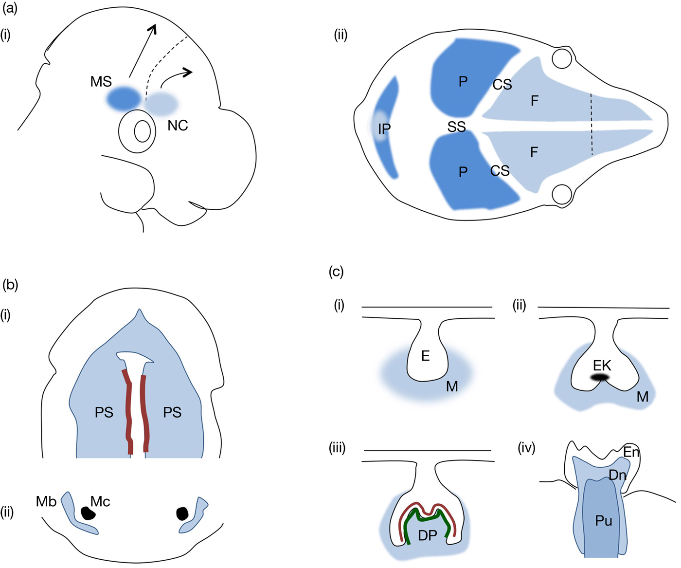
The lower jaw, the mandible, is formed by the mandibular prominences. The mandibular bone develops from neural crest-derived mesenchymal cells in proximity to a cartilaginous structure called Meckel's cartilage. Figure 1 summarises key steps of mouse craniofacial and orodental development. The largest part of the mandibular bone is created through the intramembranous ossification process. On E12.5 phosphorylated Erk1/2 is detected in the chondrocytes of Meckel's cartilage and on E13.5 in the osteogenic fronts of the mandibular bone (Ref. Reference Parada, Han, Grimaldi, Sarrion, Park, Pelikan, Sanchez-Lara and Chai16). However, by E14.5 studies report that the presence of active Erk1/2 is restricted only to the undifferentiated mesenchymal cells of the jaw, while the perichondrium of Meckel's cartilage displays also an amount of phosphorylated kinase that is detected specifically on the side of the tongue (Ref. Reference Cho, Cho, Lee, Lee, Gang and Jung17). These data indicate the existence of a defined spatiotemporal pattern of ERK1/2 activation during orofacial development.
Mouse embryonic craniofacial and orodental development. The scheme summarises main events of mouse craniofacial, orofacial and dental development described in the review. (a) Developmental origin of calvarial bones. (I) Populations of mesoderm-derived mesenchymal cells (shown in dark blue) and neural crest-derived mesenchymal cells (shown in light blue) accumulate at the supraorbital regulatory centre at E10.5, from which they migrate to the sites of the future parietal and frontal bones, respectively, shown in (II). MS, mesoderm-derived mesenchymal cells; NC, neural crest-derived mesenchymal cells; F, frontal bone; P, parietal bone; IP, interparietal bone; CS, coronal suture; SS, sagittal suture. (b) Development of the oral cavity and jaw. (I) Oral view of secondary palate at E14.5. Palatal shelves consisting of neural crest-derived mesenchymal cells are depicted in light blue. Epithelial cells that will constitute the MEE (medial edge epithelium) are shown in dark red. (II) Mandibular bone is formed by differentiating neural crest-derived mesenchymal cells (light blue) in proximity to Meckel's cartilage depicted in black. PS, palatal shelf; Mb, mandibular bone; Mc, Meckel's cartilage. (c) Tooth development from (I) bud through (II) cap and (III) bell stage until (IV) eruption. Neural crest-derived structures are indicated by blue colour. At the bell stage, ameloblasts and odontoblasts are shown in red and green, respectively. E, epithelium; M, mesenchyme; EK, enamel knot; DP, dental papilla; En, enamel; Dn, dentine; Pu, pulp.

Human orofacial disorders and associated animal studies
Mutations in members of the RAS/ERK signalling pathway have been identified as a cause of certain human orofacial disorders, while ERK signalling appears to also mediate the effects of mutations in factors that were previously not connected directly to the ERK kinase. Orofacial clefts constitute the most common facial disorder with an occurrence rate of about 1–700 births and among them cleft lip and cleft palate appear to be the malformations most often observed among individuals. Genomic studies on cleft patients indicate the presence of ERK1 (MAPK3) duplication as the sole gene alteration observed in some cases (Ref. Reference Conte, Oti, Dixon, Carels, Rubini and Zhou18), while individuals that carry loss-of-function mutations in SPROUTY2 (SPRY2) gene coding for an inhibitor of the RAS/ERK pathway display also cleft palate (Ref. Reference Vieira, Avila, Daack-Hirsch, Dragan, Felix, Rahimov, Harrington, Schultz, Watanabe, Johnson, Fang, O'Brien, Orioli, Castilla, Fitzpatrick, Jiang, Marazita and Murray19). Of note, Sprouty2−/− (for the MGI nomenclature of the genetically modified mouse strains reviewed herein, see Box 1) mice exhibit the cleft phenotype with a prevalence of 22% (Ref. Reference Matsumura, Taketomi, Yoshizaki, Arai, Sanui, Yoshiga, Yoshimura and Nakamura20). Human craniofrontonasal syndrome (CFNS) is caused by mutations in EPHRINB1 gene, and studies on EphrinB1+/− mice with a CFNS-like phenotype including frontonasal dysplasia, abnormalities of thoracic skeleton and cleft palate reveal that elevated Erk signalling provokes the increase in cellular proliferation observed in the affected palates (Ref. Reference Bush and Soriano21).
MGI nomenclature of reviewed mouse strains

Surprisingly, similar phenotypes are also derived by mutations that result in the overall decrease of ERK signalling. Haploinsufficiency of ERK2 has been shown to be the cause of the facial phenotype in patients with 1Mb microdeletion of the distal 22q11.2 locus encompassing ERK2 (MAPK1) gene (Ref. Reference Newbern, Zhong, Wickramasinghe, Li, Wu, Samuels, Cherosky, Karlo, O'Loughlin, Wikenheiser, Gargesha, Doughman, Charron, Ginty, Watanabe, Saitta, Snider and Landreth22). The deleted region is different from the common DiGeorge syndrome region which includes many genes, yet ERK2 is located outside this area. These patients display a craniofacial phenotype similar to those observed in DiGeorge cases and exhibit among others mandibular hypoplasia, bifid uvula as well as cardiac anomalies. Moreover, there is evidence supporting the hypothesis that ERK2 inadequacy accounts for the pathologic phenotype in the case of Pierre–Robin syndrome, in which individuals display micrognathia (short mandible), incorrect tongue position and cleft palate (Ref. Reference Parada, Han, Grimaldi, Sarrion, Park, Pelikan, Sanchez-Lara and Chai16).
Mice in which the Erk2 gene has been eliminated in neural crest-derived cells under the control of Wnt1-Cre transgene – Wnt1-Cre;Erk2LoxP/LoxP mice – display complete cleft palate, hypoplasia of maxilla and mandible as well as tongue malformations including microglossia and incorrect tongue position (Refs Reference Parada, Han, Grimaldi, Sarrion, Park, Pelikan, Sanchez-Lara and Chai16, Reference Newbern, Zhong, Wickramasinghe, Li, Wu, Samuels, Cherosky, Karlo, O'Loughlin, Wikenheiser, Gargesha, Doughman, Charron, Ginty, Watanabe, Saitta, Snider and Landreth22). These phenotypic characteristics present remarkable overlap with the human phenotypes observed in patients with the 1Mb 22q11.2 microdeletion and Pierre–Robin syndrome. Detailed analysis of the orofacial defects in Wnt1-Cre;Erk2LoxP/LoxP mice revealed that the cleft palate is not a primary but rather a secondary disease trait since isolated palatal shelves are able to fuse normally in ex vivo cultures. In support of this finding, Osr2-Cre;Erk2LoxP/LoxP mice in which Erk2 is deleted specifically in E12.5 palatal shelves but not in the tongue or mandible do not exhibit the cleft phenotype (Ref. Reference Parada, Han, Grimaldi, Sarrion, Park, Pelikan, Sanchez-Lara and Chai16). The tongue malformation seems to be also a secondary defect in Wnt1-Cre;Erk2LoxP/LoxP mice since the excision of the mandible restores the normal phenotype. Interestingly, however, the elimination of Erk2 seems to primarily affect the formation of the lower jaw in these animals. Diminished condyle size and decreased osteogenic differentiation of the mandibular bone were observed in mutant mice, indicating the significance of Erk signalling in mandibular development. Consistent with the previous findings, conditional deletion of Ptpn11 gene that codes for Shp2, a tyrosine phosphatase required for the activation of the Ras/Erk cascade, results in severe malformation of the mandibular condyle in Col2a1-Cre;Shp2LoxP/LoxP mice (Ref. Reference Kamiya, Shen, Noda, Kitami, Feng, Chen and Komatsu23). Taken together, both activating and suppressive alterations in the ERK pathway seem to lead to similar phenotypes in the affected individuals implying that possibly orofacial development is sensitive to the exact location, the timing or even the activation level of this particular signalling cascade.
Molecular mechanisms
Although the amount of information on the downstream targets of the RAS/ERK cascade involved in the pathogenesis of orofacial malformations is limited, there are some studies that shed light on the possible molecular mechanisms underlying specific phenotypic outcomes. In mandibular formation, Erks seem to act at least in part through the serum response factor (Srf), a downstream target of MAPKs, since Wnt1-Cre;SrfLoxP/LoxP mice exhibit the same mandibular phenotype as the one observed in the case of Wnt1-Cre;Erk2LoxP/LoxP mice (Ref. Reference Newbern, Zhong, Wickramasinghe, Li, Wu, Samuels, Cherosky, Karlo, O'Loughlin, Wikenheiser, Gargesha, Doughman, Charron, Ginty, Watanabe, Saitta, Snider and Landreth22). A study on palatal mesenchymal cells isolated from E13.5 embryos revealed that Srf regulates the expression of genes particularly involved in cytoskeletal organisation via MRTF (myocardin-related transcription factors) recruitment depending on differential response to extracellular signals, thus playing a crucial role in the migration and proliferation of orofacial neural crest cells (Ref. Reference Vasudevan and Soriano24). In another model organism, zebrafish, the migration of neural crest-derived mesenchymal cells is shown to be dependent on Gata6 phosphorylation by Erks (Ref. Reference Kelly, Astsaturov, Rhodes and Chernoff25), indicating the importance of Erk signalling and its downstream target, Gata6, in craniofacial development.
In mandibular condyles, Shp2 via Erk1/2 phosphorylation is proposed to induce the expression of the intraflagellar transport complexes (Ref. Reference Kamiya, Shen, Noda, Kitami, Feng, Chen and Komatsu23). These complexes are necessary for the formation of the primary cilia and the differentiation of chondrocytes. Consequently, Shp2 via Erk signalling positively regulates condyle formation in the jaw.
One of the cellular mechanisms underlying the emergence of cleft palate is the inability of MEE cells to stop proliferation and enter apoptosis. In the EGF-induced cleft palate model, increased levels of phosphorylated Erk1/2 were detected in the nuclei of MEE cells in palate organ cultures treated with EGF, while non-treated palatal cells displayed cytoplasmic kinase activity (Ref. Reference Yamamoto, Cui and Shuler26). Activated nuclear Erk1/2 was proposed to be responsible for the sustained DNA replication and proliferation of MEE cells, since the addition of U0126, inhibitor of MEK1/2, was shown to stop proliferation and restore normal palatal fusion. However, no downstream targets have been identified in this particular cleft palate model so far.
RAS/ERK pathway in craniofacial malformations
Craniofacial development
Although the facial skeleton is derived primarily by neural crest mesenchymal stem cells, the bones of the cranial vault originate from two sources: both neural crest- and mesoderm-derived mesenchymal cells. More specifically, the frontal bones and the squamous temporal bones come from the cranial neural crest cells, while the parietal bones and the occipital one come primarily from mesoderm-derived mesenchymal stem cells (Refs Reference Morriss-Kay and Wilkie27, Reference Richtsmeier and Flaherty28). In mice, at around E10 mesenchymal cells of neural crest and mesoderm origin complete their migration towards the first and second branchial arches and the positions of the future skull bones (Ref. Reference Yoshida, Vivatbutsiri, Morriss-Kay, Saga and Iseki29). In the case of the parietal and frontal bones, groups of mesoderm- and neural crest-derived cells accumulate initially in a place above the embryonic eye called the supraorbital regulatory centre at E10.5 (Fig. 1), from which they migrate to the sites of the future parietal and frontal bones, respectively, between E11.5 and E13.5 (Refs Reference Twigg and Wilkie30, Reference Deckelbaum, Holmes, Zhao, Tong, Basilico and Loomis31). Proliferation and condensation take place, while from E13.5 and on, the expression of osteogenic markers is evident leading to the formation of a calcified extracellular matrix between E15.5 and E17.5. At the same time, the formation of cranial sutures is completed. Sutures comprise the fibrous tissues existing between the bones of the skull and have crucial role in the maintenance of the coordinated growth between calvarial bones and the brain. Non-committed mesenchymal stem cells are found to be located in the middle of the sutures (Ref. Reference Lana-Elola, Rice, Grigoriadis and Rice32), often expressing the markers Twist1 and Gli1. More differentiated cells are found to populate the region surrounding the osteogenic fronts. Although the suture between the frontal bones – interfrontal suture – and the one between the parietal bones – sagittal suture – are mainly populated by neural crest-derived mesenchymal cells, the sutures between parietal and frontal bones – coronal sutures – contain exclusively mesoderm-derived mesenchymal cells (Ref. Reference Jiang, Iseki, Maxson, Sucov and Morriss-Kay33). The flat bones of the skull – the parietal and frontal bones – along with the majority of the facial bones are formed through the intramembranous ossification process, thus the sutures have generally been characterised as intramembranous bone growth sites (Ref. Reference Opperman34). Yet there are studies in rodents revealing that the posterior part of the interfrontal suture fuses through a cartilaginous template, thereby displaying endochondral ossification ability (Refs Reference Sahar, Longaker and Quarto35, Reference James, Xu, Lee, Wang and Longaker36).
WNT signalling through β-catenin, TCF and TWIST1, and BMP signalling through MSX2 and FGF signalling through the RAS/ERK cascade (reviewed in (Ref. Reference Katsianou, Adamopoulos, Vastardis and Basdra37)) seem to have important roles in the regulation of craniofacial development. These roles are constantly being uncovered and studied particularly due to the variety of human craniofacial syndromes that arise upon mutations in components of these pathways.
Human craniofacial disorders and associated animal studies
Craniosynostosis
Craniosynostosis is the condition in which one or more of the cranial sutures ossify prematurely resulting in abnormal head shapes, facial deformities and vision, hearing defects or even mental impairment. Craniosynostosis cases have an occurrence rate of about one in 2500 births and can be divided into non-syndromic and syndromic cases according to the presence of additional phenotypic traits beyond the fused sutures. In the case of Apert craniosynostosis syndrome, gain-of-function mutations in FGFR2 account for the majority of incidences (Ref. Reference Wilkie, Slaney, Oldridge, Poole, Ashworth, Hockley, Hayward, David, Pulleyn, Rutland, Malcolm, Winter and Reardon38). Increased ERK1/2 signalling has been shown to mediate the appearance of the abnormal phenotypes in a significant amount of studies on Apert syndrome mouse models such as the Fgfr2P253R/+ mice displaying craniosynostosis, cranial base abnormalities and defects in the growth plates of long bones (Ref. Reference Yin, Du, Li, Xu, Chen, Su, Zhao, Qi, Li, Xue, Yang, Jin, Deng and Chen39), and the Fgfr2S252W/+ mice that exhibit craniosynostosis, hypertelorism and midface hypoplasia (Ref. Reference Chen, Li, Li, Engel and Deng40), having significant overlap with the human Apert features. The administration of MEK/ERK inhibitors starting on embryonic day 13 (E13) alleviates or, in some cases, even completely inhibits the premature fusion of the cranial sutures in Fgfr2S252W/+ animals (Ref. Reference Shukla, Coumoul, Wang, Kim and Deng41). Further studies on mice carrying the same mutations show however that increased p38 MAPK signalling in neurocranial tissues is also observed along with the increase in the phosphorylation level of Erk1/2 (Ref. Reference Wang, Sun, Uhlhorn, Zhou, Peter, Martinez-Abadias, Hill, Percival, Richtsmeier, Huso and Jabs42), indicating that possibly additional pathways play roles in the development of skull phenotype in Apert syndrome. A different group of mutations in FGFR2 cause Crouzon syndrome in humans (Ref. Reference Reardon, Winter, Rutland, Pulleyn, Jones and Malcolm43). Chick embryos infected with a virus expressing the Crouzon-related FGFR2-Cys278Phe mutant develop craniofacial malformations and exhibit decreased levels of phosphorylated Erk1/2 in frontonasal prominences, possibly due to increased expression of the Sprouty inhibitors via negative feedback mechanisms (Ref. Reference Li, Young, Tropp, Hu, Xu, Hallgrimsson and Marcucio44). Mice carrying the Crouzon gain-of-function mutation Cys342Tyr, Fgfr2cC342Y/+ mice, display severe coronal synostosis along with elevated levels of phosphorylated Erk1/2 in coronal sutures from E18.5 to postnatal day 1 (P1) (Ref. Reference Pfaff, Xue, Li, Horowitz, Steinbacher and Eswarakumar45). Interestingly, although Fgfr2c−/− mice exhibit decreased mineralisation of the calvarial bones and lower levels of phosphorylated Erk1/2, they also develop the craniosynostosis phenotype, which features the complexity of the role of the ERK pathway in cranial suture formation and maintenance.
Loss-of-function mutations in a variety of negative regulators of the RAS/ERK cascade, such as SPRY1, SPRY4, RASAL2 and ARAP3, have also been associated with a particular number of midline non-syndromic craniosynostosis cases (Ref. Reference Timberlake, Furey, Choi, Nelson-Williams, Loring, Galm, Kahle, Steinbacher, Larysz, Persing and Lifton46), yet the causative role of these mutations in the development of the disease is not well defined so far. However, the strongest link between ERK1/2 signalling and cranial suture development was established when loss-of-function mutations in Ets2 repressor factor (ERF), a downstream target of ERKs, were shown to be the cause for ERF-complex craniosynostosis disorder (Refs Reference Twigg, Vorgia, McGowan, Peraki, Fenwick, Sharma, Allegra, Zaragkoulias, Sadighi Akha, Knight, Lord, Lester, Izatt, Lampe, Mohammed, Stewart, Verloes, Wilson, Healy, Sharpe, Hammond, Hughes, Taylor, Johnson, Wall, Mavrothalassitis and Wilkie47, Reference Chaudhry, Sabatini, Han, Ray, Forrest and Bowdin48) as well as for some non-syndromic cases of premature suture fusion in humans (Ref. Reference Clarke, Fok, Gustafson, Smyth, Timms, Frazar, Smith, Birgfeld, Lee, Ellenbogen, Gruss, Hopper and Cunningham49). Unpublished data of our laboratory reveal that Erk1/2 inhibition starting on postnatal day 5 (P5) is able to prevent the premature suture ossification observed in ErfloxP/− mice having about 30% of functional Erf protein, indicating that cranial suture closure can possibly be sensitive to subtle changes in ERK signalling mediated by ERF.
RASopathies
Germ-line mutations in components of the RAS cascade lead to a group of developmental disorders called RASopathies which often present overlapping characteristics such as craniofacial malformations, skin and skeletal abnormalities, heart defects and an increased cancer risk. Noonan syndrome is caused by gain-of-function mutations in PTPN11 – coding for SHP2 – in about half of the syndrome cases, while mutations in SOS1, KRAS and RAF1 have also been reported to lead to a Noonan phenotype in humans (Ref. Reference Bezniakow, Gos and Obersztyn50). Mouse models that recapitulate many characteristics of Noonan syndrome patients have been constructed through the years. However, the features they display often depend on the genetic background of the animals and attention should be paid during experimentation and phenotypic evaluation (Ref. Reference Hernandez-Porras and Guerra9). Increased levels of phosphorylated Erk1/2 are detected in the frontal bones of Wnt1-Cre;Shp2Q79R mice displaying growth retardation and craniofacial abnormalities (Ref. Reference Nakamura, Gulick, Pratt and Robbins51) as well as in hearts of Raf1L613V/+ mice that exhibit short stature, craniofacial dysmorphia and cardiac hypertrophy (Ref. Reference Wu, Simpson, Hong, Kim, Thavarajah, Backx, Neel and Araki52). In this latter case, MEK/ERK inhibition leads to the restoration of the normal cardiac and facial phenotypes starting the treatment either at 4 weeks of age or on postnatal day 0 (P0), respectively (Ref. Reference Wu, Simpson, Hong, Kim, Thavarajah, Backx, Neel and Araki52). Additionally, in Ptpn11D61G/+ Noonan mice, elevated Erk1/2 activation is shown to be responsible for the decrease in the levels of insulin-like growth factor 1 (Igf-1), thus promoting the growth retardation evident in both mouse and human patients (Ref. Reference De Rocca Serra-Nedelec, Edouard, Treguer, Tajan, Araki, Dance, Mus, Montagner, Tauber, Salles, Valet, Neel, Raynal and Yart53). In the above studies, animals of mixed 129/SV × C57BL/6 background were utilised. Finally, in zebrafish, the expression of the Noonan-associated KRAS-Asn116Ser mutant results in heart developmental defects reminiscent of those observed in human patients, while knock-down of the normal kras gene causes craniofacial and heart malformations as well (Ref. Reference Razzaque, Komoike, Nishizawa, Inai, Furutani, Higashinakagawa and Matsuoka54).
Although gain-of-function mutations in PTPN11 lead to Noonan syndrome, loss-of-function mutations in the same gene account for the diseased phenotype in the majority of incidences with LEOPARD syndrome (Lentigines, Electrocardiographic conduction abnormalities, Ocular hypertelorism, Pulmonic stenosis, Abnormal genitalia, Retardation of growth and sensorineural Deafness). In ptpn11a−/−;ptpn11b−/− zebrafish embryos, craniofacial defects are evident by day 4 postfertilisation (4dpf) accompanied by decreased Erk1/2 phosphorylation levels at 5dpf (Ref. Reference Bonetti, Rodriguez-Martinez, Paardekooper Overman, Overvoorde, van Eekelen, Jopling and Hertog55). In heart lysates of Ptpn11Y279C/+ LEOPARD mice, decreased Shp2 and Erk1/2 activity is detected along with elevated Akt and mTOR activation (Ref. Reference Marin, Keith, Davies, Conner, Guha, Kalaitzidis, Wu, Lauriol, Wang, Bauer, Bronson, Franchini, Neel and Kontaridis56). However, a downregulation in the activity of Erk kinases cannot be commonly confirmed by different studies so far. Conflicts regarding the changes in Erk1/2 signalling exist also in the case of cardio-facio-cutaneous (CFC) syndrome, which is caused by gain-of-function mutations in BRAF, MEK1/2 and KRAS. Although the majority of mutations lead to increased kinase activity, there are three particular modifications in BRAF (E501G, G469V and D638E) that result in a decline in its action (Ref. Reference Rodriguez-Viciana and Rauen57). Mice carrying the BrafQ241R allele, corresponding to the CFC human mutation Q257R, display craniofacial, heart and lymphatic defects and are embryonic/neonatal lethal (Ref. Reference Inoue, Moriya, Watanabe, Miyagawa-Tomita, Niihori, Oba, Ono, Kure, Ogura, Matsubara and Aoki58). Increased phosphorylation of Erk1/2 is detected in the hearts of these mice accompanied by elevated levels of Etv1, Etv4 and Etv5 expression. Administration of the MEK inhibitor PD0325901 from E10.5 to E18.5 although rescuing lethality, is unable to restore the normal craniofacial morphology. In zebrafish embryos, the continuous application of the same inhibitor at low doses for the first 5 days of development is able to alleviate the CFC-like phenotypes caused by the expression of the human BRAFQ257R allele (Ref. Reference Anastasaki, Rauen and Patton59). Collectively, although the majority of RASopathies involve an upregulation of ERK1/2 signalling, suppressive mutations leading to pathway downregulation result in similar phenotypes, suggesting that further studies are required in order to discriminate between primary and secondary phenotypic traits and specify the structures, the developmental stage and the dosage of ERK1/2 action for proper craniofacial formation.
Rare craniofacial syndromes
In addition to the craniosynostosis and RASopathies cases, there are some rare craniofacial conditions that seem to arise by changes in the ERK1/2 pathway. Faciogenital dysplasia or Aarskog–Scott syndrome is an X-linked disease caused by loss-of-function mutations in FGD1 encoding a GEF protein (Ref. Reference Pasteris, Cadle, Logie, Porteous, Schwartz, Stevenson, Glover, Wilroy and Gorski60). In vitro studies on human mesenchymal stem cells as well as on human cell lines showed that FGD1 along with CDC42 activate MLK3 (mixed lineage kinase 3) which in turn leads to ERK1/2 phosphorylation and subsequent RUNX2 activation (Ref. Reference Zou, Greenblatt, Shim, Kant, Zhai, Lotinun, Brady, Hu, Gygi, Baron, Davis, Jones and Glimcher61). Mlk3−/− mice exhibit defective skeletal mineralisation, reminiscent of the bone phenotype of patients, while they also display reduced Erk1/2 phosphorylation levels in calvarial osteoblasts undergoing osteogenic differentiation.
A distinct mutation in ERF, Tyr89Cys, was recently found to be the cause of Chitayat syndrome, a rare condition in which individuals display facial malformations along with bronchomalacia and hyperphalangism, without however any sign of premature cranial suture fusion (Ref. Reference Balasubramanian, Lord, Levesque, Guturu, Thuriot, Sillon, Wenger, Sureka, Lester, Johnson, Bowen, Calhoun, Viskochil, Bejerano, Bernstein and Chitayat62). Current studies aim to decipher the result of this mutation on ERF protein functionality and investigate the role of this particular effector of ERK pathway in the regulation of craniofacial development.
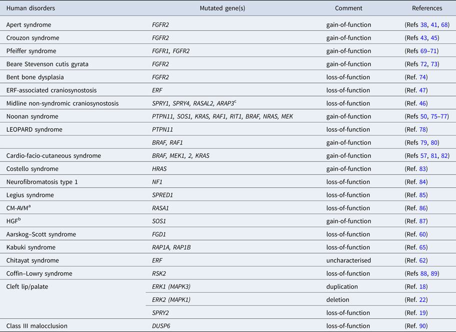
Kabuki syndrome is another disorder affecting craniofacial, mental and heart development and it is usually caused by loss-of-function mutations in KMT2D and KDM6A genes coding for two chromatin regulators, lysine-specific methyltransferase 2D and lysine-specific demethylase 6A, respectively (Refs Reference Ng, Bigham, Buckingham, Hannibal, McMillin, Gildersleeve, Beck, Tabor, Cooper, Mefford, Lee, Turner, Smith, Rieder, Yoshiura, Matsumoto, Ohta, Niikawa, Nickerson, Bamshad and Shendure63, Reference Lederer, Grisart, Digilio, Benoit, Crespin, Ghariani, Maystadt, Dallapiccola and Verellen-Dumoulin64). Recently, two novel alterations were reported to lead to Kabuki syndrome, one in RAP1A and one in RAP1B, both belonging to the RAS family of small GTPases (Ref. Reference Bogershausen, Tsai, Pohl, Kiper, Beleggia, Percin, Keupp, Matchan, Milz, Alanay, Kayserili, Liu, Banka, Kranz, Zenker, Wieczorek, Elcioglu, Prontera, Lyonnet, Meitinger, Stewart, Donnai, Strom, Boduroglu, Yigit, Li, Katsanis and Wollnik65). MEK/ERK signalling is shown to mediate the diseased condition since inhibition of this particular cascade at the one- to four-cell stage embryo restores normal phenotype in zebrafish models, despite however the fact that RAP1 is reported to have contradictory effects on the activation of the pathway in ex vivo and in vivo studies (Refs Reference Bogershausen, Tsai, Pohl, Kiper, Beleggia, Percin, Keupp, Matchan, Milz, Alanay, Kayserili, Liu, Banka, Kranz, Zenker, Wieczorek, Elcioglu, Prontera, Lyonnet, Meitinger, Stewart, Donnai, Strom, Boduroglu, Yigit, Li, Katsanis and Wollnik65–Reference Hu, Kariya, Kotani, Shirouzu, Yokoyama and Kataoka67). All in all, further research is needed in order to clarify the role of the RAS/ERK cascade in these rare syndromes. A list including known human craniofacial disorders that are caused by mutations in the components of the RAS/ERK signalling cascade is provided in Table 1.
Human cranio-oro-dento-facial disorders associated with alterations of the RAS/ERK signalling cascade

a Capillary malformation–arteriovenous malformation.
b Hereditary gingival fibromatosis.
c The causative role of the mutations is still not well defined.
Molecular mechanisms
Although a great number of animal models for craniofacial disorders have been generated and phenotypically well characterised during the last decades, the knowledge of the precise molecular mechanisms downstream of the affected signalling pathways is still limited. In the case of craniosynostosis disorders, both the complexity in the origin of the calvarial cells and the heterogeneity of cellular populations make the discovery of the implicated targets even more difficult. In a study on calvarial pre-osteoblasts, Fgf2 was shown to activate Erk1/2 via Frs2, leading to an increase in the transcriptional activity of Msx2 and Runx2 (Ref. Reference Li, Liu, Hudson, Kim and Hatch91). According to this study, Msx2 and Runx2 bind then to the promoter of the Pc1 (Enpp1) gene coding for the generator of pyrophosphate (PPi) which is further hydrolysed by alkaline phosphatase (TNAP) towards inorganic phosphate. Upon aberrant FGFR-ERK1/2 signalling, increased expression of PC1 is considered to drive the accumulation of PPi levels, leading to the formation of calcium pyrophosphate dehydrate crystals instead of hydroxyapatite crystals and the subsequent calcification of soft tissues (Refs Reference Thouverey, Bechkoff, Pikula and Buchet92, Reference Terkeltaub93). Consistent with this hypothesis, Tnap−/−;Pc1−/− double knock-out mice display normal mineralisation of the calvarial bones (Ref. Reference Hessle, Johnson, Anderson, Narisawa, Sali, Goding, Terkeltaub and Millan94).
In a study on MC3T3 murine calvarial cell line, stimulation of ERK1/2 by FGF2 was shown to increase the expression of activator protein 1 (AP1), which subsequently induces the expression of osteopontin and the onset of osteogenic differentiation (Ref. Reference Kim, Lee, Park, Park, Lee, Kim, Choi, Shin and Ryoo95). Apart from that, in C3H10T1/2 murine mesenchymal cell line and in human primary adipose-derived mesenchymal stem cells (hASCs), TAZ (transcriptional coactivator with PDZ-binding motif) is found to play a central role downstream of ERK1/2 signalling cascade (Refs Reference Byun, Kim, Hwang, Kim, Hwang and Hong96, Reference Quarto, Senarath-Yapa, Renda and Longaker97). More specifically, in hASCs, suppression of TWIST1 is shown to activate both BMP and FGF/ERK pathways which lead to an increase in the osteogenic differentiation of these cells mediated by TAZ upregulation (Ref. Reference Quarto, Senarath-Yapa, Renda and Longaker97). These data suggest that ERK1/2 signalling could potentially mediate some of the effects of TWIST1 loss-of-function mutations in the case of Saethre–Chotzen craniosynostosis syndrome by controlling TAZ activity. Furthermore, in an independent study on calvarial explants isolated from E15 mouse embryos, FGF2 is shown to drive Twist upregulation in suture mesenchymal cells (Ref. Reference Rice, Aberg, Chan, Tang, Kettunen, Pakarinen, Maxson and Thesleff98). In turn Twist suppresses Fgfr signalling and subsequent osteoblast differentiation, indicating that feedback mechanisms and complex integration of molecular pathways such as those of FGF and TWIST can regulate calvarial suture formation and maintenance.
In opposition to the studies showing that RAS/ERK cascade has a positive effect on the osteogenic differentiation of calvarial cells, there are reports indicating that increased ERK1/2 signalling may have an overall inhibitory act upon calvarial osteoblast mineralisation. In primary osteoblasts isolated from calvarial bones, addition of FGF1 is shown to drive SHP2-FRS2-GRB2 complex formation, resulting in the activation of ERK1/2 signalling followed by an increase in the proliferation of cells and a decrease in their mineralisation ability (Ref. Reference Mansukhani, Bellosta, Sahni and Basilico99). Furthermore in the same study, OB1 osteoblastic cell lines modified to express the Crouzon-related C342Y-FGFR2 mutant or the Apert-related S252W-FGFR2 mutant display diminished calcification upon in vitro osteogenic differentiation conditions. In bone marrow mesenchymal cells derived from the Crouzon mouse model Fgfr2C342Y/+, increased early-stage osteogenic differentiation is evident upon in vitro induction conditions, followed however by decreased late-stage mineralisation in comparison to that of wild-type cells (Ref. Reference Liu, Kwon, Nam and Hatch100). Last but not least, in an independent study, administration of a virus carrying a dominant negative form of Ras gene into the skull of newborn mice accelerated calvarial calcification, while administration of a virus carrying a constitutively active form of Mek1 repressed it, indicating an inhibitory effect of Erk pathway on matrix mineralisation (Ref. Reference Kono, Oshima, Hoshi, Bonewald, Oda, Nakamura, Kawaguchi and Tanaka101). However, until now there are no reports regarding downstream molecular effectors that could account for these phenotypes. Unpublished data of our laboratory indicate that cranial suture cells isolated from ErfloxP/− craniosynostotic mice display diminished mineralisation upon in vitro osteogenic conditions, featuring ERF as an effector that could potentially account for the inhibitory act of ERK cascade on matrix calcification. All in all, although a great number of studies have been conducted in animal models and a group of potential downstream targets have already been explored, there is still a long way to go for a full understanding of how RAS/ERK pathway and its effectors act on primitive as well as on differentiating cells during the different steps of craniofacial construction and development. Thus, similar to orofacial disorders, alterations in the spatiotemporal pattern of ERK1/2 activation or even in the strength of signalling appear to affect the overall phenotypic outcome.
RAS/ERK pathway in dental malformations
Dental development
Four types of teeth are commonly observed in mammals: incisors, canines, premolars and molars. Rodents however, lack canines and display an empty space between the incisor and the molar instead, called diastema. In mice, one incisor and three molars are observed in each side of the jaw. Interactions between neural crest-derived mesenchymal cells and epithelial cells take place during tooth development (Refs Reference Thesleff102). Initially, at the site of the future tooth, the oral epithelium becomes thicker and finally buds towards the underlying mesenchymal tissue. This is called ‘the bud stage’. Condensation of mesenchymal cells takes place around the bud, followed by a sequence of morphogenetic processes during the cap and bell stages of tooth development. At the cap stage, a small group of epithelial cells aggregate and form the primary enamel knot which serves as a signalling centre required for the development of tooth cusps (Fig. 1). In mouse embryos the formation of the enamel knot is observed at around E14.5 and a wide range of signalling molecules are found to be expressed such as Bmp2, 4, 7, Shh, Fgf4 and Spry2 (Refs Reference Thesleff and Sharpe103, Reference Klein, Minowada, Peterkova, Kangas, Yu, Lesot, Peterka, Jernvall and Martin104). At the end of the cap stage, apoptosis becomes evident among the cells of the enamel knot and finally at the late bell stage, the knot does not exist anymore. Ameloblasts and odontoblasts are driven into terminal differentiation at this stage too. Ameloblasts are the epithelial cells that produce the enamel of teeth, while odontoblasts originate from neural crest-derived mesenchymal cells and form the dentine matrix of teeth. Neural crest mesenchymal cells differentiate and form the dentin pulp and cementum too. Tooth eruption comes in the end. During dental development, Erk1/2 is shown to be active in the tooth germ on E13.5 in both epithelial and mesenchymal cells (Ref. Reference Cho, Cho, Lee, Lee, Gang and Jung17). Studies on molar teeth isolated from mice reveal the presence of phosphorylated Erk1/2 at the dental epithelium and cervical loop on E15.5 and E16.5 (Ref. Reference Cho, Cai, Kim, Hosoya, Ohshima, Choi and Jung105), while on E18.5 and later on postnatal day 2 (P2), phosphorylated Mek1/2 and Erk1/2 are highly expressed in ameloblasts and odontoblasts in mandibular molars and incisors (Refs Reference Cho, Cai, Kim, Hosoya, Ohshima, Choi and Jung105, Reference Lee, Cai, Kwak, Cho, Harada and Jung106).
Human dental abnormalities and associated animal studies
A variety of dental malformations are often observed in individuals with orofacial and craniofacial disorders associated with the RAS/ERK pathway. In patients displaying cleft lip and/or cleft palate, deformities in crown and root are usually observed along with hypodontia or supernumerary teeth. Enamel hypoplasia is another feature in some cases (Ref. Reference Akram, McKnight, Bellardie, Beale and Evans107). Individuals with RASopathies usually present dental characteristics such as missing teeth, often observed in LEOPARD and neurofibromatosis type 1 syndrome, and malocclusion with anterior open bite and posterior crossbite in Noonan, Costello and CFC syndromes. All these phenotypic traits are already reviewed in detail (Ref. Reference Cao, Alrejaye, Klein, Goodwin and Oberoi108). The mineralisation of enamel is severely affected in Costello syndrome patients (Refs Reference Goodwin, Oberoi, Landan, Charles, Massie, Fairley, Rauen and Klein109, Reference Goodwin, Tidyman, Jheon, Sharir, Zheng, Charles, Fagin, McMahon, Diekwisch, Ganss, Rauen and Klein110). In the mouse model of Costello syndrome, HrasG12V mice, diminished enamel mineralisation is detected along with increased levels of phosphorylated Erk1/2 protein in the incisor (Ref. Reference Goodwin, Tidyman, Jheon, Sharir, Zheng, Charles, Fagin, McMahon, Diekwisch, Ganss, Rauen and Klein110). Ameloblasts exhibit higher growth rate than normal controls and their cell polarity is destroyed. Inhibition of Mek1/2 in 12-week-old mice results in the restoration of the normal phenotype in ameloblasts while inhibition of PI3K is not able to completely reverse the pathologic condition, indicating the role of Ras/Erk signalling in the ameloblast phenotypic outcome in Costello syndrome. On the contrary, CFC patients do not have any defects in enamel mineralisation; however, malocclusion and dental crowding are often observed (Ref. Reference Goodwin, Oberoi, Landan, Charles, Groth, Martinez, Fairley, Weiss, Tidyman, Klein and Rauen111). Among the syndromic craniosynostosis cases, individuals with Apert syndrome display also enamel defects as well as missing or supernumerary teeth. Furthermore, increased growth rate of isolated dental pulp and enamel organ epithelia cells from Apert patients is observed in this case (Ref. Reference Lu, Huguley, Cui, Cabaniss, Waite, Sarver, Mamaeva and MacDougall112). Finally, individuals carrying the missense substitution of serine residue 182 by phenylalanine on DUSP6 exhibit Class III malocclusion (Ref. Reference Nikopensius, Saag, Jagomagi, Annilo, Kals, Kivistik, Milani and Metspalu90), a situation in which incorrect alignment of teeth happens, mainly due to maxilla and/or mandible growth defects.
Another link between dental development and the RAS/ERK signalling pathway is provided by studies on mutant mice that lack expression of some inhibitory molecules of the RAS cascade. Sprouty2−/− mice display supernumerary teeth in the mandible, while Sprouty4−/− mice share also the same defect, yet this abnormal phenotype is observed in both the upper and the lower jaw (Ref. Reference Marangoni, Charles, Tafforeau, Laugel-Haushalter, Joo, Bloch-Zupan, Klein and Viriot113). Finally in the same study mice that lack Rsk2, an ERK1/2 substrate, mutations of which are associated with Coffin–Lowry syndrome in humans exhibit supernumerary teeth either in the upper or in the lower jaw with a prevalence of 14%.
Molecular mechanisms
Studies on ex vivo cultures of ameloblasts, odontoblasts and stem cells from either dental pulp or periodontal ligament have improved the understanding of mechanisms by which the RAS/ERK signalling cascade might act on the differentiation and growth of these particular dental cell types. In ameloblasts isolated from rat mandible incisors, Erk1/2 activation results in significant increase in Amelogenin mRNA expression (Ref. Reference Abe, Miyoshi, Muto, Ruspita, Horiguchi, Nagata and Noma114). Amelogenin is an extracellular matrix protein involved in enamel mineralisation. Consequently, according to that particular study, Erk signalling is considered to have a positive effect on ameloblast differentiation. On the contrary, however, ameloblasts isolated from Costello syndrome mouse models (CS mice) display significantly increased levels of phosphorylated Erk1/2 along with diminished enamel mineralisation (Ref. Reference Goodwin, Tidyman, Jheon, Sharir, Zheng, Charles, Fagin, McMahon, Diekwisch, Ganss, Rauen and Klein110), indicating that further studies are needed in order to decipher the role of RAS/ERK pathway in the differentiation of this specific cell type and the formation of enamel.
In primary cultures of murine pulp cells, exposure to Ffg2 is shown to have a dual effect on the differentiation towards odontoblasts, with early and short exposure to enhance while long-term exposure to inhibit odontoblastic differentiation (Ref. Reference Sagomonyants and Mina115). In any case, the effect of Fgf2 is shown to be mediated by Erk1/2 signalling since the addition of U0126, inhibitor of Mek1/2, is able to restore the normal phenotype (Ref. Reference Sagomonyants, Kalajzic, Maye and Mina116). Further study on the stimulatory mechanism in the case of the short-term exposure reveals that Erk1/2 signalling along with Bmp signalling induces the expression of dentin matrix acidic phosphoprotein 1 (Dmp1) and dentin sialophosphoprotein (Dspp) – both required for proper dentin mineralisation – thus enhancing odontoblastic differentiation (Ref. Reference Sagomonyants, Kalajzic, Maye and Mina117). Dentin phosphoprotein (Dpp) is the proteolytic product of Dspp. In dental pulp mesenchymal stem cells, Dpp is shown to interact with integrins and drive focal adhesion kinase phosphorylation which subsequently activates Erk1/2 kinases (Ref. Reference Eapen, Ramachandran and George118). This event leads to the activation of Elk1, an Ets-domain transcription factor, and the induction of genes facilitating odontogenic differentiation such as Dmp1. The stimulatory effect of Erk1/2 signalling on the differentiation of dental pulp stem cells towards odontoblasts is confirmed by further studies on human cultures upon binding of extracellular calcium and strodium to the calcium-sensing receptor in stem cells (Ref. Reference Mizumachi, Yoshida, Tomokiyo, Hasegawa, Hamano, Yuda, Sugii, Serita, Mitarai, Koori, Wada and Maeda119).
Another study on human adult dental pulp mesenchymal stem cells reveals that upon EphrinB binding to the EphB receptor, Erk1/2 signalling is activated leading to a block in the migration of dental pulp stem cells in ex vivo cultures (Ref. Reference Stokowski, Shi, Sun, Bartold, Koblar and Gronthos120). The reduction in migratory ability is rescued after treatment with specific Mek1/2 inhibitors indicating the importance of Erk in adult pulp stem cell mobilisation.
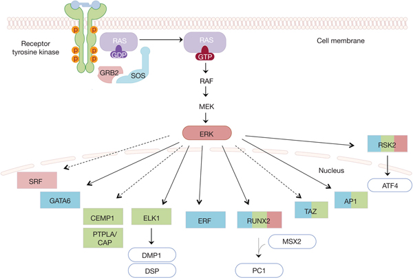
Finally, in ex vivo cultures of periodontal ligament stem cells isolated from humans and dogs, elevated extracellular calcium levels are observed to drive phosphorylation of Erk1/2 kinases and the subsequent increase in the expression of cementum-associated proteins, such as cementum protein 1 (CEMP1) and protein tyrosine phosphatase-like, member A/cementum attachment protein (PTPLA/CAP) (Ref. Reference Paula-Silva, Ghosh, Arzate, Kapila, da Silva and Kapila121). As a result, the periodontal ligament stem cells are induced to acquire a cementoblastic phenotype. All in all, during the last decades the isolation and the successful cultivation of dental stem cells from a variety of sources, such as the dental pulp, mandibular bone and the periodontal ligament in studies as those described above, have provided a deeper understanding of the mechanisms underlying dental development and disease. Figure 2 collectively depicts the downstream molecular players of the ERK cascade that regulate dental, craniofacial and orofacial formation analysed in this review.
Molecular targets of the RAS/ERK cascade in craniofacial and orodental development. A collective illustration of the effectors of the ERK kinase found to play crucial roles during craniofacial, orofacial and dental development is given by colour-coded boxes (blue for craniofacial, red for orofacial and green for dental development). RUNX2 and RSK2 are implicated in all three aspects of head development. In addition to their role in craniofacial development, TAZ and AP1 are found to have an effect in tooth formation (Refs Reference Wang and Martin122, Reference Matsushita, Motani, Sakuta, Yamaguchi, Koga, Matsuo, Nagaoka, Abeyama, Maruyama and Torii123). FGF1, FGF2 and EphrinB are among the ligands that activate the RTKs, as described in the text. Dashed arrows indicate targets that are activated by ERK, yet no direct phosphorylation is reported so far. In the case of SRF, ERK phosphorylates ternary complex factors (TCFs) which in turn activate SRF (Refs Reference Vasudevan and Soriano24, Reference Esnault, Gualdrini, Horswell, Kelly, Stewart, East, Matthews and Treisman124, Reference Buchwalter, Gross and Wasylyk125). Common arrows are used in cases in which the corresponding effectors are known substrates of ERK1/2.

Is it a matter of neural crest cells?
It is known that the majority of facial structures and a considerable number of cranial bones and sutures are derived from mesenchymal stem cells of neural crest origin. Descendants of neural crest cells contribute to both craniofacial and orofacial development as well as tooth formation. But would it be wise to conclude a neural crest cell-autonomous effect of ERK1/2 signalling in the development of head structures? On the one hand, there is strong evidence that the Erk1/2 pathway regulates neural crest cell migration and differentiation, with the majority of the respective publications already described in this review including studies performed on crest-specific Wnt1-Cre mice that recapitulate a number of the human phenotypes (Refs Reference Parada, Han, Grimaldi, Sarrion, Park, Pelikan, Sanchez-Lara and Chai16, Reference Newbern, Zhong, Wickramasinghe, Li, Wu, Samuels, Cherosky, Karlo, O'Loughlin, Wikenheiser, Gargesha, Doughman, Charron, Ginty, Watanabe, Saitta, Snider and Landreth22). A set of studies report the role of this cascade in neural crest induction and proliferation too, being however less clear about a direct or indirect mode of Erk function on this particular cell type, as recently reviewed (Ref. Reference Dinsmore and Soriano126). On the other hand, however, during development, neural crest cells interact extensively with neighbouring paraxial mesoderm cells and these interactions have been shown to guide the movements of neural crest cells (Refs Reference Vitelli, Morishima, Taddei, Lindsay and Baldini127–Reference Noden and Trainor129), resulting finally in the creation of well-defined head structures originating from the one of the two sources either the neural crest- or the mesoderm-derived progenitors. Furthermore, the branchial arch ectoderm, the endoderm and the paraxial mesoderm as well have been shown to play crucial roles in the determination of neural crest cell fate, providing signalling molecules that control the patterning of craniofacial structures. During mandibular formation, the oral ectoderm is the source of FGF and BMP signals that in turn regulate the expression of a variety of transcription factors in neural crest mesenchymal cells thus determining the polarity along the proximal–distal axis (Refs Reference Trumpp, Depew, Rubenstein, Bishop and Martin130–Reference Liu, Selever, Murali, Sun, Brugger, Ma, Schwartz, Maxson, Furuta and Martin132). Regarding tooth formation, the proximal domain of the mandibular ectoderm that produces FGF8 specifies the neural crest mesenchyme to form molars, while the distal domain which produces BMP4 instructs the neural crest mesenchyme to form incisors (Refs Reference Tucker, Matthews and Sharpe133–Reference Chai and Maxson135). Consequently, signals arising from paraxial mesodermal populations, the endoderm and ectoderm could guide and potentially affect the location, the final size and the fate of the neural crest populations. Therefore, we should not exclude the possibility that in some craniofacial disorders and even in some cases of neurocristopathies, particular malformations could spring from a primary defect in mesoderm-derived mesenchymal populations or even in the endodermal and ectodermal layers. The study of particular pathways in certain cell types in combination with cell tracing techniques would possibly provide answers to these questions.
Concluding remarks
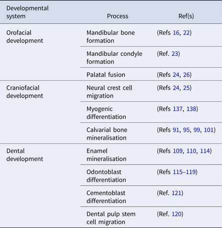
RAS/ERK signalling has a major role in craniofacial and orodental development as evidenced by the plethora of human disorders triggered by mutations in the components of this particular pathway. Animal models and ex vivo studies have already revealed structures, cell types and stages of Erk1/2 action during development (summarised in Table 2); however, the precise molecular and cellular mechanisms are still under investigation. A number of features increase the difficulty in understanding the mechanisms underlying the pathophysiology of these disorders. First, there are cases in which a single mutation in one component of the pathway results in different phenotypic outcomes and consequently, in different syndromes in the affected individuals. For example, the E433K substitution in SOS1 that was initially discovered to lead to Noonan syndrome is also reported in patients with CFC syndrome and reversely, the K499E substitution in BRAF, although previously being reported only in CFC cases, is currently also found in patients with Noonan (Ref. Reference Nystrom, Ekvall, Berglund, Bjorkqvist, Braathen, Duchen, Enell, Holmberg, Holmlund, Olsson-Engman, Anneren and Bondeson136).
Overview of head developmental processes regulated by RAS/ERK pathway activity

Furthermore, different mutations in a single component often cause syndromes with features that are distinct, yet overlap in some cases. Noonan syndrome is caused by gain-of-function mutations in PTPN11 (Ref. Reference Tartaglia, Mehler, Goldberg, Zampino, Brunner, Kremer, van der Burgt, Crosby, Ion, Jeffery, Kalidas, Patton, Kucherlapati and Gelb75), while loss-of-function mutations in the same gene lead to LEOPARD syndrome (Ref. Reference Digilio, Conti, Sarkozy, Mingarelli, Dottorini, Marino, Pizzuti and Dallapiccola78) which shares common craniofacial features with Noonan. Heterozygous inactivating mutations in ERF cause complex craniosynostosis in humans along with facial malformations (Ref. Reference Twigg, Vorgia, McGowan, Peraki, Fenwick, Sharma, Allegra, Zaragkoulias, Sadighi Akha, Knight, Lord, Lester, Izatt, Lampe, Mohammed, Stewart, Verloes, Wilson, Healy, Sharpe, Hammond, Hughes, Taylor, Johnson, Wall, Mavrothalassitis and Wilkie47), while novel still uncharacterised mutations in the same gene cause craniofacial abnormalities without any evidence for craniosynostosis in the case of the newly discovered Chitayat syndrome (Ref. Reference Balasubramanian, Lord, Levesque, Guturu, Thuriot, Sillon, Wenger, Sureka, Lester, Johnson, Bowen, Calhoun, Viskochil, Bejerano, Bernstein and Chitayat62).
The administration of MEK/ERK inhibitors is efficiently used for the restoration of the normal phenotype in some animal models of RASopathies and craniosynostosis disorders, indicating the importance of rebalancing RAS/ERK signalling for normal development. A variety of compounds that inhibit different components of the RAS/ERK cascade have reached clinical trials for the treatment of specific types of cancer, such as PD184352 (MEK1/2 inhibitor) and Ulixertinib (ERK1/2 inhibitor), while others have already gained approval, as is the case of Trametinib, inhibitor of MEK1/2, for BRAF-mutant melanoma (Ref. Reference Garcia-Gomez, Bustelo and Crespo139). The experience in drug discovery obtained through years of research on this molecular pathway holds promise for a potential therapeutic application in craniofacial disorders (Ref. Reference Wilkie140). However, since similar phenotypes are sometimes derived from either activating or overall suppressive mutations for this cascade, as observed for instance in some cases of orofacial clefts and RASopathies, further research is needed in order to gain an insight into the precise structures, developmental stages and molecular targets that are specifically involved each time. Variations in signal initiation and intensity can also lead to altered transcriptional outputs (Ref. Reference Patel and Shvartsman141). Additionally, cross-talks between ERK and other signalling pathways such as TGF-β/BMP (Refs Reference Guo and Wang142, Reference Rahman, Akhtar, Jamil, Banik and Asaduzzaman143) and WNT pathway (Ref. Reference Cervenka, Wolf, Masek, Krejci, Wilcox, Kozubik, Schulte, Gutkind and Bryja144) taking place during head formation increase the complexity surrounding the developmental outcome of particular modifications. Consequently, current studies aim to shed light on the accurate networks of players that regulate these processes downstream of the RAS/ERK cascade.
The greatest difficulty in understanding these mechanisms springs from the fact that calvarial and brain development, facial development as well as oral, jaw and dental formation are tightly connected and affected by each other. This could be the underlying reason for the opposing results observed for many of the involved factors and Erk activity per se. It is important to establish the exact cell that is affected by the specific genetic changes and the cell-autonomous or intercellular mechanisms causing the defects. Dental, oral and craniofacial manifestations often co-occur and attention should be also paid to the precise phenotypic characterisation of disease animal models. Although they recapitulate many disease features, in some cases they either display additional traits or miss some others, while particular phenotypes depend on the genetic background (Ref. Reference Hernandez-Porras and Guerra9). The need to separate primary from secondary effects of each mutation and study the specific phenotypes in isolated cell- or organ-culture systems is inevitable. In this direction, the isolation of specific populations of mesenchymal stem cells that give rise to craniofacial and dental structures is currently being explored with promising results (Refs Reference Mao and Prockop145–Reference Maruyama, Jeong, Sheu and Hsu148). Organoid cultures have already been established for modelling microcephaly, Crown's and Alagille syndrome in addition to cancer and infectious diseases (Ref. Reference Dutta, Heo and Clevers149) and could potentially serve as a useful tool for studying tooth development as well (Ref. Reference Athanassiou-Papaefthymiou, Papagerakis and Papagerakis150). On the whole, stem cell-based technologies have a lot to offer in unravelling the precise molecular background of craniofacial and dental defects. Without this information, the extensive pipeline of RTK/RAS/ERK pathway-targeting drugs for cancer and other pathologies will be very difficult to be extrapolated to these genetic, early-onset developmental disorders.
Author ORCIDs
George Mavrothalassitis, 0000-0001-6214-3179
Acknowledgments
We apologise to researchers whose work was not cited because of space limitations or was cited indirectly through other publications. We would like to thank professor Andrew OM Wilkie for helpful comments and suggestions on the manuscript.
Financial support
This work was supported by an IKY-SIEMENS #AP2788 and a GSRT #KA4937 grant to GM and an Onassis Foundation Fellowship #GZM 008-1 to AV.
Conflict of interest
None.