Introduction
Pre-eclampsia (PE) is a complication of hypertension in pregnancy after the 20th week of gestation (Cheng et al., Reference Cheng, Jiang, Chen, Li, Ao and Zhang2019). It is characterized by high blood pressure (≥140/90 mmHg) for non-PE women before pregnancy and proteinuria (urine collection: ≥300 mg/24 h or random urine protein: positive), and divided into early-onset and late-onset PE before and after the 34th week of gestation according the occurrence time of symptoms of PE (Sakowicz et al., Reference Sakowicz, Bralewska, Pietrucha, Habrowska-Gorczynska, Piastowska-Ciesielska, Gach, Rybak-Krzyszkowska, Witas, Huras, Grzesiak and Biesiada2020). The growth and differentiation of the placenta are directly related to maternal physiological adaptations to ensure proper growth and development of fetuses (Huppertz, Reference Huppertz2018). Trophoblast cells are pivotal components of placental tissues for placental development and angiogenesis (Rana et al., Reference Rana, Lemoine, Granger and Karumanchi2019). The insufficient maternal immunological response to the allogenic fetus and abnormal trophoblast invasion into the spiral arterioles are mainly attributed to maternal hypertension and PE pathogenesis (Sakowicz et al., Reference Sakowicz, Pietrucha, Rybak-Krzyszkowska, Huras, Gach, Sakowicz, Banaszczyk, Grzesiak and Biesiada2017). Therefore, investigating the biological episodes of trophoblast is imperative for the understanding of PE pathogenesis and development of treatment methods.
Long non-coding RNAs (lncRNAs), types of molecules of over 200 nucleotides in length, mainly regulate cell growth, differentiation and apoptosis, as well as affect the pathogenesis of many diseases (Safa et al., Reference Safa, Gholipour, Dinger, Taheri and Ghafouri-Fard2020). Tissue differentiation-induced non-coding RNA (TINCR) is located on human chromosome 19p13.3 (Kretz et al., Reference Kretz, Siprashvili, Chu, Webster, Zehnder, Qu, Lee, Flockhart, Groff, Chow, Johnston, Kim, Spitale, Flynn, Zheng, Aiyer, Raj, Rinn, Chang and Khavari2013). TINCR expression is related to shorter gestation lengths at delivery (Burris et al., Reference Burris, Just, Haviland, Neo, Baccarelli, Dereix, Brennan, Rodosthenous, Ralston, Hecht and Hacker2018). lncRNA TINCR is reported to contribute to a variety of cellular processes, including apoptosis, proliferation, migration, metastasis, invasion, and autophagy, which promote the migration of rat mesenchymal stem cells (Zheng J et al., Reference Zheng, Huang, Li, Lai, Chen, Yi, Pang, Lan, Xu and Fang2020). Cell cycle assay is important for understanding the cellular changes associated with the progress of epithelial–mesenchymal transition (EMT) (Jordan et al., Reference Jordan, Johnson and Abell2011, Ma et al., Reference Ma, Long, Wang, Lou, Yuan, Duan, Yang, Li, Qian, Zeng, Lin, Shen and Lin2021). lncRNA TINCR acts as ceRNA to regulate the migration of mesenchymal stem cells through miR-761 (Zheng J et al., Reference Zheng, Huang, Li, Lai, Chen, Yi, Pang, Lan, Xu and Fang2020). Overexpression of TINCR and cyclin D1 causes an increased rate of cell proliferation (Hou et al., Reference Hou, Zhang, Zheng, Fan, Liu and Zhou2019). Trophoblasts have been recognized to share histological and behavioural characteristics with cancer cells (Xie et al., Reference Xie, Zhu, Liu, Li, Song, Wang, Zhou, Jia and Li2019), therefore it is crucial to explore the effect of lncRNA TINCR on the trophoblast of placenta tissues for PE patients.
lncRNA TINCR affects ischaemia-reperfusion injury by regulating key molecules of related pathways (Wang M et al., Reference Wang, Liu, Wang, Tang and Chen2020). The Wnt/β-catenin pathway has been identified as the target of lncRNA TINCR in many cancers (Ghafouri-Fard et al., Reference Ghafouri-Fard, Dashti, Taheri and Omrani2020). The Wnt/β-catenin pathway, a canonical Wnt pathway, regulates several biological processes, such as proliferation, migration, invasion and apoptosis (Zhang Z et al., Reference Zhang, Wang, Zhang, Shi, Wang and Yan2017). There are many similarities between the invasion of trophoblast and tumour invasion. The Wnt pathway is involved in the proliferation, differentiation and invasion of trophoblasts (Li N et al., Reference Li, Huang, Li, Chen, Yang, Hou and Qiao2020). It may have a crucial role in PE pathogenesis and act as a prospective therapeutic target for the prevention and treatment of PE (Zhang Z et al., Reference Zhang, Wang, Zhang, Shi, Wang and Yan2017). Therefore, we speculated that TINCR might also regulate the biological function of trophoblasts through the Wnt/β-catenin pathway. However, little information is known about the role of TINCR in the placenta tissues of PE through activation of the Wnt/β-catenin pathway. This study investigated how lncRNA TINCR activates the Wnt/β-catenin pathway and affects the biological episodes of Htr-8/Svneo cells, which may provide a reference for the management of PE.
Materials and Methods
Clinical samples
From January 2017 to September 2019, 77 patients with PE admitted to Puren Hospital, affiliated to Wuhan University of Science and Technology, and who met the inclusion criteria were selected as research subjects. Among them, there were 34 patients with severe PE (SPE group) and 33 patients with mild PE (MPE group). The diagnostic inclusion criteria were based on the Guidelines for the Diagnosis and Treatment of Hypertension in Pregnancy (Garovic, Reference Garovic2000). In the MPE group, the following criteria needed to be met at the same time: (1) blood pressure ≥ 140/90 mmHg (1 mmHg = 0.133 kPa); (2) 0.3 g ≤ 24 h urine protein < 2.0 g; (3) random urine protein was positive. In the SPE group, one or more of the following criteria were met: (1) blood pressure: systolic blood pressure ≥ 160 mmHg or diastolic blood pressure ≥ 110 mmHg; (2) 24 h urine protein ≥ 2.0 g; (3) persistent headache, visual disturbance or other cranial nerve symptoms; (4) persistent pain of upper abdomen, hepatic subcapsular hematoma or liver rupture; (5) abnormal liver enzymes: elevated levels of alanine aminotransferase (ALT) or aspartate aminotransferase (AST); (6) abnormal renal function: oliguria (24 h urine output < 400 ml or hourly urine output < 17 ml) or blood creatinine >106 μmol/l; (7) hypoproteinaemia with ascites or pleural effusion; (8) abnormal blood system: continuously decreased platelet, less than 100 × 109/l; intravascular haemolysis, anaemia and jaundice or elevated levels of lactate dehydrogenase (LDH); (9) heart failure, pulmonary oedema; (10) fetal growth restriction or oligohydramnios; and (11) onset before 34 weeks of pregnancy. In addition, 33 pregnant women with normal third trimester during the same period were selected as the control group. All cases were singleton pregnancy without other maternal complications, such as multiple pregnancy, fetal abnormalities, and premature rupture of membranes, maternal infection, or previous history of hypertension, heart disease, kidney disease, and diabetes. Sterile placental tissues were selected (2–3 cm away from the umbilical cord attachment on the maternal surface of placenta to avoid organized foci, calcification foci, bleeding foci, etc.) from the MPE group, SPE group, and non-pre-eclamptic control group after caesarean delivery, and the sterile saline was used to wash them repeatedly. All patients and healthy volunteers signed a written informed consent.
Cell culture and transfection
Htr-8/Svneo cells were purchased from the Shanghai Institute of Biochemistry and Cell Biology, Chinese Academy of Sciences. The cells were seeded in 25 cm2 culture flasks with RPMI-1640 medium (Gibco, Carlsbad, CA, USA) containing 10% fetal bovine serum, 100 U/ml streptomycin and 100 U/ml penicillin. Cells were passaged when c. 75% of the bottom of the flask was filled. The cells were detached with 0.25% trypsin and their morphologies were observed using an inverted microscope (Olympus Optical Co., Ltd, Tokyo, Japan). When the cells became rounded and the intercellular space increased, an equal volume of RPMI-1640 medium was added to inactivate the trypsin and disperse the cells, therefore making the cells fall off the bottle wall. Then, the suspended cells were pipetted using a graduated pipette and subcultured at a ratio of 1:3. After subculture, the medium of cells was replaced every 2 d.
pcDNA-TINCR, pcDNA-negative control (NC), small interfering RNA (siRNA)-TINCR, and siRNA-NC were purchased from Sangon Biotech Co., Ltd (Shanghai, China). After cell transfection using Lipofectamine 2000 (Invitrogen, Carlsbad, CA, USA), the cells in each group were cultured in an incubator for subsequent experiments. Cells were transfected with si-TINCR for 24 h, then cells were treated with Wnt inhibitor FH535 (HY-15721, 20 μM, MCE, Monmouth Junction, NJ, USA). The cells in the control group were treated with the same amount of DSMO.
Reverse transcription-quantitative polymerase chain reaction (RT-qPCR)
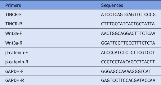
RNA was extracted using the TRIzol method (Ambion, Austin, TX, USA), and cDNA was obtained by reverse transcription using M-MLV and random primer methods. Nucleic acid ethidium bromide staining was used to assess the integrity of RNA before agarose gel electrophoresis. A reverse transcriptase M-MLV kit (Shanghai LMAI Bio Co., Ltd, Shanghai, China) was used to prepare complementary DNA and the Ultra SYBR Mixture (CoWin Biosciences Co., Ltd, Beijing, China) was adopted to perform amplification on a quantitative PCR instrument (ABI 7500, Applied Biosystems, Foster City, CA, USA). The relative expression of genes was calculated using the 2−ΔΔCt method (Livak and Schmittgen, Reference Livak and Schmittgen2001) with GAPDH as an internal reference. The primer sequences for related genes are shown in Table 1.
Primer sequences for RT-qPCR

Western blot analysis
Protein in Htr-8/Svneo cells was extracted and protein concentration was determined according to the Pierce™ bicinchoninic acid protein detection kit (Thermo Fisher, San Jose, CA, USA). The extracted protein was added to the loading buffer, then heated to 95°C for 10 min, and then separated on a 10% polyacrylamide gel. The voltage for gel electrophoresis was controlled at 80–120 V, and the voltage of wet transport and membrane transfer was controlled at 100 mV, lasting 45–70 min. Protein was transferred to membranes using polyvinylidene fluoride, sealed in 5% bovine serum albumin for 1 h, and cultured with primary antibodies E-cadherin (1:500, ab15148, Abcam, Cambridge, MA, USA), vimentin (1:500, ab137321), N-cadherin (1:5000, ab76011, Abcam), CDK2 (1:1000l, ab32147, Abcam), p21 (1:1000, ab109520, Abcam), cyclin D1 (1:200, ab16663, Abcam), Wnt3a (1:1000, ab219412, Abcam), β-catenin (1:5000, ab32572, Abcam), and β-actin (1:5000, ab8227, Abcam) overnight at 4°C. Protein was washed three times using membrane washing buffer (5 min each time), and then incubated with secondary antibody (ZSGB Bio Co., Ltd, Beijing, China) for 1 h. Membranes were washed three times, then a chemiluminescence reagent was added to develop the protein. β-actin was taken as the internal reference. A Bio-Rad Gel DolEZ imager was used to image the protein and ImageJ software (Media Cybernetics, Silver Spring, Maryland, USA) was adopted to analyze the grey intensity of the target band.
3-(4,5-Dimethylthiazol-2-yl)-2,5-diphenyltetrazolium bromide (MTT) method
Each group set had six replicate wells, and each well was seeded in 96-well plates at 5 × 10⁴ cells. Each group was incubated at 37°C in 5% CO2 in air. At 0, 24, 48, and 72 h, 20 μl of 5 mg/ml MTT solution (Sigma-Aldrich, Merck KGaA, Darmstadt, Germany) was added to each well for a further 4 h incubation. Next, the culture supernatant in the well was carefully aspirated. Each well was supplemented with 200 μl dimethyl sulfoxide, and shaken for 10 min to fully dissolve the crystals. Optical density (OD) at 490 nm wavelength (OD490) for each well was measured using a microplate reader, and the average value of six wells was calculated.
Transwell assays
For the cell invasion experiment, Matrigel was thawed at 4°C overnight to maintain equilibrium. Matrigel (200 μl) was diluted with 200 μl serum-free medium at 4°C, and then 50 μl diluted Matrigel was added to the apical chamber of a plate for Transwell assay and incubated at 4°C for 2–3 h until solidified. Cells were separated and counted, then cells were resuspended in complete medium. The cell suspension (100 μl, cell density of 2 × 10⁵ cells/ml) was placed in the apical chamber and 250 μl medium containing 10% fetal bovine serum was add to the basolateral chamber. Cells were cultured in an incubator at 37°C in 5% CO2 in air for 48 h, then cells in the apical chamber were wiped using a cotton swab. The apical and basolateral chambers were carefully rinsed twice with phosphate-buffered saline (PBS). Cells were fixed in 4% paraformaldehyde and stained for 15 min with crystal violet. Transwell chambers were rinsed with PBS. When dry, cells were observed under an inverted microscope (Olympus).
For cell migration experiments, in the apical Transwell chamber without Matrigel, 100 μl cell suspension (2 × 10⁵ cells/ml) was added according to the grouping. The remaining procedure was the same as described for the above Transwell invasion experiment.
Flow cytometry
Htr-8/Svneo cells were detached, the suspended cells were mixed and centrifuged. The supernatant was discarded and resuspended in PBS. The cell concentration was adjusted to 1 × 10⁶ cell/ml. Cold ethanol (70%) was added to cells of each group, which were fixed for 2 h to overnight at 4°C; the fixative solution was discarded, and PBS was added to the cells, followed by centrifugation. Next, the supernatant was removed, and RNase A was added to the cells placed in a 37°C water bath for 30 min. Then the cells were subjected to propidium iodide (PI) staining and mixed. The cells were tested on a flow cytometer for 30 min at 4°C in the dark.
Statistical analysis
All data were processed using SPSS 21.0 statistical software (IBM Corp. Armonk, NY, USA) and GraphPad Prism v.8.0.1. Measurement data were expressed as mean ± standard deviation. The normality and homogeneity of variance tested whether the data conformed to the normal distribution with uniform variance. T-test was used for the comparison between the two groups, and one-way analysis of variance (ANOVA) was adopted for the comparison among multiple groups. Tukey’s multiple comparisons test was used for the post-hoc test. If the data did not conform to the normal distribution or the homogeneity of variance, a rank sum test would be performed. A P-value < 0.05 indicated that the difference was statistically significant.
Results
LncRNA TINCR was elevated in the placenta of PE patients
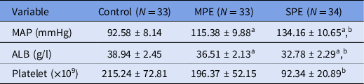
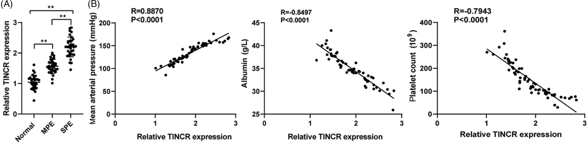
TINCR is abnormally expressed in most malignant tumours and affects the proliferation and invasion of tumour cells (Guo et al., Reference Guo, Zhu, Zhao and Huang2020). The biological behaviour of placental trophoblasts is similar to that of tumour cells. To study the role of TINCR in PE patients, 67 patients with PE were assigned into two groups. Pregnant women in the SPE group were 28–43 years old, with an average age of 35.92 ± 5.59 years old and 34–37 weeks of gestation. The pregnant women in the SPE group were 29–45 years old with an average age of 35.95 ± 5.71 years old, and 35–37 weeks of gestation (the average gestation is 35.83 ± 1.61 weeks). There was no significant difference in age and gestation for the two groups (P < 0.05). Then the clinicopathological characteristics of 33 pregnant women with normal third trimester, 33 patients with MPE and 34 patients with SPE were compared and analyzed. There were statistically significant differences in mean arterial pressure, albumin and platelet levels (Table 2; all P < 0.05). RT-qPCR detected the expression of TINCR in placenta tissues of the three groups of pregnant women. TINCR in the placenta tissue of MPE and SPE groups was higher than that of the control group (all P < 0.01), and TINCR expression in the placenta of SPE group was higher than that of the MPE group (P < 0.01) (Figure 1A). Finally, pregnant women in the MPE and SPE groups were analyzed together. Pearson analysis results showed that the TINCR level in the placenta tissue of PE group was positively correlated with the mean arterial pressure and negatively correlated with albumin and platelet levels (Figure 1B; P < 0.0001).
Comparison of clinical characteristics of pregnant women in three groups

Note: ALB, albumin; MAP, mean arterial pressure; PE, pre-eclampsia.
a Compared with the control group, P < 0.05
b Compared with the MPE group, P < 0.05.
lncRNA TINCR is highly expressed in the placenta tissue of patients with pre-eclampsia. (A) RT-qPCR detects the expression of TINCR in the placenta tissues of the MPE group, SPE group and control group. (B) Pearson analysis of the correlation of TINCR with mean arterial pressure, albumin and platelet levels in patients with pre-eclampsia; data in the figure are all measurement data. One-way ANOVA is used in (A), Tukey’s multiple comparisons test is used for post-hoc test, and Pearson analysis is used for correlation analysis in (B). **P < 0.01.

TINCR downregulation promoted the proliferation and invasion of Htr-8/Svneo cells
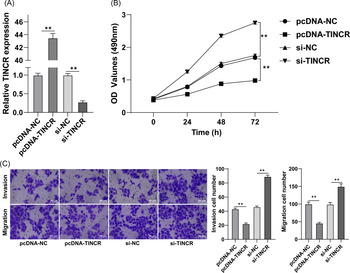
Abnormal proliferation, migration and invasion of placental trophoblast cells are the main pathological changes of PE (Zheng W et al., Reference Zheng, Chen, Yang and Hong2020). To study the specific role of TINCR in Htr-8/Svneo cell episodes, si-NC, si-TINCR, pcDNA-NC, and pcDNA-TINCR were transfected into Htr-8/Svneo cells to silence TINCR expression. RT-qPCR showed that transfection was successful (Figure 2A). Compared with the control group, cell proliferation ability was significantly improved in the si-TINCR group and clearly reduced in the pcDNA-TINCR group (P < 0.05) (Figure 2B). Compared with the control group, the number of migrating cells was significantly increased in the si-TINCR group but clearly decreased in the pcDNA-TINCR group (P < 0.01). Matrigel invasion experiments showed that inhibition of TINCR accelerated the invasion ability of cells (P < 0.01) (Figure 2C). From the above-mentioned results, it can be seen that TINCR downregulation promoted the proliferation, invasion and migration of Htr-8/Svneo cells.
TINCR downregulation promotes the proliferation, migration and invasion of Htr-8/Svneo cells. si-TINCR, pcDNA-TINCR and their controls were transfected into Htr-8/Svneo cells. (A) RT-qPCR detects the expression of TINCR in the cells. (B) MTT method detects the proliferation ability of cells. (C) Transwell assays detects the migration and invasion abilities of cells. In the cell experiment, three independent repeated tests were carried out. Data are expressed as mean ± standard deviation. Data analysis was all performed using one-way ANOVA. Post-hoc test was implemented using Tukey’s multiple comparisons test; *P < 0.05, **P < 0.01.

TINCR downregulation induced the EMT in Htr-8/Svneo cells
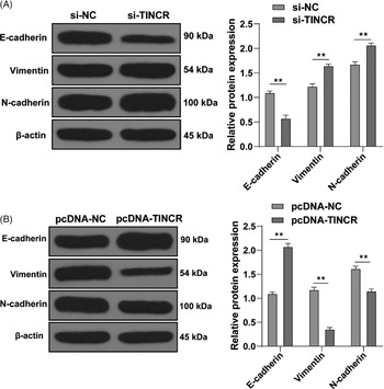
From the above experimental results, we found that silencing TINCR promoted the episodes of Htr-8/Svneo cells. EMT is related to changes in cell migration and invasion (Zhou et al., Reference Zhou, Sun, Chi, Gao and Wang2020), therefore we speculated that TINCR regulated the invasion and migration of Htr-8/Svneo cells by modulating EMT. First, western blot was used to determine the levels of EMT-related proteins. Compared with the control group, the level of epithelial marker epithelial cadherin (E-cadherin) was downregulated after TINCR downregulation, while expression of interstitial markers vimentin and N-cadherin was upregulated (all P < 0.01) (Figure 3A). In addition, the variation in EMT-related proteins in cells overexpressing TINCR was opposite (all P < 0.01) (Figure 3B). The above results proved that TINCR downregulation could promote EMT.
TINCR downregulation promotes the EMT in Htr-8/Svneo cells. si-TINCR, pcDNA-TINCR and their controls were transfected into Htr-8/Svneo cells. (A, B) Western blot detects the protein levels of E-cadherin, vimentin and N-cadherin in cells, and the GAPDH protein was used as an internal control. In the cell experiment, three independent repeated tests were carried out, and the data are expressed as mean ± standard deviation. Data analysis was all performed using one-way ANOVA. Post-hoc test was conducted using Tukey’s multiple comparisons test, **P < 0.01.

TINCR downregulation induced Htr-8/Svneo cells to transit from G0/G1 to S phase
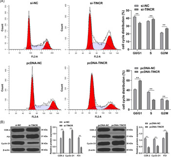
Cell cycle was further measured using flow cytometry to clarify changes in the distribution of the trophoblast cell cycle. Compared with the control group, the number of cells in the pc-TINCR group was increased clearly in the G0/G1 phase, and decreased significantly in S phase. This indicated that cells were blocked in S phase, DNA synthesis was blocked, and cell growth stagnated, so the number of cells decreased (P < 0.01) (Figure 4A). However, after TINCR downregulation, the ratio of HTR-8/SVneo cells was increased in S phase and decreased in G0/G1 (P < 0.01) (Figure 4A), indicating that downregulation of INCR induced Htr-8/Svneo cells to transit from G0/G1 phase to S phase. Western blot analysis showed that protein levels for CDK-2 and cyclin D1 increased after TINCR downregulation, while the expression of p21 decreased significantly. After overexpression of TINCR, protein levels for CDK-2 and cyclin D1 decreased, and p21 increased (P < 0.01) (Figure 4B).
TINCR downregulation induces Htr-8/Svneo cells to transit from G0/G1 to S phase. si-TINCR, pcDNA-TINCR and their controls were transfected into Htr-8/Svneo cells. (A) Flow cytometry detects the HTR-8/SVneo cell cycle. (B) Western blot detects the expression levels of cell cycle-related proteins p21, cyclin D1 and CDK-2. In the experiment, three independent repeated tests were carried out, and data are expressed as mean ± standard deviation. One-way ANOVA was used for all data analysis, and the Tukey’s multiple comparisons test was used for post-hoc test. **P < 0.01.

TINCR downregulation activated the Wnt/β-catenin pathway
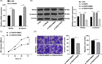
The Wnt/β-catenin pathway is closely related to the proliferation and invasion of trophoblasts (Chen et al., Reference Chen, Wang, Fan, Zhang, Zhoua, Li and Wang2020). We further speculated that TINCR promoted the trophoblasts episodes through the Wnt/β-catenin pathway. First, RT-qPCR and western blot methods were adopted to examine the activation status of the Wnt/β-catenin pathway. Wnt3a/β-catenin levels were elevated significantly after TINCR downregulation (both P < 0.05) (Figure 5A, B). Then, inhibitor FH535 was further used to inhibit the Wnt/β-catenin pathway (Figure 5B) and to observe the biological episodes of cells. Compared with the control group, the proliferation, migration and invasion ability of cells in the si-TINCR + FH535 group was clearly reduced (all P < 0.05) (Figure 5C, D).
TINCR downregulation activates the Wnt/β-catenin signalling pathway and promotes the proliferation, invasion and migration of trophoblast cells. The inhibitor FH535 was used to inhibit the Wnt/β-catenin signalling pathway in Htr-8/Svneo cells. (A) RT-qPCR detects mRNA levels of Wnt and β-catenin. (B) Western blot analysis detects protein levels of Wnt3a and β-catenin. (C) MTT method detects the proliferation ability of cells of each group. (D) Transwell experiments detect the migration and invasion ability of cells. In the experiment, three independent repeated tests were carried out, and the data are expressed as mean ± standard deviation. One-way ANOVA was used for all data analysis, and Tukey’s multiple comparisons test was used for post-hoc test. *P < 0.05; ***P < 0.001.

Discussion
Pre-eclampsia is a devastating complication of pregnant women that seriously threatens the health and safety of mothers and infants (Rana et al., Reference Rana, Burke and Karumanchi2020), but the aetiology of PE has not been fully elucidated, and no effective treatments are currently available (Liu et al., Reference Liu, Guo, Gong and Wang2021). Previous microarray analyses have highlighted some differentially expressed lncRNAs in PE placentas (He et al., Reference He, He, Xi, Zheng, Zeng, Cai, Ouyang, Wang, Zhou, Huang, Deng, Xin, Huang and Liu2013; Long et al., Reference Long, Rui, Song, Dai, Xue, Lu, Shen, Li, Li and Ding2016). This study elucidated the mechanism of TINCR in biological functions of trophoblasts via the Wnt/β-catenin pathway.
lncRNAs have profound impacts on PE development through regulating the biological functions of trophoblasts (Yang and Meng, Reference Yang and Meng2019). TINCR is abnormally expressed in most malignant tumours and affects the biological episodes of tumour cells (Guo et al., Reference Guo, Zhu, Zhao and Huang2020). The biological behaviour of placental trophoblasts is similar to that of tumour cells. In this study, TINCR in the placenta tissue of MPE and SPE patients was higher than that of controls, and TINCR expression in the placenta of SPE patients was higher than that of MPE patients. Overexpressing lncRNA TINCR was associated with high-grade, aggression and recurrence of bladder urothelial carcinoma (Han and Sun, Reference Han and Sun2020). Additionally, significant differences were noticed in mean arterial pressure, albumin and platelet levels between non-PE women and patients with PE. Pearson analysis showed that the TINCR level in placenta tissue of the PE group was positively correlated with mean arterial pressure and negatively correlated with albumin and platelet levels. High circulating TINCR is closely related to worse clinicopathological features and clinical prognosis of breast cancer (Wang M et al., Reference Wang, Liu, Wang, Tang and Chen2020), therefore we identified that TINCR expression was implicated in PE progression.
Trophoblast cells are critical components of placental tissue and participate in placental development and angiogenesis, and the abnormal function of trophoblast cells is closely related to PE pathogenesis (Rana et al., Reference Rana, Lemoine, Granger and Karumanchi2019). The migration and invasion abilities of trophoblast cells are primary in placental formation and maternal–fetal circulation (Ji et al., Reference Ji, Brkic, Liu, Fu, Peng and Wang2013; Zhang Y et al., Reference Zhang, Cao, Jia, Ye, Wang, Zhou and Zhou2019). To study the specific role of TINCR in Htr-8/Svneo cells, si-NC, si-TINCR, pcDNA-NC and pcDNA-TINCR were transfected into Htr-8/Svneo cells. Compared with control cells, cells with silenced TINCR showed evidently improved proliferation, migration and invasion abilities, and cells overexpressing TINCR showed obvious reduced abilities. The decreased proliferation and invasion of trophoblasts is the important pathogenesis of PE (Alasztics et al., Reference Alasztics, Kukor, Panczel and Valent2012; Fu et al., Reference Fu, Ye, Nadeem, Ji, Manchanda, Wang, Zhao, Qiao, Wang, Lye, Yang and Peng2013; Xue et al., Reference Xue, Fan, Diao, Li, Kong, Dai, Peng, Chen, Wang, Hu and Hu2020). TINCR overexpression promotes breast cancer cell proliferation and invasion, and inhibits apoptosis (Guo et al., Reference Guo, Zhu, Zhao and Huang2020). There is little information about the expression and role of TINCR in trophoblasts, which in turn indicated the novelty of our study. In brief, TINCR downregulation promoted the episodes of Htr-8/Svneo cells.
After TINCR downregulation, the ratio of HTR-8/SVneo cells was increased in S phase and decreased in G0/G1. Cell cycle progression is controlled by CDKs and CDK inhibitors, especially proteins p21, cyclin D1 and CDK-2 (Sze et al., Reference Sze, Wong, Liu, Ng, Wong, Cheung, Yow, Chu, Qing, Hu, Tsang, Lee and Yao2011). Western blot showed that the protein levels of CDK-2 and cyclin D1 were increased after TINCR downregulation, while p21 was decreased notably. TINCR downregulation induced Htr-8/Svneo cells to transit from G0/G1 to S phase. In the transitional phase between G1 and S, cyclin D1 and CDK-2 are first expressed in G1 phase (Adon et al., Reference Adon, Zeng, Harrison, Sannem, Kiyokawa, Kaldis and Saavedra2010). Cell cycle is arrested due to reduced cyclin expression and CDK activity (Liu et al., Reference Liu, Zhou, Zhou, Liu, Zhang, Xia, Liu, Chen, Li and Zhu2014). p21 is a well recognized CDK inhibitor, which can block cell cycle G1/S and G2/M transitions under various stimuli (Karimian et al., Reference Karimian, Ahmadi and Yousefi2016). Briefly, we highlighted that silencing TINCR induced the transition from G0/G1 to S phase in HTR-8/SVneo cells. EMT is involved in the differentiation of placental cytotrophoblasts to invasive extravillous trophoblasts in early pregnancy (DaSilva-Arnold et al., Reference DaSilva-Arnold, Zamudio, Al-Khan, Alvarez-Perez, Mannion, Koenig, Luke, Perez, Petroff, Alvarez and Illsley2018). EMT is related to changes in cell migration and invasion (Zhou et al., Reference Zhou, Sun, Chi, Gao and Wang2020). Our results demonstrated that the expression of E-cadherin was downregulated after TINCR silencing, while interstitial markers vimentin and N-cadherin were upregulated. Brown and colleagues reported an increase in E-cadherin in the trophoblasts of PE placenta, suggesting a pivotal role for E-cadherin-mediated promoted proliferation of trophoblasts (Brown et al., Reference Brown, Lacey, Baker and Crocker2005). The above results proved that TINCR downregulation could promote EMT.
The Wnt/β-catenin pathway is closely related to the proliferation and apoptosis of trophoblast cells and the pathogenesis of PE (Zhang Z et al., Reference Zhang, Li, Zhang, Jia and Wang2013, Reference Zhang, Wang, Zhang, Shi, Wang and Yan2017; Chen et al., Reference Chen, Wang, Fan, Zhang, Zhoua, Li and Wang2020). We noticed that Wnt/β-catenin levels were elevated significantly after TINCR downregulation. The inhibitor FH535 was further used to block the Wnt/β-catenin pathway. Compared with the control cells, the biological episodes of cells in the si-TINCR + FH535 group were clearly reduced. The DKK1/Wnt/β-catenin pathway is activated to promote cell proliferation and invasion in PE pathogenesis (Li N et al., Reference Li, Huang, Li, Chen, Yang, Hou and Qiao2020). In summary, TINCR downregulation activated the Wnt/β-catenin pathway to promote trophoblast proliferation, invasion and migration.
This study supported the idea that TINCR regulates the proliferation, migration and invasion of trophoblasts through the Wnt/β-catenin pathway. Research work at this level is relatively scarce, and is expected to provide a new theoretical method for treatment of PE patients. In this study, we examined the biological characteristics of trophoblast using TINCR at the cellular level, but did not look at mechanisms in animal experiments and clinical studies, which need further subsequent verification. The Wnt/β-catenin pathway is a very conservative pathway in biological evolution. In normal somatic cells, β-catenin acts only as a cytoskeletal protein to prevent cell movement. When the Wnt signalling is activated, β-catenin exerts other effects by nuclear importation. Previous studies have shown that there are many pathways involved in regulating the biological function of trophoblasts in the process of placenta formation such as MAPK, Notch and PI3K/Akt (Ou et al., Reference Ou, Zhu, Wei, Bai, Chen, Chen and Zhang2020; Li Z et al., 2021; Qi et al., Reference Qi, Yao, Xing and Qin2021), which also play an important role in the migration, invasion and EMT mechanisms of trophoblasts. The Wnt/β-catenin pathway can regulate cell proliferation, apoptosis, migration and invasion, and plays an important role in PE pathogenesis. Therefore, this study focused on the role of lncRNA TINCR in trophoblasts for the Wnt/β-catenin pathway. However, roles for TINCR in regulating other pathways in trophoblasts have not been explored, and need further expansion.
Data availability
All the data generated or analyzed during this study are included in this published article.
Ethical standards
All patients and healthy volunteers signed a written informed consent.
Competing interests
The authors declare that they have no competing interests.
Funding
Not applicable.