Nutritional deficiencies develop within 48 hours of admission to the ICU in up to 20% of critically ill children. Reference Krafte-Jacobs, Persinger and Carver1 Effective, early delivery of nutritional requirements is an important component in the management of critically ill patients. Reference Young, Chapman and Fraser2 Nutritional intervention can increase resistance to infection, improve wound healing, prevent organ failure, and reduce mortality. Reference Krafte-Jacobs, Persinger and Carver1 Enteral feeding is the optimal mode of nutrient delivery, with advantages over parenteral nutrition. Reference Young, Chapman and Fraser2 In recently published report, up to 30% of neonates post-cardiac surgery were discharged home or transferred to another hospital with a feeding tube. Reference McKean, Kasparian and Batra3 Nasogastric feeding tube placement is the most frequently used method for enteral feeding and drug administration in peri-operative cardiac children. Reference Gok, Kilicaslan and Yosunkaya4 A major limiting factor in the provision of enteral nutrition in critically ill patients is the feed intolerance due to high residual or persistent vomiting. Reference Young, Chapman and Fraser2
Delayed gastric emptying in the presence of preserved small bowel motility may be overcome by post-pyloric tube placement enabling enteral feeding. Reference Young, Chapman and Fraser2 Traditional approaches to positioning of small intestinal feeding tubes are under fluoroscopic, endoscopy assistance, or blind introduction with prokinetic administration or air insufflations. Reference Young, Chapman and Fraser2 Transpyloric feeding tubes placement can also be confirmed with pH assessment of tube aspirate, which may not be easy when dealing with the small tube diameter or flexible tubes that are commonly used in infants. Reference Woolgar5
Ultrasound-guided procedures have increased in the last 30 years in ICU. Reference Gok, Kilicaslan and Yosunkaya4,Reference Hamadah and Kabbani6 The data related to US-PPT (ultrasound-guided post-pyloric feeding tube insertion) placement in paediatric ICU are insufficient. Reference Gok, Kilicaslan and Yosunkaya4 Ultrasound has several advantages such as being real-time bedside examination that does not expose the patient to radiation or mobilisation. Reference Woolgar5,Reference Hamadah and Kabbani6 We aimed to present our experience and practice in (US-PPT) in peri-operative cardiac infants using newly modified technique that facilitate insertion and positioning in duodenum.
Materials and methods
After obtaining approval from the Institutional Research Board of our hospital, we performed a pilot prospective experimental observational study in peri-operative cardiac infants at King AbdulAziz Medical City, Riyadh, Saudi Arabia. Study continued between May 2019 and May 2021.
Inclusion criteria were both of the following:
-
Infants less than 6 months in paediatric cardiac ICU settings post-paediatric cardiac surgery.
-
Indication for transpyloric tube feeding: persistent high gastric residue (>25%/4 hours) or frequent vomiting despite optimised medical therapy through Nasogastric feeding tube within 1 week including minimising narcotics, optimisation of serum electrolytes levels, use of prokinetics and hydrolysed formula.
While exclusion criteria were any of the following:
-
History of gastrointestinal tract obstruction or previous gastrectomy or bowel resection.
-
Evidence of necrotising enterocolitis peri-operatively.
US-PPT procedures were performed by the paediatric cardiac ICU intensivist and confirmed initially by bedside abdominal X-ray and subsequently verified by radiologists through picture archiving and communication system.
Description of US-PPT insertion technique
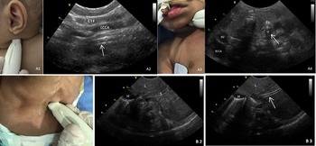
Patients were scanned initially in the supine position during quiet breathing. Studies were performed within at least 4 hours from last feeding trial. Polyurethane non-weighted enteral tube without stylet was used initially in our cases. Stylets were used only if second trial was needed. The tip of the feeding tube was lubricated and flushed with distilled normal saline before insertion. The presumed maximal total length for placing a post-pyloric tube in the small intestine was determined by measuring the distance from the bridge of the nose down to an ankle with the leg fully extended. Reference Woolgar5 We used a high-frequency transducer (8c) curvilinear probe with a frequency of 8–12 MHz. Depth was adjusted to get optimal image size (5–9 cm). US-PPT insertion continued in the following stages (Figs 1 and 2):
Pictures illustrating ultrasound (US) probe position andorientation to visualise the feeding tube within the esophagus in 2infants with feeding intolerance peri-operatively. The feeding tube in esophagus (arrow) is seen in US longitudinal plain innon-ventilated infant (A 1,2) and in short axis plain in ventilatedinfant (A3,4). Es indicates esophagus; ETT: endotracheal tube; RCCA: right common carotid artery; RIJ: right internal jugular; LCCA: left common carotid artery; LIJ: left internal jugular. B1-3: Pictures illustrating US probe position and orientation to visualise the feeding tube within the stomach in infant with feeding intolerance peri-operatively. B1: The US probe is orientated in subxiphoid area toward the right shoulder; B2: US visualisation of the stomach; B3: US visualisation of the feeding tube insertion in the stomach lumen as a double line artifact (arrow). H indicates heart; L: liver; S: stomach.

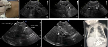
A1-4: Pictures illustrating ultrasound (US) probe positionand orientation to visualise the insertion of feeding tube within the pylorusin infant with feeding intolerance peri-operatively; A1: The US probe is placedin the longitudinal plain toward left shoulder, 2 cm below the costal marginright to subxiphoid area; A2: US visualisation of the pylorus; A3: US visualisationof dynamic fogging in stomach using air-saline mixture (arrow); A4: USvisualisation of the feeding tube passing the pylorus as a double line artefact (arrow). GB indicates gallbladder; P: pylorus; S: stomach. B1-3 Picturesillustrating (US and X-ray) to visualise the post-pyloric feeding tube ininfant with feeding intolerance peri-operatively. B1: feeding tube passingpylorus in trans-liver US window; B2: US visualisation of post-pylorus tubeinsertion in distal second segment of duodenum (arrow); C: Abdominal X-ray withcontrast confirms post-pyloric feeding tube in distal second segment ofduodenum without intestinal perforation. GB indicates gallbladder; L: liver; PN: pancreas; SMV: superior mesenteric vein.

-
A. Visualisation of the feeding tube insertion in the oesophagus lumen.
-
B. Visualisation of the feeding tube insertion in the stomach cavity.
-
C. Visualisation of the feeding tube insertion in the pylorus, agitated saline is infused.
-
D. Visualisation of the feeding tube in the post-pylorus.
The patient was kept initially in supine position during feeding tube insertion in the oesophagus and stomach cavity and then was elevated in right lateral decubitus at 45° during feeding tube insertion in the pylorus and the small intestines. Gastric insufflation with air-saline mixture was used in the third stage (Fig 2, A3), 3–5 ml/kg of agitated saline was instilled in stomach cavity to facilitate feeding tube passage through pylorus, while feeding tube was advanced guided by ultrasound screening. Two operators were needed during procedure. The first operator (intensivist) will handle the ultrasound probe and feeding tube, while the second operator (nurse or doctor) will handle the ultrasound machine, inject the agitated saline, position the baby during procedure, and follow vital signs.
Plain and contrast abdominal X-rays were both done to confirm appropriate feeding tube placement and to rule out complications such as kinking of feeding tube or intestinal perforation (Fig 2, B3).
Results
During study period of 2 years between May 2019- May 2021, 500 infants less than 6 months underwent cardiac surgery. Fifteen needed US-PPT insertion with incidence of 3%. The average age and weight were 2.6 ± 0.5 month and 3 ± 0.2 kg, respectively. Feeding tube was inserted on median 15 postoperative day (interquartile range IQR 11–21). The median time of procedure was 15 minutes (interquartile range IQR 12–25, range 10–40). The depth of tip of post-pyloric tube was 36 ± 1.2 cm from nostril. No complications were encountered. We completed procedures successfully in 13 patients from first attempt using non-stylet tube (first pass success rate 87%). A second attempt of insertion was needed in 2 cases using stylet tube (13%). Once insertion was confirmed, we initiated gradual feeding to achieve the targeted feeding. Target daily calorie intake was achieved within average of 3.5 ± 0.4 days. Post-pyloric tube stayed of average of 20 ± 3 days before removal. The median Risk Adjustment for Congenital Heart Surgery Categories in the 15 patients was 4 (46% were Risk Adjustment for Congenital Heart Surgery of 4). Out of 15 infants, 3 patients died, 1 patient needed gastrostomy tube, and 11 patients were discharged home on oral feeds. Continuous feeding was continued till tube removal in all cases. Subjects were monitored until approximately 48 hours following removal of the enteric feeding tube with no adverse events.
Discussion
Feeding difficulties are not uncommon following paediatric cardiac surgery. Reference McKean, Kasparian and Batra3 Early enteral nutrition is believed to optimise gut function. Slow gastric emptying frequently hampers feeding, and thus, post-pyloric tube insertion can provide adequate nutrient delivery. Reference Young, Chapman and Fraser2
The main barrier to post-pyloric feeding has been the technical difficulty in placing a tube beyond the pylorus. Reference Young, Chapman and Fraser2 Many approaches were used for placement of post-pyloric feeding tubes including surgical insertion, fluoroscopic, blind technique, gastric insufflation, and endoscopy. Reference Young, Chapman and Fraser2,Reference Spalding, Sullivan and Soremi7 The optimal method remains controversial. US-PPT technique provides a simple bedside and feasible tool to identify the tube position without need to utilise multiple ionising radiation or transport to the medical imaging department for fluoroscopic positioning. Reference Woolgar5,Reference Hamadah and Kabbani6 Ultrasound allows us to follow the trail of feeding tube by using the landmarks. The data related to US-PPT placement in paediatric ICU are still insufficient and not well described. Reference Gok, Kilicaslan and Yosunkaya4 We presented our experience in illustrated materials. In our technique, we used gastric insufflation with agitated normal saline (3–5 ml/kg of air-saline mixture) as soon as the feeding tube is seen by ultrasound in the stomach near pylorus to obtain good view and to enhance progression of the tube through pylorus. Agitated saline has the advantages of being echogenic, relatively safe, non-allergenic, and easily detectable by ultrasound. Its preparation through three-way stopcock has been described in many procedures related to critical care setup. Reference Blanco and Bello8 Feeding tubes can be weighted or unweighted and can be co-administered with medicines to promote passage through pylorus. Reference Woolgar5 Stylet tubes or weighted tip feeding tubes were recommended by some clinicians to facilitate transpyloric placement, Reference Krafte-Jacobs, Persinger and Carver1 but they may have some rare complications as they increase rigidity and risk of injury or perforation, Reference Vigneau, Baudel and Guidet9,Reference Prabhakaran, Doraiswamy and Nagaraja10 so we elected in our cases to use initially non-weighted tip feeding tubes without stylets with 87% success rate in the first attempt. In the remained 2 patients, we used stylet tubes in the second attempt with 100% success rate. We observed no complications during insertion or thereafter, and the reason for death in 3 patients who were deceased was due to cardiac or respiratory problems not related to tube placement. Our median time needed for US-PPT insertion was 15 minutes which is comparable to other advanced techniques such as The Cathlocator™; Reference Young, Chapman and Fraser2 however, ultrasound is increasingly available worldwide. Our findings compare favourably with other bedside techniques such as the blind technique or gastric insufflation of air. Reference Young, Chapman and Fraser2,Reference Spalding, Sullivan and Soremi7 Although the gastric insufflation with air technique took only two to four minutes to perform, it was less successful (60% success rate). Most techniques for placement of post-pyloric feeding catheters are complex, time-consuming, or both. Reference Young, Chapman and Fraser2 US-PPT is time saver and avoids the risk of mobilising critical ill cardiac infants to another department. Additionally, cost of performing bedside ultrasound without need for transportation or booking in radiology suite favour the use of bedside ultrasound-guided procedure once the intensivists learn and master the skill needed for US-PPT. In our pilot study, we included the infants less than 6 months of age as the incidence of feeding intolerance is reported more in this age group. Reference McKean, Kasparian and Batra3 Nevertheless, the principle of procedure and its application can be extended to older children when indicated.
Our study has several limitations being a pilot study in single-centre experience with small sample size that reflects local experience with the need to verify its results by other institutional experiences. It is also confined to the paediatric cardiac surgery population. Nevertheless, the present study highlights the importance and emerging role of bedside ultrasound for intensivist for management of peri-operative cardiac infants with feeding intolerance. Additional investigation with a larger pool of operators and randomised controlled patient assignment is required.
Conclusion
US-PPT insertion using gastric insufflation with air-saline mixture technique in peri-operative cardiac infants is used safely and successful in our study. It allows placement and location of an enteral feeding tube in real time in critically ill patients with feeding difficulties. These findings warrant further studies to ensure safety and applicability of this newly modified technique for placement of post-pyloric feeding tubes.
Acknowledgements
This manuscript describes novel work and is not under consideration for publication/published by any other journal. All the author(s) have approved the manuscript and this submission.
Financial Support
This research received no specific grant from any funding agency or commercial or not-for-profit sectors.
Conflicts of Interest
None.
Ethical Standards
The manuscript was completed according to good clinical practice and approved by Institutional Research Board.