INTRODUCTION
The family Molidae comprises species of epipelagic fish commonly known as molas or ocean sunfish, which are distributed in warm, tropical waters in all oceans (Matsuura, Reference Matsuura and Carpenter2002). Only three species were described for this family: Ranzania laevis (Pennant, 1776), Mola mola (Linnaeus, 1758) and Masturus lanceolatus Liénard, 1840. A recent genetic and biogeographical study confirmed the existence of a fourth species, Mola ramsayi (Giglioli, 1883), which is restricted to the southern hemisphere (Parenti, Reference Parenti2003; Bass et al., Reference Bass, Dewar, Thys, Streelman and Karl2005). Despite their broad distribution, studies on the biology of these species are rare in Brazil and the majority involve the analysis of individuals stranded on the shore either dead or severely debilitated (Lopes et al., Reference Lopes, Oliveira-Silva, Batista, Esquivel and Olavo2004; Ahid et al., Reference Ahid, Filgueira, Fonseca, Soto-Blanco and Oliveira2009).
Morphologically, molas are considered the heaviest living marine teleosts and have a very tall, laterally flat body. The dorsal and anal fins are used for propulsion and the caudal fin is modified in a structure called a clavus. The mouth is very small and the teeth are united in the form of a beak (Parenti, Reference Parenti2003). Molidae feed mainly on zooplankton, but also jellyfish, algae, crustaceans and fish (Parenti, Reference Parenti2003); juveniles feed on benthic invertebrate (Matsuura, Reference Matsuura and Carpenter2002). These fish are frequently infected with the larvae of worms (Arru et al., Reference Arru, Garippa and Sanna1991) and are well-known for their impressive parasite load. Forty different genera of parasites have been reported in a single species (Volgelinest, Reference Volgelinest2006).
Masturus lanceolatus, commonly known as the sharptail mola, has broad distribution in the western Atlantic, occurring from Nova Scotia to south-eastern Brazil (Klein-MacPhee, Reference Klein-MacPhee, Collette and Klein-MacPhee2002; Menezes, Reference Menezes, Menezes, Buckup, Figueiredo and Moun2003). Records of occurrence in Brazil, however, are rare and the species is only cited for the states of São Paulo, Ceará (Figueiredo & Menezes, Reference Figueiredo and Menezes2000) and Bahia. Only two specimens are deposited in ichthyologic collections in Brazil (Lopes et al., Reference Lopes, Oliveira-Silva, Batista, Esquivel and Olavo2004).
Recently Ahid et al. (Reference Ahid, Filgueira, Fonseca, Soto-Blanco and Oliveira2009) published an article on a specimen of Mola mola stranded in the Brazilian State of Rio Grande do Norte. However, the photograph clearly shows the clavus in the shape of a lance, which characterizes it as M. lanceolatus.
The aims of the present study are to analyse the possible causes of the stranding of a sharptail mola and record the first occurrence of M. lanceolatus for the coast of Pernambuco State, Brazil.
MATERIALS AND METHODS
Characterization of the area
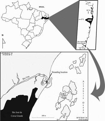
The estuary of the Una River is located at São José da Coroa Grande on the southern coast of the State of Pernambuco, 123 km from Recife (State capital). Its margins are colonized by mangrove forests. Tidal plains form at the mouth of the river, along with a sandbar parallel with the coast. Reefs are abundant throughout the coast and are located approximately 400 m from the mouth of the river. The stranding occurred on the left margin of the delta approximately 120 m from the coastline at the coordinates 8°52′42.18″S 35°08′17.85″W (Figure 1).
Location of stranding of Masturus lanceolatus in the estuary of the Una River, Pernambuco, Brazil.

Description of specimen and external injuries
The examination of the external injuries was initially performed at the stranding site and subsequently through photographs taken on the day of the capture. Investigations were made into the causes of the injuries through a comparative analysis of data published in scientific journals. For the identification of shark bites, specialist Dr Paulo Guilherme Vasconcelos de Oliveira, adjunct professor of the Universidade Federal Rural de Pernambuco, was consulted, along with bibliographic resources.
Preparation of histological slides
After being collected and fixed in 10% formalin, fragments were removed from the oesophagus, stomach, intestine, liver and heart (auricle and ventricle). The samples were submitted to routine histological techniques. Tissues were dehydrated in an increasing concentration of alcohol, diaphanized in xylol, embedded in paraffin, cut on a microtome to micro-sections 5 µm in thickness and stained with haematoxylin–eosin (HE) (Maia, Reference Maia1979) for mounting on slides.
RESULTS AND DISCUSSION
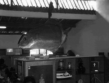
The specimen of Masturus lanceolatus was found by three local fishermen in the estuary of the Una River at about 0900 on 15 September 2006. The fish was being tossed by the waves in the surf zone. It was quite debilitated, but still alive. The fish was unknown to the fishermen and their colleagues, who wanted to either burn or bury it, thinking that it was a ‘bad omen’. Native fishermen called the team from the Tropical Marine Ichthyology Group (IMAT) to help them. Two researchers of IMAT, coordinated by M.E. Araújo, together with other researchers, including the authors, went to the site to: (1) identify the specimen; (2) reassure the population; and (3) conserve the fish on ice until the arrival of the taxidermist (I.R. Joca). The IMAT acquired the specimen and donated it to the Una Museum, where it is currently on display (Figure 2).
Specimen of Masturus lanceolatus on display at Una Museum (São José da Coroa Grande, PE) following taxidermy.

The occurrence was reported by national newspapers and television news due to the fact that this fish is rare, not only to the local population of the small beach, but also to the scientific community.
It is possible that the specimen described here died prematurely, as its age was estimated at 20 years, using a calculation based on the von Bertalanffy curve presented by Liu et al. (Reference Liu, Lee, Joung and Chang2009). According to the authors, M. lanceolatus longevity is estimated at 105 and 82 years for females and males, respectively. While molas have k-strategie characteristics, such as slow growth and late maturation, they also have the greatest fecundity among existing vertebrates, with as many as 300,000,000 eggs (Parenti, Reference Parenti2003).
The specimen described here was male, measured 170 cm and weighed 78 kg. The three other specimens found in Bahia (Lopes et al., Reference Lopes, Oliveira-Silva, Batista, Esquivel and Olavo2004), São Paulo (Figueiredo & Menezes, Reference Figueiredo and Menezes2000) and Rio Grande do Norte (Ahid et al., Reference Ahid, Filgueira, Fonseca, Soto-Blanco and Oliveira2009) measured 112 cm, 170 cm and 93 cm and weighed 50 kg, 119 kg and 24 kg, respectively. Comparing the body mass index (BMI) of these specimens, the fish from Pernambuco was the thinnest (BMI = 27) and the specimen from São Paulo was the fattest (BMI = 41.2).
Along with the low body weight, a set of factors suggest the reasons for the stranding: (1) high parasite load in the liver; (2) net entanglement followed by stress; (3) susceptibility to shark attacks, with mutilation of the fins; (4) locomotion difficulty stemming from mutilated fins; and (5) body striking the reefs that form a barrier before arriving at the site where the specimen was found.
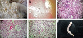
The histopathological study of fragments from the oesophagus, stomach, intestine and heart revealed no noteworthy alterations. The liver had numerous cists from nematodes and cestodes from the order Trypanorhyncha in the hepatic parenchyma, with perforation of the stroma of the organ, allowing a view of the cysts externally (Figure 3A). On average, each cyst contained one parasite, as reported by Rodrigues et al. (Reference Rodrigues, Ranzani Paiva and Santos2002) in fish from the family Characidae. The liver exhibited parenchyma loss due to the presence of worm polycytosis (Figure 3B). Several transversal concentric hyaline formations similar to nematodes were observed (Figure 3E). The cysts were enveloped by a fibrous capsule of conjunctive tissue and some exhibited invasion of the hepatic parenchyma, leukocyte infiltrates (Figure 3C) and dilation of arteries (Figure 3D).
Some results of the histopathological study of the specimen of Masturus lanceolatus, stranding on Una River, Pernambuco: (A) macroscopic view of liver with numerous cysts from parasites; (B) worm polycytosis in hepatic parenchyma (magnification: 10×); (C) leukocyte infiltrate (magnification: 10×); (D) dilation of artery (left side) and encapsulated parasite (right side) (magnification: 10×); (E) transversal concentric hyaline formation similar to encapsulated nematode (magnification: 40×); (F) macroscopic view of cestode from the order Trypanorhyncha.

Encysted nematodes inhabit different tissues in fish, causing cell disorganization and a variable amount of phagocyte cells and lymphocyte infiltrates (Eiras & Rego, Reference Eiras and Rego1989). The erratic migration of nematode larvae in viscera such as the liver is not uncommon (Roberts, Reference Roberts2001). In the intermediate host, the larvae are found either floating freely or encapsulated in the abdominal cavity or adhered to the liver (Moravec, Reference Moravec1998), as found in the liver of the specimen described here. Post mortem studies of the Mola mola revealed multifocal hepatic and renal granulomas and hepatic cysts of cestodes (Volgelnest, 2006). Digenetic hemiurid and plerocercoid parasites from Trypanorhyncha were recorded in the specimen of M. lanceolatus mistakenly identified as M. mola (Ahid et al., Reference Ahid, Filgueira, Fonseca, Soto-Blanco and Oliveira2009). Adults and larvae of species from this order are often found in the gastrointestinal apparatus of elasmobranchs and a number of teleost species (Campbell & Beveridge, Reference Campbell, Beveridge, Khalil, Jones and Bray1994; Knoff et al., Reference Knoff, Clemente, Pinto, Lanfredi and Gomes2004, São Clemente et al., Reference Clemente, Knoff, Padovani, Lima and Gomes2004, Dias et al., Reference Dias, Clemente and Knoff2009), as found in the specimen of M. lanceolatus described here.
Although Campos et al. (Reference Campos, Moraes and Moraes2008) state that extensive eosinophilia is not usual with the inflammatory reaction of host fish to parasites, the intensity of encapsulated parasites in the specimen of M. lanceolatus led to various points of leukocyte infiltrates, thereby characterizing an inflammatory process. This condition may have led to the initial debility of the specimen, making it an easy target for predation or fishing nets.
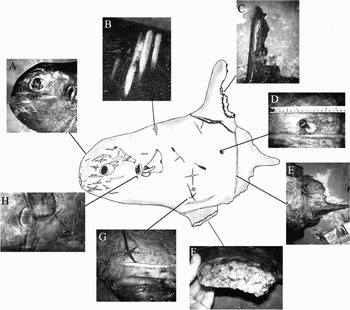
Fishing net marks in the form of an ‘x’ were found on the epidermis of the specimen (Figure 4G), revealing that the fish must have been imprisoned in a fishing net and suffered stress and trauma, which further aggravated its state of health and enabled attacks from predators and parasites. Accidental entanglement in fishing gear is one of the main threats to mola populations, as species from the family are often reported as by-catch in lone-line fisheries (Joung et al., Reference Joung, Liu, Liao and Hsu2005) and drift nets (Akiol et al., Reference Akiol, Erdem, Unal and Ceyham2005).
External injuries on specimen of Masturus lanceolatus captured live in the estuary of Una River (São José da Coroa Grande, PE). (A, B) Scratches; (E & H) rashes; (G) x-shape mark and linear cut with knife; (C, D & F) shark bites.

Both recent and old shark bites of different shapes and origins were found during the external analysis of the specimen. One bite mark was typical of some species of cigar shark from the genus Isistius. The specimen had a circular orifice that was apparently recent, as the skin that covered the area was still attached to the upper margin of the injury (Figure 4D). The concentration of bites on the sides of the body was likely due to the fact that these areas are the largest and most easily accessed in this type of attack (Souto et al., Reference Souto, Oliveira, Nunes, Maia-Nogueira and Sampaio2007). These sharks may be considered temporary ectoparasites of oceanic fish, pinnipids and cetaceans (Perrin & Hohn, Reference Perrin, Hohn, Ridgway and Harrison1994; Miyazaki & Perrin, Reference Miyazaki, Perrin, Ridgway and Harrison1995; Heithaus, Reference Heithaus2001; Silva Jr et al., Reference Silva, Silva, Sazima and Sazima2007).
Other marks from sharks were much more violent: two on the dorsal fin, with the basal bite more recent thare the upper (Figure 4C); and another mutilating bite on the anal fin (Figure 4F). In a period of 14 years, there have been 53 shark attacks on humans along the coast of Pernambuco by the bull shark (Carcharhinus leucas) and tiger shark (Galeocerdo cuvier) that attacked the legs and buttocks of swimmers (Hazin et al., Reference Hazin, Burgess and Carvalho2008). It is likely that the attack on the mola was from these species, as they are aggressive and territorial and are found throughout the entire coast of Pernambuco.
With its dorsal and anal fins mutilated by shark attacks, the mola had swimming difficulties that caused profound rashes in the areas of the insertion of the caudal and pectoral fins (Figure 4E & H). Under these morpho-physiological conditions, the specimen must have remained adrift and, upon approaching the coast, suffered a number of scratches (Figure 4A, B ) from the reefs near the estuary.
Although it can live at depths of 670 m (Lopes et al., Reference Lopes, Oliveira-Silva, Batista, Esquivel and Olavo2004), M. lanceolatus is a pelagic species that spends most of its time in depths of less than 200 m and prefers waters with temperatures greater than 20°C (Seitz et al., Reference Seitz, Weng, Boustany and Block2002). It is in this marine zone where reefs are concentrated, over which molas pass when needing to clean off ectoparasites, which is performed by fish from the families Chaetodontidae and Pomacanthidae (Konow et al., Reference Konow, Fitzpatrick and Barnett2006). Although the coast of Pernambuco has a large area of reefs, this is the first record of this species for this area. Thus, it is believed that the injuries supposedly caused by the specimen striking the reefs occurred after the shark bites, which is the main factor attributed to its stranding.
ACKNOWLEDGEMENTS
The authors are grateful to the fishermen Amaro Tenório, Amaro Júnior and Valnencir Ferreira; to Luciano Silva who notified us of the stranding, thereby keeping the mola from being buried or burned; to IMAT biologists Leonardo Bruto and Fernando Melo for having conserved the specimen, photographed it on the day of capture and interviewed the fishermen; to Bertrando Bernardino, Ricardo G. Ferreira, J. Barbosa de Andrade and his wife for the logistic and financial support; to J. Carlos Tenório, Diogo Tenório, Jailton Batista and Lucilo Marinho for help with the transportation, maintenance and taxidermy of the specimen; and to the Brazilian fostering agency CNPq for the grant given to the first author.