INTRODUCTION
Studies of individuals with intractable temporal lobe epilepsy (TLE) have demonstrated a consistent relationship between the integrity of left mesial temporal lobe structures and verbal memory function (Griffith et al., 2003, Lencz et al., 1992; Rausch & Babb, 1993; Sass et al., 1992). This has made it possible to predict the likely effect of left temporal lobectomy on verbal memory on the basis of presurgical investigations including MRI hippocampal volumetrics (Trenerry et al., 1993), baseline verbal memory testing (Chelune & Najm, 2001), and the intracarotid amobarbital procedure (IAP; Kneebone et al., 1995; Loring et al., 1995).
In contrast to the established connection between the left temporal lobe and verbal memory, the identification of a reliable neuropsychological marker of right mesial temporal integrity has proven elusive. This has particularly been the case with commonly used commercially available tests of nonverbal/visuospatial memory such as the Rey Complex Figure (RCF) and various Wechsler Memory Scale subtests and indices (Barr et al., 1997; Kilpatrick et al., 1997; Kneebone, 2001; Lacritz et al., 2004; Lee et al., 2002; McDermid Vaz, 2004). This has lead some to argue against the lateralized model of material-specific memory function (Saykin et al., 1992) whereby verbal and nonverbal aspects of memory are supported by analogous (homotopic) regions within the left and right temporal lobes, respectively. It has been suggested that the functional correlates of the right hemisphere may not be as tightly associated with its anatomical substrates as those of the left hemisphere (Rausch, 1991). Others instead have argued that the construction of the nonverbal/visuospatial tests and/or the scoring systems used simply do not capture that aspect of performance to which the right temporal lobe makes a substantial and unique contribution (Barr et al., 1997; Lee et al., 1989). In this regard, Barr et al. (1997) point out that the development of nonverbal/visuospatial tests has proceeded with too little consideration of the conceptual advances made in neuroscientific studies of the visual processing system thereby obfuscating the relationship between structure and function.
In a study of nonverbal memory in temporal lobe epilepsy surgery candidates, Brier et al. (1996) attempted to accommodate contemporary theory of visual cognition by devising a scoring system for the RCF with the intent of obtaining separate indices of figural and spatial memory. This modification was based on knowledge of the presence of independent visual pathways that process figural or “what” information [occipitotemporal (ventral) pathway] and spatial or “where” information [occipitoparietal (dorsal) pathway] in parallel (Haxby et al., 1991, 1993). Brier et al. hypothesized that a decrease in either figural or spatial memory should be associated with the presence of right hippocampal atrophy if the right hippocampus is critical for that process. They found both figural and spatial memory to be sensitive to the presence of right hippocampal atrophy with a nonsignificant trend towards spatial memory being the more sensitive of the two. In their concluding remarks, they suggested differences between figural and spatial memory might be too subtle to detect presurgically and speculated that a more robust dissociation might emerge following anterior temporal lobectomy.
Using the same modified RCF scoring system adopted by Brier et al., we attempt to replicate and extend on their study by examining not only presurgical performance in figural and spatial memory in TLE but also the changes that occur in both following right and left anterior temporal lobectomy (RATL, LATL). Consistent with the findings of Brier et al. and the general notion of lateralized material-specific memory function (Saykin et al., 1992), we hypothesized that individuals with RTLE would be inferior to those with LTLE on both figural and spatial aspects of RCF preoperatively and that they would exhibit the largest decrement postoperatively, especially in the absence of histopathologically confirmed hippocampal sclerosis (HS).
In relation to the potential differential impact of temporal lobectomy on the figural and spatial aspects of RCF recall two competing hypotheses emerge. First, based on a number of studies that found memory and perception of form (principally faces) to be a more sensitive indicator of right temporal lobe pathology and subsequent resection than memory for location (Barr, 1997; Baxendale et al., 1998; Donofrio et al., 1999; Hermann et al., 1993) it is feasible to anticipate that memory for the figural aspects of RCF recall would be more vulnerable to RATL than memory for spatial aspects. However, this hypotheses implies anatomical separability of the ventral and dorsal visual systems at the level of the hippocampus when in fact these pathways are known to converge on the entorhinal cortex before entering the hippocampus, which is believed to mediate the association between object and location. (Sutherland & Rudy, 1989). This being the case hippocampal disease and resection should impact equally on the figural and spatial aspects of memorization. We tested these hypotheses in a retrospective study combining the samples of two international epilepsy surgery centers in an effort to enhance the generalizability of the findings.
METHODS
Participants
From an initial pool of 126 participants with IAP defined exclusive left hemisphere speech and language representation who underwent unilateral temporal lobectomy for intractable epilepsy at either the Medical College of Georgia (MCG; n = 93) or the South Australian Comprehensive Epilepsy Program (SACEP; n = 33), a final combined sample of 80 (MCG n = 57; SACEP n = 23) participants met the following study selection criteria: age ≥ 16 years, WAIS-R FSIQ >70, no evidence of structural lesion on MRI other than hippocampal atrophy, RCF copy scores >26 (corresponding to 10th percentile for the combined samples) and, postoperative testing within 3 years of surgery. The Medical College of Georgia participants' data were gathered with MCG Human Assurance Committee approval in accordance with the United States Department of Health and Human Services (DHHS) policy and the institutional assurance on file with the US DHHS. For SACEP participants, approval was obtained by the Central North Adelaide Health Service Ethics of Human Research Committee.
The site of seizure focus in participants from both institutions was determined by 24-hour EEG telemetry monitoring, MRI, and many underwent interictal and ictal single photon emission computed tomography (SPECT) as well. Intracranial recording was undertaken in some MCG (but not SACEP) cases. The surgical procedures at both institutions were similar. Temporal lobectomy consisted of subpial dissection/aspiration of the anterior temporal lobe and 3–4 cm of the hippocampus. All participants underwent electrocorticography at the time of surgery to detect epileptogenic tissue. Segments of the lateral temporal lobe, amygdala, and hippocampus were removed separately as en bloc specimens. Portions of the adjacent parahippocampal and fusiform gyri were removed in all cases.
There were some minor differences in the demographic characteristics of the two institutional samples. The SACEP sample was older [36.2 ± 10.5 yrs. vs. 29.1 ± 9.5 yrs.; t(78) = −2.96, p < .01], had fewer years education [11.43 ± 1.4 vs. 12.7 ± 2.0; t(78) = 2.83, p < .01] and had a longer duration of seizure disorder [26.1 ± 13.1 yrs. vs. 19.13 ± 10.2 yrs.; t(78) = −2.57, p < .05]. The MCG sample also comprised a significantly larger proportion of individuals with left temporal seizure foci (64.9% vs. 21.7%; χ2 = 12.25, p = .001). However, the two samples were comparable in terms of gender distribution, age of seizure onset, postsurgical seizure outcome, and presence of histopathologically confirmed hippocampal sclerosis. In addition, there were no between-sample differences on baseline WAIS-R IQ scores or RCF performance (copy and recall). The demographic and seizure data for the final combined sample are presented in Table 1. As can be seen, no significant differences were found between individuals undergoing LATL or RATL on any variable. Furthermore a series of Laterality × Pathology [hippocampal sclerosis present/absent (HS+/HS−)] ANOVAs and Chi-square tests for categoric data also failed to disclose any significance between group differences on demographic and seizure variables except for HS+ participants having a significantly longer duration of seizure disorder than HS− participants [23.3 ± 1.6 yrs. vs. 17.9 ± 2.1 yrs.; F(1,76) = 4.16, p < .05]. Although statistically significant it was felt unlikely that this relatively small difference would have any systematic effect upon RCF performance.
Demographic, seizure variables and test-retest interval by laterality

MATERIALS AND PROCEDURE
Each participant was administered the RCF as part of larger pre- and postsurgical test batteries. As shown in Table 1, the average pretest to surgery interval was approximately 6 months with the average surgery to retest interval being approximately 15 months. Participants were first asked to make a drawn copy of the RCF without warning that they would later be required to redraw the figure from memory. For the MCG sample both immediate and 30 minute delayed recall trials were undertaken whereas SACEP participants only undertook the 30-minute trial, which served as the primary measure for this study. Although this represents a systematic procedural difference between centers, importantly it did not significantly bias the presurgical 30-minute delayed recall scores between the two samples [t(78) = 1.21, p = .23].
Three RCF delayed recall measures were calculated, the composite, figural and spatial memory indices. The composite method of scoring was the commonly used 18 element, 36 point approach described in Lezak et al. (2004), which combines aspects of figural and spatial memory into a single score. Separate figural and spatial memory indices were derived using the scoring methodology devised by Brier et al. (1996). Again, the figure was scored according to its 18 elements, however the traditional 2, 1, 0.5 points awarded for each element was modified to emphasize either accuracy in reproduction (figural memory) or accuracy in location (spatial memory). For a full description of the scoring criteria readers should consult Brier et al. (1996). Briefly however, scoring proceeded as follows:
Figural memory
2 points: Correctly rendered element regardless of placement. 1 point: Distorted or incomplete (but recognizable) element regardless of placement. 0 points: Element absent or unrecognizable.
Spatial memory
The RCF was divided into 5 sectors defined by the vertical and horizontal bisectors and the right edge of the large rectangle. 2 points: Correct placement of a correctly rendered, incomplete or distorted element. 1 point: Placement of a correctly rendered, incomplete or distorted element in the wrong place but within the correct sector. 0 points: Element absent or in incorrect sector. We made one minor modification to Brier's scoring of spatial memory. Because some elements (numbers 3–6 and 17) straddled more than one sector, a score of 0.5 points was awarded if that element, or part of it, was present in only one of the correct sectors.
Each participant's RCF was independently scored by a qualified examiner from MCG (L.T.W.) and SACEP (A.C.K). The SACEP examiner was blind to the seizure laterality of MCG participants, and vice versa. The scores of the two examiners were compared, and if the difference between the scores was ≤2 points, the mean of the two scores was used. If the scores differed by >2 points a third examiner (G.P.L.) scored the figure. If this score was within 2 points of one of the other examiners, the mean of the two scores was used. On the two occasions that all three examiners disagreed by >2 points, discussion ensued until consensus was reached. Using this approach we achieved a high degree of inter-rater reliability (r) ranging from .944 (baseline copy) to .996 (pre- and postsurgical composite memory).
RESULTS
Preoperative Scores
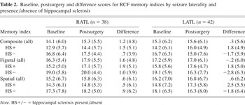
Presurgical, postsurgical, and pre-post differences on the composite, figural, and spatial memory indices are presented in Table 2. Scores are grouped by seizure laterality and the presence or absence of hippocampal sclerosis. In order to first test for seizure laterality effects on baseline memory performance independent of pathology, figural and spatial memory indices were analyzed using a repeated measures ANOVA with Index (figural memory index, spatial memory index) as the within-subjects factor and seizure laterality (RATL, LATL) as the between-subjects factor. There was a significant effect for Index [F(1,78) = 14.21, p < .001] reflecting lower spatial index scores compared to figural index scores. However, although RATL participant's scores were generally lower than those of LATL participants there was no effect for seizure laterality [F(1,78) = .49], and there was no Index × Laterality interaction [F(1,78) = .03]. This indicates that neither memory index was differentially sensitive to right or left temporal seizure foci.
Baseline, postsurgery and difference scores for RCF memory indices by seizure laterality and presence/absence of hippocampal sclerosis

Because of the importance of identifying lateralized temporal lobe dysfunction in individual cases in presurgical neuropsychological evaluations (often without prior knowledge of EEG and MRI findings) we calculated effect sizes (Cohen's d; Zakzanis, 2001) to further examine the extent to which presurgical performance on each of the memory indices discriminated between participants with right and left seizure foci. For the composite, figural and spatial indices these were .20, .16, and .15, respectively representing overlaps (OL) of between 85% and 92%. These small effect sizes fell well short of the Cohen's d = 3.0 (OL 7.2%) suggested as a cut-off for a useful clinical marker in neuropsychological research (Zakzanis, 2001).
Effect of hippocampal sclerosis
In order to ascertain whether potential laterality effects on baseline memory scores could be disclosed by taking into account the presence or absence of hippocampal sclerosis (HS+/HS−), another Index × Laterality repeated measures ANOVA was performed incorporating pathology (HS+/HS−) as an additional between-subjects factor. Within-subjects analyses once again revealed a significant effect for Index [F(1,76) = 14.15, p < .001] with lower spatial memory scores relative to figural memory scores. However, there were no Index × Laterality [F(1,76) = 0.19], Index × Pathology [F(1,76) = 0.24], or Index × Laterality × Pathology [F(1,76) = 0.50] interactions. Between-subjects analyses disclosed a significant main effect for Pathology [F(1,76) = 5.42, p = .02] in the absence of an effect for Laterality [F(1,76) = 0.12] or Laterality × Pathology interaction [F(1,76) = .001]. These results indicate that even with the additional consideration of the presence or absence of HS, laterality effects on figural and spatial memory indices were not found. However, regardless of their seizure laterality, the performance of participants without HS on both memory indices was superior to that of participants with HS. This suggests that the left as well as the right hippocampus might contribute to both the figural and spatial aspects of RCF memorization. If this were the case, it would be anticipated that surgical resection of a non-sclerotic right or left hippocampus would result in performance decrements on figural and spatial memory indices.
Pre- to postoperative change
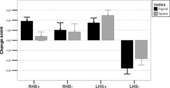
Change scores on each of the memory indices were calculated by subtracting presurgical scores from postsurgical scores (see Table 2). These change scores were then subjected to an Index × Laterality × Pathology repeated measures ANCOVA with presurgical composite memory score as a covariate to control for regression to the mean. Within-subjects contrasts revealed no effects for Index [F(1,75) = 0.63] although a significant Index × Laterality interaction was found [F(1,75) = 5.87, p = .02] reflecting a decrement in figural memory following LATL. However, no Index × Pathology [F(1,75) = 1.63] or Index × Laterality × Pathology [F(1,75) = .62] interactions were found. No between-subjects effects were found for Laterality [F(1,75) = .63], and contrary to the hypothesis that HS− participants would exhibit greater postsurgical memory decrement than HS+ participants regardless of their seizure laterality, no main effect for Pathology was found [F(1,75) = .80]. However, there was a significant Laterality × Pathology interaction [F(1,75) = 4.20, p = .044]. As depicted in Figure 1 these results indicate that LATL HS− participants exhibit the greatest postsurgical decrement in the figural and spatial aspects of RCF recall.

RCF recall: Pre- to postoperative change scores as a function of laterality and presence or absence of hippocampal sclerosis (HS+/HS−). Error bars show the Standard Error of the Mean.
Effect of RCF copy quality
Because the quality of the initial copy of the RCF has been found to influence subsequent delayed recall (Newman & Krikorian, 2001) all analyses were rerun using percent delayed recall scores expressed as a percentage of the copy score [(delayed recall/copy score) ×100]. This did not alter any of the significant main effects or interactions.
DISCUSSION
In this study we investigated whether the utility of the RCF as an index of right temporal lobe function in temporal lobe surgery candidates is enhanced by using a modified scoring methodology, which separates the figural and spatial aspects visuospatial memorization (Brier et al., 1996). We extended on earlier presurgical work by examining changes in memory following right and left temporal lobectomy. In keeping with the lateralized model of memory function we first hypothesized that participants with RTLE would be inferior to LTLE participants on figural and spatial aspects of RCF recall preoperatively and that they would exhibit the largest decrement postoperatively, especially in the absence of histopathologically confirmed HS. Second we tested two competing hypotheses in relation to the impact of temporal lobe disease and resection on figural and spatial memory. On the one hand previous research with TLE samples suggested that figural memory would prove to be the more vulnerable of the two given its dependence on the occipitotemporal (“what”) network (Barr, 1997; Baxendale et al., 1998; Donofrio et al., 1999; Hermann et al., 1993). On the other hand given that both the occipitotemporal and occipitoparietal pathways are known to converge upon the hippocampus via the entorhinal cortex (Sutherland & Rudy, 1989) it was believed that hippocampal disease and resection should impact equally on the figural and spatial aspects of memorization.
Laterality Effects
Contrary to expectation regardless of the scoring methodology used (figural, spatial, or composite) we did not find RCF recall to be differentially sensitive to RTL seizure onset, hippocampal sclerosis, or ultimate resection. This is consistent with the large multi-center study by Barr et al. (1997) as well as the failure of presurgical RCF recall to correlate with right hippocampal MRI volumetric data (Griffith et al., 2003; Trenerry et al., 1993) and cell densities (Rausch & Babb, 1993). Our presurgical results indicated that the presence of hippocampal sclerosis, either right or left, was associated with poorer RCF recall with the greatest postsurgical decrement in RCF recall being sustained by LATL participants without HS. This not only suggests that RCF recall is a poor marker of right temporal lobe function but is possibly a “surrogate” measure of left temporal lobe function. Such a view is consistent with the finding by Kilpatrick et al. (1997) that the presence of left but not right hippocampal atrophy is associated with poorer presurgical RCF recall.
A common criticism leveled against figural reproduction tasks as measures of visuospatial memory is the ease with which they can be verbally encoded (Lee et al., 1989). This being the case it follows that the more verbalizable the material (regardless of the modality in which it is presented) the more its memorization will be supported by left temporal lobe structures. This was well demonstrated by Helmstaedter et al. (1995) who established the level of visuospatial complexity beyond which verbal encoding of the Benton Visual Retention Test was possible. They found that memory for items that exceeded the verbal memory span discriminated between presurgical right and left TLE participants whereas less complex (i.e., verbally encodable) items did not. Furthermore, given the well established link between the absence of hippocampal sclerosis and severity of verbal memory decline following left sided resections (Chelune & Najm, 2001; Hermann et al., 1992; Rausch & Babb, 1993; Sass et al., 1994; Trenerry et al., 1993) our finding that LATL HS− participants showed the largest postsurgical decrement in RCF recall lends support to the view that memorization of the figure is achieved primarily through verbal encoding. If this is the case, and that RCF recall is essentially a surrogate measure of left temporal lobe function, a positive correlation should exist between pre- to postsurgical change in RCF recall and change on putative verbal memory measures. Unfortunately, because of cross-institutional differences in verbal memory test selection we were unable to test for this association.
Our findings clearly challenge the lateralized material-specific model of memory function in its most stringent form. That is, the Laterality/Independence assumption that the systems of verbal and visuospatial memory are independently supported by homologous left and right temporal lobe regions (Saykin et al., 1992). Nevertheless there is ample evidence in the literature that material-specific memory deficits are associated with unilateral temporal lobe dysfunction. In particular several studies have identified robust deficits among individuals with RTLE on non-commercial, laboratory generated tests of visuospatial memory (Abrahams et al., 1997; Baxendale et al., 1998; McDade & Jones-Gotman, 2001; Nunn et al., 1998). Citing the fact that individuals with normal temporal lobes having undergone commissurotomy demonstrate verbal memory deficits Dobbins et al. (1998) reconciles these viewpoints by suggesting some degree of interdependence between the medial temporal lobe memory systems in which “communication” between hemispheres is required to attain optimal memorization. Implicit in this suggestion is that the extent to which performance on a given memory task is more dependent on one temporal lobe than the other can be conceived of as being on a continuum. This continuum is likely to involve not only degree of verbalizability of the test material but also its complexity and novelty (Redoblado et al., 2003). In the case of the RCF it is has been shown that many of its 18 scoreable components are highly verbalizable and familiar thereby engaging a considerable amount of left hemisphere processing (McConley et al., 2006).
Figural versus spatial memory
Given that our hypothesis regarding right hemisphere specialization for RCF recall was rejected, it is somewhat superfluous to discuss the possible existence of separate right hemisphere neural correlates of figural and spatial memory. We found LATL generally to have a more detrimental effect upon recall of the figural aspects of RCF components than it did upon recall of their location. More interestingly, in the case of removal of a non-sclerotic and presumably functional left hippocampus there was no effect for Index suggesting that figural and spatial forms of information were verbally encoded. If this were the case then it remains uncertain whether both forms of information were encoded simultaneously or whether the figural aspects were verbally encoded first thereby preferentially engaging a subsequent similar verbal encoding strategy for location. Either way there is evidence from incidental learning experiments in normals that identification processes involved in object naming boosts memory for not only object identity but for absolute object location as well (Köhler et al., 2001). Thus, when an entire object is selected and attended to, the activated perceptual representation includes all of its perceived attributes, including its location (O'Craven et al., 1999). Whereas our results and those of others from functional imaging studies (Köhler et al., 1998) suggest that the left hippocampus plays a central role in memory for object and location, other functional imaging studies have suggested that the neural correlates supporting both forms of memory are inter- and intra-hemispherically distinct (Moscovitch et al., 1995; Nyberg et al., 1996; Owen et al., 1996).
CONCLUSION
Using the same modified RCF scoring system adopted by Brier et al. (1996) we aimed to replicate and extend their study by examining both pre- and postsurgical figural and spatial memory in individuals with TLE. To the extent that this scoring system accurately captured the figural and spatial aspects of RCF recall we found neither to be differentially sensitive to right temporal lobe disease or resection. Instead our findings indicated that left temporal lobectomy produced the greatest decrement in recall, particularly in the absence of hippocampal sclerosis.
The failure of the present RCF scoring methodology to serve as reliable clinical marker of right temporal lobe integrity as well as that of conventional (Barr et al., 1997; Griffith et al., 2003; Kilpatrick et al., 1997; Rausch & Babb, 1993; Trenerry et al., 1993) and other modified RCF scoring systems (McConley et al., 2006; Piguet et al., 1994) seriously brings into question the utility of this instrument but not necessarily the concept of nonverbal memory in epilepsy surgery evaluations. Although trite to say, we believe that the concept of nonverbal verbal memory exists in so far as to-be-remembered material is not verbalizable. Whether memory for such material is critically dependent on right-sided mesial temporal lobe structures remains to be determined. This being the case, it would appear to be time to more vigorously pursue cross-institutional clinical trials of experimentally generated tests shown to hold some promise in differentiating right from left temporal lobe dysfunction (Abrahams et al., 1997; Baxendale et al., 1998; Helmstaedter et al., 1995; McDade & Jones-Gotman, 2001; Nunn et al., 1998). For each test this would involve the establishment of satisfactory pre- and post-test data normative bases to identify not only the specificity and sensitivity of the test in its presurgical detection of right temporal lobe abnormality but to also reliably identify “true” change postoperatively (Sawrie et al., 1996).
ACKNOWLEDGMENT
This study was undertaken without financial support to any of the authors.