Introduction
High-frequency ultrasound is a common imaging modality used to examine the thyroid, salivary glands, lymph nodes, and soft tissues in the head and neck.Reference Gritzmann1 However, a transcervical approach does not generally allow complete sonographic evaluation of the larynx, due to ossification and intraluminal air.Reference Arens, Eistert, Glanz and Waas2 For this reason, computed tomography (CT) and magnetic resonance imaging (MRI) are used to supplement microlaryngoscopy as additional diagnostic tools in the investigation of more extensive laryngeal lesions.Reference Arens and Glanz3 Endoscopic versions of ultrasound technology have been developed and successfully applied in the fields of cardiology and gastroenterology.Reference Bose, von Birgelen and Erbel4, Reference Caddy and Chen5 Recent investigations of laryngeal endosonography have demonstrated a penetrating depth of 10 to 25 mm (depending on the applied frequency) and a high tissue resolution.Reference Zech, Scheer, Maier and Heppt6–Reference Tamura, Kitahara and Kohno9 Therefore, endolaryngeal ultrasound may be able to predict the exact extension of a laryngeal tumour before surgery is performed, and to assist in finding the best therapeutic solution for the patient.Reference Arens and Glanz3 Thus, this non-invasive method could represent an additional diagnostic tool in the field of laryngology.
There is currently no precise description in the literature of how to perform in vivo endolaryngeal ultrasound imaging of the larynx. Therefore, the objective of the present study was to fill this gap, and also to offer a number of practical suggestions on the use of this technology.
Methods
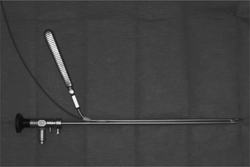
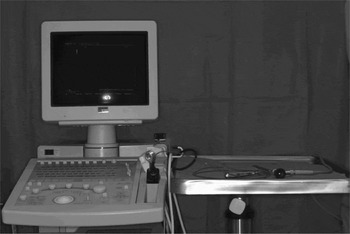
At our institution, each patient with a laryngeal lesion undergoes indirect laryngoscopy, videostroboscopy and voice analysis before surgery is performed. During microlaryngoscopy, the larynx is exposed using surgical laryngoscopes with suspension. First, the lesion is inspected under the operating microscope, palpated and documented digitally. Then, a Hitachi EUB 525 ultrasonograph (Hitachi Medical Systems, Wiesbaden, Germany) with a 360° radial scanning 12 MHz probe (SP-501, Fujinon Europe GmbH, Willich, Germany) is used to investigate extensive tumours before surgery is performed. The flexible probe is inserted through a specially designed applicator and fixed by means of Steri-Strips (3M Health Care, St Paul, Minneapolis, USA) (Figure 1). Alternatively, the probe can be taped directly to a rigid 30° endoscope (commonly used for bronchoscopy). All equipment is placed on a single table in order to avoid damage to the sensitive probe (Figure 2).
The special applicator. An oesophageal aspirator with an internal diameter of 2 mm is shortened to a length of 21 cm. After drilling out an opening on its upper side to allow for probe insertion, the aspirator and a guide jacket from a rigid 30° endoscope are welded together.

Layout of endolaryngeal ultrasound device. All equipment is placed on a single table to avoid possible damage to the sensitive ultrasound probe. The area under the probe is covered with a sterile drape; additional protection can be provided by a piece of foam-rubber.

In general, two persons are necessary for endolaryngeal ultrasound monitoring. The surgeon positions and holds the probe steady in the centre of the laryngeal lumen, while the assistant starts and stops the scanning process, saves data and labels the images. Good communication between the two operators results in rapid acquisition of data, generally requiring less than 10 minutes of additional operating time.
First, the patient is positioned in the anti-Trendelenburg position, and a normally blocked orotracheal tube (Safety-Flex; Mallinckrodt, Athlone, Ireland; 6.5 mm for men, 6.0 mm for women) is placed distally in the trachea just above the carina. After re-exposing the larynx using a Lindholm laryngoscope (Karl Storz, Tuttlingen, Germany), physiological saline is instilled into the larynx, via a 50 ml syringe, to ensure optimal ultrasound connection. To avoid artefacts, any air bubbles should be removed by suction and gentle tapping on the cricoid.
The surgeon then inserts the endolaryngeal ultrasound probe through the laryngoscope, under endoscopic control, and places its tip as far distally as possible. Systematic imaging of the larynx is performed in real-time mode, usually starting in the trachea and slowly moving upwards. The actual position of the probe is controlled either endoscopically or through direct observation of the sonographic image on the monitor.
To evaluate lesions located in the posterior larynx, the orotracheal tube must be loaded up by the laryngoscope.
After endosonography, the assistant evaluates the endolaryngeal ultrasound scans and communicates the results to the surgeon, while the latter carefully removes the instilled saline from the upper aerodigestive tract and continues the operation.
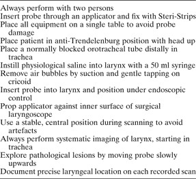
At the end of surgery, the applicator is sterilised as usual, while the probe is disinfected using alcoholic solution (Table I).
In vivo endolaryngeal ultrasound imaging: practical suggestions

Discussion
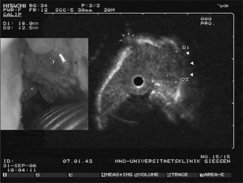
Although a single person could carry out endosonography of the larynx, there are several reasons for performing this procedure with the help of two persons. Firstly, the surgeon is fully occupied in keeping the probe steady in the centre of the laryngeal lumen, in order to obtain a high quality picture of the larynx (Figure 3). This does not allow any time for controlling the scanning process or labelling the recorded images. In our experience, rapid data acquisition (within 10 minutes) is only possible when two persons are involved in the imaging process, and when there is good communication between the two.
Endolaryngeal ultrasound scan, showing an extensive glotto-subglottic tumour with cartilage infiltration on the right (arrowheads) and extension into the pre-epiglottic space on the left anteriorly (small arrows). Although hardly visible during microlaryngoscopy (inset view), these sonographic findings were later histologically confirmed from the laryngectomy specimen.

Although their small diameter fits even paediatric larynges, flexible ultrasound catheters have the disadvantages of more difficult handling, reduced life expectancy (due to fragility) and no available colour Doppler facilities. In addition, a special adapter with an engine (SP-711, Fujinon Europe GmbH Willich, Germany) is necessary to rotate the mirror in the tip of the transducer. In the absence of small, rigid endosonography probes suitable for the larynx, taping the flexible catheter to a 30° endoscope is generally an easy way to make it more rigid. We prefer to use our specially designed applicator (which will soon be made commercially available by Karl Storz), which facilitates the handling and positioning of the probe (Figure 1). In such a manner, the latter can easily be applied under endoscopic control, and lasts for approximately 100 endolaryngeal ultrasound examinations before replacement is necessary.
As spreading laryngoscopes are not suitable, due to continuous drainage of physiological saline into the hypopharynx, we prefer a Lindholm laryngoscope to expose the larynx during endosonography. Although indispensable for optimal ultrasound connection, instillation of fluid into the larynx normally produces a certain quantity of air bubbles. As these induce severe artefacts by acoustic shadowing, it is very important to completely remove them by suction and repeated tapping on the cricoid.Reference Arens and Glanz3 Placing the patient in an anti-Trendelenburg position encourages ascension of air bubbles within the larynx and considerably facilitates their removal. In our experience, a normally blocked (25 mm H2O) orotracheal tube is generally sufficient to prevent the instilled solution from entering the lower airways. However, at the end of the procedure, all physiological saline should be carefully removed from the larynx, hypopharynx and oesophagus by suction (using an aspirator and a stomach tube), in order to prevent subsequent regurgitation and aspiration. Using this approach, we have not encountered any complications at our institution, in over 100 endolaryngeal ultrasound examinations.
Endosonography of the larynx produces horizontal slice images comparable with axial CT or MRI scans but with a higher resolution (Figure 3).Reference Arens, Eistert, Glanz and Waas2 During the imaging process, it is essential to position the probe as centrally as possible, avoiding contact with the laryngeal wall. In this manner, the antero-posterior and medio-lateral diameters of laryngeal neoplasms can directly be measured on the ultrasound scan, while the cranio-caudal diameter is determined by setting marks on the applicator, corresponding to the lower and upper borders of the tumour.Reference Arens and Glanz3 The location of the orotracheal tube and anatomical structures such as the cricoid and thyroid cartilage are very helpful landmarks within the larynx.Reference Arens, Eistert, Glanz and Waas2 Hence, orientation during endolaryngeal ultrasound is possible either by endoscopic control or through direct observation of the sonographic image on the monitor.
The advantages of endosonography of the larynx can be summarised as follows. Firstly, the cost of endolaryngeal ultrasound is one-third that of CT or MRI, without the need for contrast agents. Secondly, swallowing artefacts do not occur as the patient is under general anaesthesia. Thirdly, endosonography is carried out in real-time mode, with high resolution of solitary structures and the possibility of viewing critical regions repeatedly. Fourthly, as the treating physician performs and evaluates the sonographic images themself, there is a closer relation between diagnostics and therapy.Reference Arens and Glanz3
However, the technique does have some disadvantages. In general, its availability is limited to those hospitals having the necessary equipment at their disposal. Moreover, use of endolaryngeal ultrasound is restricted to the setting of microlaryngoscopy under general anaesthesia, while tumours can only be detected if larger than 3 mm (however, this is still earlier than with CT or MRI). Furthermore, the results of endolaryngeal ultrasound strongly depend on the experience of the examiner.Reference Schade, Kothe and Leuwer10 In addition, sonograms are considerably more difficult for the inexperienced viewer to interpret, compared with corresponding CT and MR images, so discussion of sonographic findings with other medical specialties (such as radiology and radio-oncology) may not be straightforward.
Today, endosonography has already replaced CT in staging oesophageal and colorectal cancer, due to a better depiction of local tumour growth and regional lymph node metastases.Reference Jones11, Reference Skandarajah and Tjandra12 Although similar results have been demonstrated for the larynx in the past, endosonography has drawn little attention from otolaryngologists thus far.Reference Arens and Glanz3, Reference Tamura, Kitahara and Kohno8 However, in a series of 74 patients undergoing surgery for laryngeal cancer at our institution, endolaryngeal ultrasound was shown to have comparable results to CT and MRI, and significantly lower costs, with the advantages of earlier detection of smaller tumours as well as better demonstration of the exact extension, midline crossing and cartilage infiltration of laryngeal lesions. Therefore, this simple and non-invasive method also qualifies for use in the larynx.
Conclusions
Endosonography may constitute an additional imaging tool for the investigation of more extensive laryngeal lesions, and one which can easily be used by the otolaryngologist themself during microlaryngoscopy. Good technique and close cooperation between surgeon and assistant are essential for rapid acquisition of data and high quality images of the inspected sites.
Acknowledgements
We would like to thank Warren Jacobs for the correction of the manuscript, and Hans-Joachim Jung for the construction of our special applicator.