Introduction
The hyper diverse Usnea Adans. is a fruticose genus of lichenized Ascomycetes with the second highest number of species in the Parmeliaceae after the genus Xanthoparmelia (Thell et al. Reference Thell, Crespo, Divakar, Kärnefelt, Leavitt, Lumbsch and Seaward2012). However, although various accounts have been proposed (Wirtz et al. Reference Wirtz, Printzen, Sancho and Lumbsch2006; Kirk et al. Reference Kirk, Cannon, Minter and Stalpers2008; Thell et al. Reference Thell, Crespo, Divakar, Kärnefelt, Leavitt, Lumbsch and Seaward2012; Clerc Reference Clerc2016), the exact number of species worldwide is still unknown. This is due to the fact that several species have been synonymized over the last decades (Swinscow & Krog Reference Swinscow and Krog1978; Stevens Reference Stevens1990; Clerc Reference Clerc1997, Reference Clerc2004, Reference Clerc2011a, Reference Clerc2016; Clerc & Herrera-Campos Reference Clerc and Herrera-Campos1997; Ohmura Reference Ohmura2001; Mark et al. Reference Mark, Saag, Leavitt, Will-Wolf, Nelsen, Tõrra, Saag, Randlane and Lumbsch2016), or newly described (Zhao et al. Reference Zhao, Hsu and Sun1975; Swincow & Krog Reference Swinscow and Krog1979; Clerc & Herrera-Campos Reference Clerc and Herrera-Campos1997; Herrera-Campos & Clerc Reference Herrera-Campos and Clerc1998; Stevens Reference Stevens1999; Halonen Reference Halonen2000; Clerc Reference Clerc2006, Reference Clerc, Nash, Gries and Bungartz2007, Reference Clerc2011a, Reference Clerc, Thell and Mobergb; Wirtz et al. Reference Wirtz, Printzen and Lumbsch2008; Truong et al. Reference Truong, Bungartz and Clerc2011; Truong & Clerc Reference Truong and Clerc2012, Reference Truong and Clerc2013, Reference Truong and Clerc2016; van den Boom et al. Reference van den Boom, Clerc and Ertz2015; Mark et al. Reference Mark, Saag, Leavitt, Will-Wolf, Nelsen, Tõrra, Saag, Randlane and Lumbsch2016; Gerlach et al. Reference Gerlach A da, Clerc and da Silveira2017; Bungartz et al. Reference Bungartz, Truong, Herrera-Campos and Clerc2018; Clerc & Otte Reference Clerc and Otte2018). Moreover, the use of molecular data seems to reveal a much higher species diversity than previously thought (Wirtz et al. Reference Wirtz, Printzen, Sancho and Lumbsch2006; Gerlach et al. Reference Gerlach A da, Zeynep, Naciri, Araujo Caviro, da Silveira and Clerc2018). In the context of the tremendous impact of molecular data on systematics, one of the main remaining questions concerns the congruence between morphological characterization of species and phylogenetic delimitation based on DNA sequence data.
Furthermore, it is well known that there is a huge problem of taxonomic misidentifications in public DNA databases today (Bridge et al. Reference Bridge, Roberts, Spooner and Panchal2003; Vilgalys Reference Vilgalys2003; Nilsson et al. Reference Nilsson, Ryberg, Kristiansson, Abarenkov, Larsson and Kõljalg2006; Bidartondo et al. Reference Bidartondo, Bruns, Blackwell, Edwards, Taylor, Horton, Zhang, Koljalg, May and Kuyper2008; Kang et al. Reference Kang, Mansfield, Park, Geiser, Ivors, Coffey, Grünwald, Martin, Lévesque and Blair2010; Ko et al. Reference Ko, Stephenson, Bahkali and Hyde2011; Hofstetter et al. Reference Hofstetter, Buyck, Eyssartier, Schnee and Gindro2019). As a consequence, we sometimes have to deal with questionable interpretations of molecular clades, as shown for instance in the genus Usnea by Clerc & Otte (Reference Clerc and Otte2018).
In Usnea, almost all studies so far have shown that the traditional species concept based on morphology, anatomy and chemistry (Clerc Reference Clerc1998) is generally confirmed (Ohmura Reference Ohmura2001; Kelly et al. Reference Kelly, Hollingsworth, Coppins, Ellis, Harrold, Tosh and Yahr2011; Saag et al. Reference Saag, Tõrra, Saag, Del Prado and Randlane2011; Truong et al. Reference Truong, Rodriguez and Clerc2013; Clerc & Otte Reference Clerc and Otte2018; Gerlach et al. Reference Gerlach A da, Zeynep, Naciri, Araujo Caviro, da Silveira and Clerc2018). In their important and innovative molecular study based on a six-locus dataset, Mark et al. (Reference Mark, Saag, Leavitt, Will-Wolf, Nelsen, Tõrra, Saag, Randlane and Lumbsch2016) evaluated the congruence of morphological and molecular data in delimiting species, using a set of 19 phenotypically delimited Usnea species (144 specimens) collected in Europe and North America, which resulted in a Bayesian 50% consensus tree inferred by BEAST (Mark et al. Reference Mark, Saag, Leavitt, Will-Wolf, Nelsen, Tõrra, Saag, Randlane and Lumbsch2016: fig. 1). They found contrasting results: on the one hand, the morphology of eight species was congruent with molecular data; on the other, the delimitation of 11 species based on the traditional species concept was not confirmed by the phylogenetic tree. However, among these 11 hypothesized non-monophyletic species, three had already been synonymized with other taxa in previous studies: Usnea subfloridana Stirt. with U. florida (L.) F. H. Wigg. (Articus et al. Reference Articus, Mattsson, Tibell, Grube and Wedin2002), and Usnea diplotypus Vain. and U. chaetophora Stirt. with U. dasopoga (Ach.) Nyl. (Clerc Reference Clerc, Thell and Moberg2011b; see ‘Systematics of the Taxa Involved in This Paper’ below). The first part of the tree (Mark et al. Reference Mark, Saag, Leavitt, Will-Wolf, Nelsen, Tõrra, Saag, Randlane and Lumbsch2016: fig. 1a) contains species that are most of the time well characterized by their secondary chemistry, and molecular data therefore agree quite well with the current traditional species concept, secondary chemistry being a good specific marker. With one exception (Clerc & Otte Reference Clerc and Otte2018), the second part of the tree (Mark et al. Reference Mark, Saag, Leavitt, Will-Wolf, Nelsen, Tõrra, Saag, Randlane and Lumbsch2016: fig. 1b) contains species that cannot be separated on the basis of their chemistry. In this tree, two features deserve attention: 1) the separation of Usnea barbata (L.) F. H. Wigg. and U. dasopoga seems not to be supported by molecular data; 2) the synonymization of U. substerilis with U. perplexans (syn. U. lapponica) seems to be supported by molecular data. Clerc (Reference Clerc, Thell and Moberg2011b) discussed the taxonomy of the last four species and considered U. barbata, U. dasopoga, U. perplexans and U. substerilis as forming distinct species based mainly on morphological and anatomical characters. For this reason, the first author requested the loan of the voucher specimens used in the Mark et al. (Reference Mark, Saag, Leavitt, Will-Wolf, Nelsen, Tõrra, Saag, Randlane and Lumbsch2016) study in order to re-evaluate the identification of these specimens. The present study is the result of this re-evaluation. To avoid identifications based solely on expert opinion, we conducted a discriminant analysis using previous measurements of the thickness of the cortex, the medulla and the central axis in U. barbata and U. dasopoga since these characters are known to be diagnostic and can be used to distinguish the two species (Clerc Reference Clerc, Thell and Moberg2011b).
We conclude with a plea for more accurate specimen identifications in molecular phylogenetic studies and finish with four suggestions which, if complied with, should help achieve this goal in lichens in general, and in Usnea in particular.
Material and Methods
Morphological, anatomical and chemical analyses
The voucher specimens of the U. barbata-dasopoga group analyzed by Mark et al. (Reference Mark, Saag, Leavitt, Will-Wolf, Nelsen, Tõrra, Saag, Randlane and Lumbsch2016) and deposited in TU were requested on loan and studied morphologically, anatomically and chemically. Morphology and anatomy were studied with a Leitz stereomicroscope. According to Clerc (Reference Clerc, Thell and Moberg2011b), anatomical characters are important in distinguishing the two species. Anatomical measurements of the cortex, medulla and central axis were carried out in longitudinal sections of branches at ×40 magnification. The percentage thickness of cortex/medulla/axis of the total branch diameter (CMA), as well as the ratio axis/medulla (A/M) of all the cited specimens were calculated according to Clerc (Reference Clerc1987). A new indicator, the ratio medulla/cortex (M/C), is introduced and used here.
Chemical analyses were performed by thin–layer chromatography (TLC) following Culberson & Ammann (Reference Culberson and Ammann1979), with solvent B modified according to Culberson & Johnson (Reference Culberson and Johnson1982).
Identification of the voucher specimens used in Mark et al. (Reference Mark, Saag, Leavitt, Will-Wolf, Nelsen, Tõrra, Saag, Randlane and Lumbsch2016)
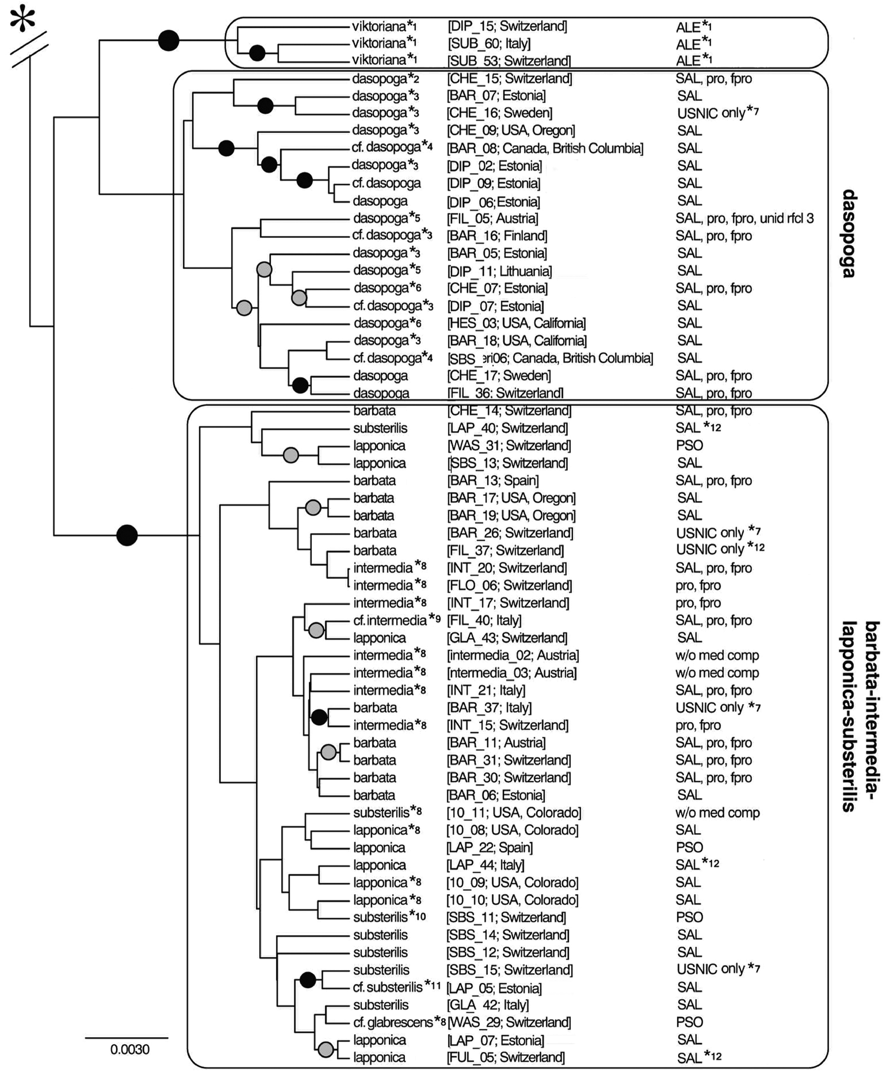
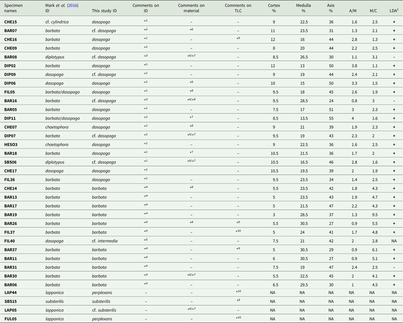
Identification of the 31 loaned voucher specimens was conducted blindly without knowing the specimen's position on the phylogenetic tree of Mark et al. (Reference Mark, Saag, Leavitt, Will-Wolf, Nelsen, Tõrra, Saag, Randlane and Lumbsch2016: fig. 1b). Our identifications are reported in Fig. 1 and Table 1. The voucher specimens were labelled reporting the code of the specimens analyzed by Mark et al. (Reference Mark, Saag, Leavitt, Will-Wolf, Nelsen, Tõrra, Saag, Randlane and Lumbsch2016: fig. 1b).
The phylogenetic tree of the Usnea barbata-dasopoga group from Mark et al. (Reference Mark, Saag, Leavitt, Will-Wolf, Nelsen, Tõrra, Saag, Randlane and Lumbsch2016: fig. 1b) with the new identifications provided in this study, with permission of Springer Nature (License no. 4634240338179). Identifications in Mark et al. (Reference Mark, Saag, Leavitt, Will-Wolf, Nelsen, Tõrra, Saag, Randlane and Lumbsch2016): *1, Usnea praetervisa (Asahina) P. Clerc, see Clerc & Otte (Reference Clerc and Otte2018); *2, U. cf. cylindrica; *3, U. barbata; *4, U. diplotypus; *5, U. barbata/dasopoga; *6, U. chaetophora; *8, identification not checked by the authors; *9, U. dasopoga; *10 & *11, U. lapponica. TLC results in Mark et al. (Reference Mark, Saag, Leavitt, Will-Wolf, Nelsen, Tõrra, Saag, Randlane and Lumbsch2016): *7, fumar- and protocetraric acids as main substances; *12, pannaric and pannaric acid-6-methylester as main substances. Main substances are given in capitals and accessory substances are in lower case. For full details see Mark et al. (Reference Mark, Saag, Leavitt, Will-Wolf, Nelsen, Tõrra, Saag, Randlane and Lumbsch2016).

Specimen identifications (ID) according to Mark et al. (Reference Mark, Saag, Leavitt, Will-Wolf, Nelsen, Tõrra, Saag, Randlane and Lumbsch2016) and to this study, together with details of the main morphological characters: comments on ID, on voucher material and on TLC results, with CMA, A/M and M/C measurements and the results of the linear discriminant analysis (LDA). The order of specimens follows that of the phylogenetic tree of Mark et al. (Reference Mark, Saag, Leavitt, Will-Wolf, Nelsen, Tõrra, Saag, Randlane and Lumbsch2016: fig. 1b) from top to bottom.

1 + = a priori morphological identification confirmed a posteriori by the discriminant analysis; − = a priori morphological identification not confirmed a posteriori by the discriminant analysis; NA = not available.
*1 = CMA, M/C values and morphology in accordance with U. dasopoga.
*2 = The packet contains a mixture of both species, U. barbata and U. dasopoga. Mark et al. (Reference Mark, Saag, Leavitt, Will-Wolf, Nelsen, Tõrra, Saag, Randlane and Lumbsch2016) performed the TLC on the U. dasopoga specimen and the DNA was probably extracted from the same specimen.
*3 = CMA, M/C values not in accordance with U. dasopoga; morphology in accordance with U. dasopoga.
*4 = CMA, M/C values and morphology in accordance with U. barbata.
*5 = This specimen has no soralia or apothecia. It displays cylindrical main branches without swellings and an M/C close to U. dasopoga values. However, the pale basal part, the absence of soralia and the cortex thickness would indicate another taxon. It is tentatively interpreted here as a thin morphotype of the U. intermedia group.
*6 = Poor material, not well developed, or damaged or stressed by insects or lichenicolous fungi.
*7 = Short or juvenile specimens.
*8 = Basal part partly or totally missing.
*9 = The TLC analysis of Mark et al. (Reference Mark, Saag, Leavitt, Will-Wolf, Nelsen, Tõrra, Saag, Randlane and Lumbsch2016) could not be confirmed. Protocetraric and fumarprotocetraric acids were never found alone as main medullary substances in this group, at least in Eurasian specimens (Ohmura & Onimaru Reference Ohmura and Onimaru2010; Clerc Reference Clerc, Thell and Moberg2011b; Ohmura et al. Reference Ohmura, Skirina and Skirin2017). In this group, protocetraric acid always occurs as an accessory substance with salazinic acid. Their presence alone as main substances in the TLC analysis by Mark et al. (Reference Mark, Saag, Leavitt, Will-Wolf, Nelsen, Tõrra, Saag, Randlane and Lumbsch2016) is most probably due to a contamination with small Bryoria fuscescens (Gyeln.) Brodo & D. Hawksw. branches that are present in some of these collections.
*10 = The TLC analysis of Mark et al. (Reference Mark, Saag, Leavitt, Will-Wolf, Nelsen, Tõrra, Saag, Randlane and Lumbsch2016) could not be confirmed. Pannaric acid has thus far never been found in the genus Usnea. Our TLC analysis confirms the absence of this compound in this specimen. The result from Mark et al. (Reference Mark, Saag, Leavitt, Will-Wolf, Nelsen, Tõrra, Saag, Randlane and Lumbsch2016) is probably due to a contamination.
Statistical analyses
All former anatomical measurements were added to a database compiled over the years by the first author on the U. barbata-dasopoga group. This database initially contained measurements of 112 U. barbata and 104 U. dasopoga specimens and is available on Github (https://Github.com/YNaciri-cjbg/Usnea). The specimens of Mark et al. (Reference Mark, Saag, Leavitt, Will-Wolf, Nelsen, Tõrra, Saag, Randlane and Lumbsch2016), as identified by the first author (PC), were added and classified as barbata-PC and dasopoga-PC. Means and variance were computed for the four groups in R (R Development Core Team 2016) as well as boxplots. For mean comparisons, a Kruskal-Wallis rank test was preferred as non-normality and/or heteroscedasticity were confirmed for nearly all variables using Bartlett tests and Shapiro tests, respectively. Since Kruskal-Wallis tests give only a general statement that differences exist among groups but do not identify which groups are different from others, pairwise tests were also carried out between groups. In the latter case, the significance level was adjusted using a false discovery rate correction with n = 6 (Benjamini & Yekutieli Reference Benjamini and Yekutieli2001). We therefore used adjusted levels of 0.0204, 0.0041 and 0.0004 for nominal levels of 5%, 1% and 0.1%, respectively.
In a second step, a linear discriminant analysis was conducted on the same dataset. The initial 216 individuals were used to find the discriminant function and the newly identified specimens from Mark et al. (Reference Mark, Saag, Leavitt, Will-Wolf, Nelsen, Tõrra, Saag, Randlane and Lumbsch2016) were added a posteriori for assignment to the two species. The analysis was conducted in R using the MASS package version 7.3–51.4 (Venables & Ripley Reference Venables and Ripley2002) and the function lda. The function stepclass of the package klaR (Weihs et al. Reference Weihs, Ligges, Luebke, Raabe, Baier, Decker and Schmidt-Thieme2005) was additionally used to identify variables that better discriminate the two species in a linear discriminant analysis framework (lda). The R program is also available on Github (https://Github.com/YNaciri-cjbg/Usnea).
Systematics of the Taxa Involved in This Paper
Usnea barbata is a pendulous species characterized by the distinctly irregular branches with slightly to strongly inflated segments (the thickest branch diameter is, most of the time, distant from the basal part), the thin cortex, and the rather large and dense to lax medulla (Clerc Reference Clerc, Thell and Moberg2011b). Chemistry: usnic, ±salazinic acids.
Usnea chaetophora Stirt. is a pendulous species characterized by main branches formed by numerous, short segments separated by distinct annulations. These characters were found to be present in specimens corresponding to U. barbata, as well as in specimens of U. dasopoga, and are therefore considered to be phenotypic modifications due to environmental conditions. The type specimen of U. chaetophora shows the typical characters of U. dasopoga and is therefore considered as a synonym of the latter species (Clerc Reference Clerc, Thell and Moberg2011b). Chemistry: usnic, ±salazinic acids.
Usnea cylindrica P. Clerc is closely related to U. dasopoga. It differs from the latter species mainly by the filamentose branching type with almost no primary thicker branches (all branches are nearly of the same diameter) (Clerc Reference Clerc, Thell and Moberg2011b). Molecular studies are needed to test the validity of this species. Chemistry: usnic, salazinic acids.
Usnea dasopoga is a pendulous species characterized by the cylindrical to tapering or slightly irregular branches (the thickest branch diameter is most of the time close to the basal part), the thick cortex and the rather thin and dense medulla (Clerc Reference Clerc, Thell and Moberg2011b). Chemistry: usnic, ±salazinic acids.
Usnea diplotypus Vain. is a species based on specimens collected on rocks in Scandinavia (Vainio Reference Vainio1925). Except for U. sphacelata R. Br. and U. pyrenaica Motyka, there are no primarily saxicolous species in Europe. However, many of the European Usnea species might grow saxicolous under optimal environmental conditions (Clerc Reference Clerc, Thell and Moberg2011b). The type specimen of U. diplotypus corresponds in all its characters to a short saxicolous morphotype of U. dasopoga. This short morphotype is consistent with the fact that, due to harsher saxicolous conditions, individuals growing on this substratum might not have the opportunity to become pendant. Usnea diplotypus was therefore considered as a synonym of U. dasopoga (Clerc Reference Clerc, Thell and Moberg2011b). Chemistry: usnic, ±salazinic acids.
Usnea perplexans Stirt. (syn. U. lapponica Vain.) is a short sorediate species with irregular branches and large excavate soralia without isidiomorphs. It seems to be closely related to U. substerilis, differing from this species by the strongly excavate soralia surrounded by a disintegrating cortex. Chemistry: usnic, ±salazinic, ±barbatic, ±psoromic, ±caperatic acids.
Usnea substerilis Motyka is a short sorediate species with irregular branches, large soralia remaining more or less superficial on branches, with no disintegration of the cortex, and with (few) isidiomorphs. It seems to be closely related to U. perplexans (see under this species above) and some individuals are extremely difficult to identify with certainty. Chemistry: usnic, ±salazinic, ±barbatic, ±psoromic, ±caperatic acids.
Results
Statistical analyses
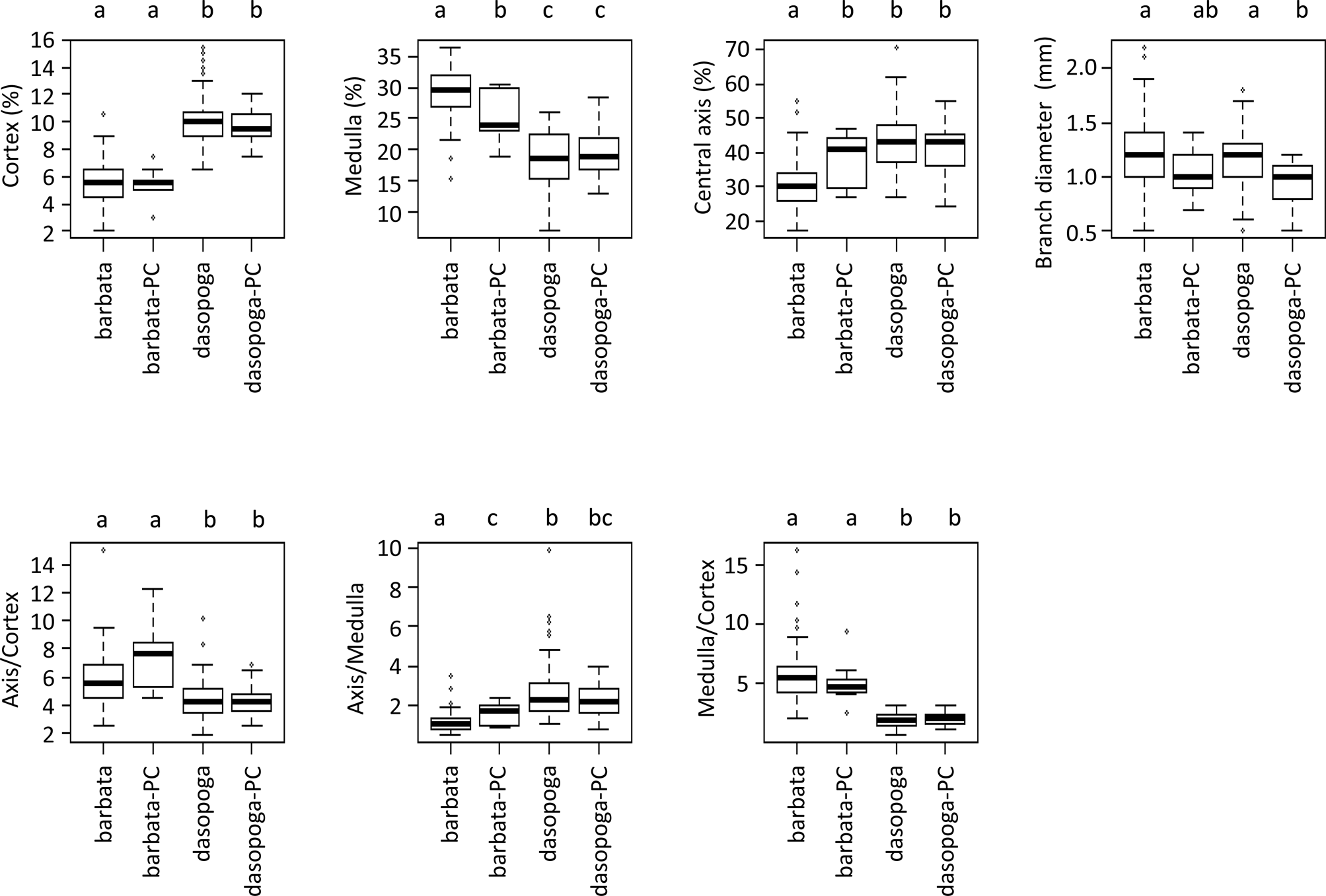
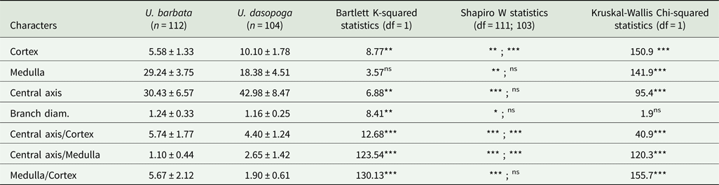
Once re-identified, the specimens from Mark et al. (Reference Mark, Saag, Leavitt, Will-Wolf, Nelsen, Tõrra, Saag, Randlane and Lumbsch2016) were labelled as dasopoga-PC or barbata-PC. The statistical analyses (Fig. 2) show that for all variables, significant differences among groups exist (P < 0.05). In the first dataset (216 specimens), U. barbata and U. dasopoga are found to be significantly different in all variables (P < 0.001, Table 2) except for branch diameter, which is similar for both species (P = 0.171). The newly identified dasopoga-PC individuals are not statistically different from U. dasopoga except in branch diameter (P = 0.0004), whereas the newly identified barbata-PC individuals are not statistically different from U. barbata except in central axis (P = 0.0058), medulla (P = 0.0128) and axis/medulla (P = 0.0096).
Boxplots for Usnea barbata (n = 112) and U. dasopoga (n = 104). The two groups dasopoga-PC (n = 19) and barbata-PC (n = 11) are the samples used by Mark et al. (Reference Mark, Saag, Leavitt, Will-Wolf, Nelsen, Tõrra, Saag, Randlane and Lumbsch2016) that were re-identified in this study. The upper letters refer to Wilcoxon pairwise tests: groups with different letters are significantly different at the adjusted level of 5% (P < 0.0204).

Mean and standard deviation on the 216 individuals assigned to the two species Usnea barbata and Usnea dasopoga for seven quantitative variables and associated tests.

n = number of individuals included in the analysis; df = degree of freedom; ns = P > 0.05; * = P < 0.05; ** = P < 0.01; *** = P < 0.001.
As branch diameter was not significantly different between the two species, this variable was discarded from subsequent analysis. Using stepclass, the best discriminant variable between U. dasopoga and U. barbata was found to be the ratio medulla/cortex (M/C) with a correctness rate of 0.94. The 216 U. dasopoga and U. barbata individuals were used to compute the linear discriminant function. Only nine samples were classified as the ‘wrong’ species, which corresponds to an error rate of 4.2%. The 30 dasopoga-PC and barbata-PC specimens were subsequently assigned to one of the two species using the previous linear function. Only three of them (10%) were misclassified according to the discriminant function (Table 1): BAR31 identified as U. barbata and a posteriori classified as U. dasopoga, and both BAR08 and BAR16 identified as U. dasopoga and a posteriori recognized as U. barbata in the discriminant analysis.
Specimen identification
Table 1 provides the results of our morphological, anatomical and chemical studies on 35 specimens identified by Mark et al. (Reference Mark, Saag, Leavitt, Will-Wolf, Nelsen, Tõrra, Saag, Randlane and Lumbsch2016) as U. barbata, U. cylindrica P. Clerc (U. dasopoga aggr.), U. dasopoga (syn.: U. chaetophora, U. diplotypus), U. perplexans (syn. U. lapponica) and U. substerilis, as well as the new identification for some specimens. The means and standard deviations for U. barbata and U. dasopoga based on new identifications are shown in Table 2.
Discussion
Usnea barbata and U. dasopoga
In this study, based on anatomical and morphological data (Figs 2–6, Tables 1 & 2), all specimens of the barbata-chaetophora-dasopoga-diplotypus clade of Mark et al. (Reference Mark, Saag, Leavitt, Will-Wolf, Nelsen, Tõrra, Saag, Randlane and Lumbsch2016: fig. 1b) were found to correspond to U. dasopoga (Fig. 1): nine specimens identified as U. barbata by Mark et al. (Reference Mark, Saag, Leavitt, Will-Wolf, Nelsen, Tõrra, Saag, Randlane and Lumbsch2016) were newly identified as U. dasopoga (we did not take into consideration here the two specimens FIL05 and DIP11 identified as U. barbata/dasopoga by Mark et al. (Reference Mark, Saag, Leavitt, Will-Wolf, Nelsen, Tõrra, Saag, Randlane and Lumbsch2016)). Five of these nine specimens (CHE16, CHE09, DIP02, BAR05 and FIL36) represented well-developed material for which the identification was straightforward, whereas the four remaining specimens (BAR07, BAR16, BAR18 and DIP07) consisted of poorly developed, juvenile or damaged material, for which the morphological identification was difficult or uncertain and needed to be backed up by the discriminant analysis based on anatomical characters, and more specifically the new character M/C. Out of the 19 specimens now identified as U. dasopoga, two were considered as being misclassified according to the discriminant function (Table 1) based on anatomical characters (CMA, A/M and M/C values). BAR08 and BAR16 were a priori identified as U. cf. dasopoga and a posteriori recognized as U. barbata in the discriminant analysis. BAR08 is a damaged specimen (Table 1) and the intermediate CMA and M/C values might well be a consequence of this damage. However, the morphological characters speak distinctly for U. dasopoga. The thallus of BAR16 is incomplete, with the basal part lacking (Table 1). Since in U. dasopoga the thickest branch diameter is close to the basal part, the CMA and M/C measurements were probably not taken at the correct place, which could explain their atypical values for U. dasopoga. Following our new identifications, the barbata-chaetophora-dasopoga-diplotypus clade of Mark et al. (Reference Mark, Saag, Leavitt, Will-Wolf, Nelsen, Tõrra, Saag, Randlane and Lumbsch2016: fig. 1b) now corresponds well to U. dasopoga and we suggest renaming it the U. dasopoga clade (Fig. 1), although it is not strongly supported in the phylogenetic tree (Mark et al. Reference Mark, Saag, Leavitt, Will-Wolf, Nelsen, Tõrra, Saag, Randlane and Lumbsch2016: fig. 1b).
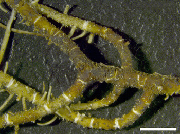
Usnea dasopoga (U. dasopoga FIL_05/Austria in Mark et al. (Reference Mark, Saag, Leavitt, Will-Wolf, Nelsen, Tõrra, Saag, Randlane and Lumbsch2016)). Cylindrical, not swollen primary and secondary branches. Scale = 1 mm. In colour online.

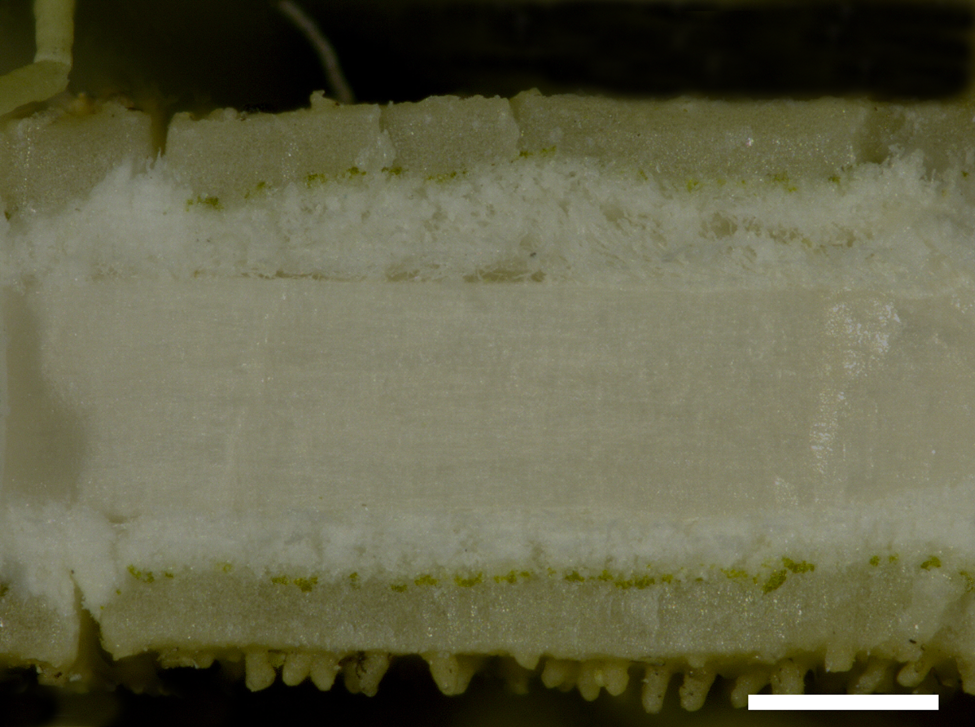
Usnea dasopoga (U. dasopoga CHE_17/Sweden in Mark et al. (Reference Mark, Saag, Leavitt, Will-Wolf, Nelsen, Tõrra, Saag, Randlane and Lumbsch2016)). Transversal section of main branch with thick cortex, thin and compact medulla and large central axis. Scale = 200 μm. In colour online.

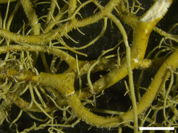
Usnea barbata (U. barbata BAR_06/Estonia in Mark et al. (Reference Mark, Saag, Leavitt, Will-Wolf, Nelsen, Tõrra, Saag, Randlane and Lumbsch2016)). Irregular and more or less swollen main branches. Scale = 1 mm. In colour online.

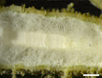
Usnea barbata (U. barbata BAR_11/Sweden in Mark et al. (Reference Mark, Saag, Leavitt, Will-Wolf, Nelsen, Tõrra, Saag, Randlane and Lumbsch2016)). Transversal section of main branch with thin cortex, large and dense to lax medulla, and thin central axis. Scale = 200 μm. In colour online.

The barbata-intermedia-lapponica-substerilis clade of Mark et al. (Reference Mark, Saag, Leavitt, Will-Wolf, Nelsen, Tõrra, Saag, Randlane and Lumbsch2016: fig. 1b) consists of several morphologically well-defined species, such as U. barbata, U. perplexans (syn.: U. lapponica) and U. substerilis (see Clerc Reference Clerc, Thell and Moberg2011b). These taxa are closely related and probably diverged recently, showing various degrees of incomplete lineage sorting (Naciri & Linder Reference Naciri and Linder2015; Mark et al. Reference Mark, Saag, Leavitt, Will-Wolf, Nelsen, Tõrra, Saag, Randlane and Lumbsch2016). This could be the reason why they are all polyphyletic in the phylogenetic tree of Mark et al. (Reference Mark, Saag, Leavitt, Will-Wolf, Nelsen, Tõrra, Saag, Randlane and Lumbsch2016).
All the specimens of this well-supported barbata-intermedia-lapponica-substerilis clade (Fig. 1) indeed belong to the U. barbata group, characterized by the irregular branches with slightly to strongly inflated segments (Fig. 5), presence or absence of soralia, a rather large and dense to lax medulla (Fig. 6), and a basal part that is often concolorous with main branches or brownish to brownish black.
If we consider only U. barbata s. str., all specimens of this clade were correctly identified by Mark et al. (Reference Mark, Saag, Leavitt, Will-Wolf, Nelsen, Tõrra, Saag, Randlane and Lumbsch2016) (see Fig. 1 of this study). In our study, only BAR31 was considered misidentified according to the discriminant function and a posteriori classified as U. dasopoga (Table 1). However, the general morphology of this specimen leaves no doubt about its identification as U. barbata (see ‘Specimen identification’ above). Under extreme environmental conditions, some specimens might display extreme CMA values, as is certainly the case here. Usnea intermedia, usually with many apothecia but without asexual propagules, forms a so-called ‘species pair’ (Poelt Reference Poelt1970) with U. barbata (Halonen et al. Reference Halonen, Clerc, Goward, Brodo and Wulff1998). It is thus not surprising that they sometimes constitute a strongly supported group (BAR37 and INT15) in the phylogeny of Mark et al. (Reference Mark, Saag, Leavitt, Will-Wolf, Nelsen, Tõrra, Saag, Randlane and Lumbsch2016: fig. 1b). The only disturbing element here is the presence of the specimen FIL40, identified as U. dasopoga by Mark et al. (Reference Mark, Saag, Leavitt, Will-Wolf, Nelsen, Tõrra, Saag, Randlane and Lumbsch2016). However, a critical analysis of its morphology (soralia absent) reveals that it does not correspond to U. dasopoga but probably to a thin morphotype without soralia of the Usnea barbata-intermedia group (see ‘Specimen identification’). A final worrying element is the presence of Usnea cf. glabrescens (Vain.) Vain. (WAS29) in this barbata-intermedia-lapponica-substerilis clade (Fig. 1). Usnea glabrescens belongs to the U. fulvoreagens-glabrescens clade in fig. 1 of Mark et al. (Reference Mark, Saag, Leavitt, Will-Wolf, Nelsen, Tõrra, Saag, Randlane and Lumbsch2016). Unfortunately we were not able to check the identity of WAS29, however, it probably corresponds either to U. perplexans or to U. substerilis. Chemistry is a good indicator here since, although occurring in specimens of U. glabrescens s. str. collected in the United Kingdom, psoromic acid was never found in Swiss specimens (WAS29 was collected in Switzerland) while it is a relatively frequent substance in U. perplexans or U. substerilis.
Finally, after a re-evaluation of the identification of specimens by Mark et al. (Reference Mark, Saag, Leavitt, Will-Wolf, Nelsen, Tõrra, Saag, Randlane and Lumbsch2016), and on the basis of a discriminant analysis, we conclude that the existing molecular data do not support the conspecificity of U. barbata and U. dasopoga. On the contrary, although the U. dasopoga clade is not strongly supported, the data of Mark et al. (Reference Mark, Saag, Leavitt, Will-Wolf, Nelsen, Tõrra, Saag, Randlane and Lumbsch2016) seem to be congruent with the morphological and anatomical data, at least for two well delimited taxa, U. barbata and U. dasopoga.
Usnea perplexans (syn.: U. lapponica) and U. substerilis
Optimally developed specimens of U. perplexans and U. substerilis can be easily separated on the basis of soralia morphology and the presence/absence of isidiomorphs (Clerc Reference Clerc, Thell and Moberg2011b). SBS15 and LAP05, respectively identified by Mark et al. (Reference Mark, Saag, Leavitt, Will-Wolf, Nelsen, Tõrra, Saag, Randlane and Lumbsch2016) as U. substerilis and U. perplexans, have a well-supported sister relationship (Mark et al. Reference Mark, Saag, Leavitt, Will-Wolf, Nelsen, Tõrra, Saag, Randlane and Lumbsch2016: fig. 1b) and for this reason these authors proposed to synonymize U. substerilis with U. perplexans. However, LAP05 is such a poor specimen, with only one small branch (2 cm long) with few thin lateral branches and few soralia, that a confident identification is difficult. The presence of small isidiomorphs on two soralia (Fig. 7) would, however, speak for U. substerilis. We therefore suggest that the synonymization of U. substerilis under U. perplexans proposed by Mark et al. (Reference Mark, Saag, Leavitt, Will-Wolf, Nelsen, Tõrra, Saag, Randlane and Lumbsch2016) should not be accepted as it is based on a poor specimen whose identification is not established with certainty.
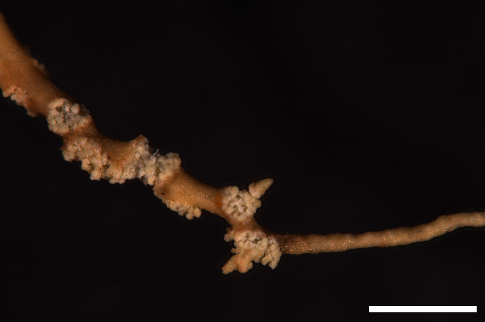
Usnea substerilis (U. lapponica_05/Estonia in Mark et al. (Reference Mark, Saag, Leavitt, Will-Wolf, Nelsen, Tõrra, Saag, Randlane and Lumbsch2016)). Soralia with isidiomorphs on thin lateral branches. Scale = 500 μm. In colour online.

A Plea for Reliable Identifications in Molecular Studies
We acknowledge the importance and the quality of the molecular work carried out by Mark et al. (Reference Mark, Saag, Leavitt, Will-Wolf, Nelsen, Tõrra, Saag, Randlane and Lumbsch2016). They used a high number of genes compared to other studies in this group and up-to-date methodologies (the multispecies coalescent and species delimitation analyses). We also acknowledge the importance of molecular data that currently revolutionize and challenge systematics as well as species delimitations based on traditional morphological data. Taxonomy, however, remains central to sequence-mediated species identification. Today, limited taxonomic expertise, as well as time and funding constraints, make barcoding approaches an efficient solution for species identification and in-depth fungal inventories, especially in difficult fungal genera. It is thus critical that DNA sequences are annotated with correct identifications. Sadly taxonomists are on the Red List now and it becomes more and more difficult to find taxonomic experts who can correctly identify species in difficult groups of organisms (Buyck Reference Buyck1999). It is currently much easier and less time consuming to train a good molecular phylogeneticist than a good and experienced classical taxonomist, since it requires years of training and experience to produce accurate identifications. This situation significantly impacts the quality of data in taxonomic identifications in public DNA databases. For example, Bridge et al. (Reference Bridge, Roberts, Spooner and Panchal2003) suggested that up to 20% of the named fungal sequences in public databases may be misidentified. This is in line with Nilsson et al. (Reference Nilsson, Ryberg, Kristiansson, Abarenkov, Larsson and Kõljalg2006), who affirmed that more than 10% of all publicly available fungal ITS sequences have incorrect taxonomic annotations, and with Ko et al. (Reference Ko, Stephenson, Bahkali and Hyde2011) who suggested that as many as 86% of fungal endophyte sequences from public databases are incorrectly named. This is confirmed in our study where we show that 28% of the specimens studied by us (n = 35) and belonging to the barbata-chaetophora-dasypoga-diplotypus and barbata-intermedia-lapponica-substerilis clades of Mark et al. (Reference Mark, Saag, Leavitt, Will-Wolf, Nelsen, Tõrra, Saag, Randlane and Lumbsch2016) were misidentified in their evaluation of traditionally circumscribed species of the genus Usnea using molecular data. We would like to point out here that in calculating this number we did not consider U. chaetophora, U. cylindrica and U. diplotypus as being wrongly identified by Mark et al. (Reference Mark, Saag, Leavitt, Will-Wolf, Nelsen, Tõrra, Saag, Randlane and Lumbsch2016) since their synonymy with U. dasopoga can be a matter of opinion. This number of 28% can, however, be reduced to 17% if we consider only the six well-developed and typical specimens for which the identification was straightforward (BAR05, CHE09, CHE16, DIP02, FIL36 and FIL40). The questionable identification by Mark et al. (Reference Mark, Saag, Leavitt, Will-Wolf, Nelsen, Tõrra, Saag, Randlane and Lumbsch2016) of the four other specimens (BAR07, BAR16, BAR18 and DIP07) was based on poor or juvenile, not well-developed material or specimens damaged and stressed by insects or lichenicolous fungi. Therefore, it is clear that their identification was difficult and made easier here by using the new M/C character and the discriminant analysis.
With 17%, Mark et al. (Reference Mark, Saag, Leavitt, Will-Wolf, Nelsen, Tõrra, Saag, Randlane and Lumbsch2016) are in the lowest part of the range when considering the data presented by the existing literature on false fungal identifications (Bridge et al. Reference Bridge, Roberts, Spooner and Panchal2003; Vilgalys Reference Vilgalys2003; Nilsson et al. Reference Nilsson, Ryberg, Kristiansson, Abarenkov, Larsson and Kõljalg2006; Bidartondo et al. Reference Bidartondo, Bruns, Blackwell, Edwards, Taylor, Horton, Zhang, Koljalg, May and Kuyper2008; Kang et al. Reference Kang, Mansfield, Park, Geiser, Ivors, Coffey, Grünwald, Martin, Lévesque and Blair2010; Ko et al. Reference Ko, Stephenson, Bahkali and Hyde2011; Hofstetter et al. Reference Hofstetter, Buyck, Eyssartier, Schnee and Gindro2019). However, even a low percentage of mistakes in specimen identifications might blur the best phylogenetic trees, as evidenced here.
When morphological and molecular data are not congruent in phylogenetic studies, it is of the highest importance that the specimen identification issue is addressed before the conclusion of non-congruence of the data. Consequently, we would like to suggest the following recommendations as a road map to avoid erroneous conclusions.
1. When evaluating traditionally circumscribed species of difficult groups using molecular data, molecular phylogeneticists should make sure that they collaborate with experts of these groups. This will ensure optimal species identification in their study.
2. For each specimen it is of course important to make sure that the material studied is not a mixture of two or more species. Herbarium packets of Usnea species often and typically contain more than one species. Two similar species can be completely entangled and it might require an expert eye to separate them. For instance, BAR07 contained both U. barbata and U. dasopoga, and BAR26, BAR 37, CHE16 and SBS15 were most probably contaminated with small branches of Bryoria spp. (presence of protocetraric as main substance) or with other unknown species as shown by the presence of pannaric acid (FIL37, FUL05 and LAP44).
3. The use of badly collected material, which makes identification much more difficult and uncertain, should be avoided. For example, in Usnea, the basal part constitutes a very important diagnostic character. In the study of Mark et al. (Reference Mark, Saag, Leavitt, Will-Wolf, Nelsen, Tõrra, Saag, Randlane and Lumbsch2016), the basal part was unfortunately lacking in BAR16, BAR26, CHE07, CHE14 and FIL05.
4. Evaluating traditionally circumscribed species of difficult groups using molecular data is not the same as using a barcoding strategy to identify poorly developed specimens. Only optimally developed specimens, easy to identify and representative of the studied species, should be used whereas badly developed or poor quality material, or stressed specimens attacked by fungi or insects (BAR07, BAR08, BAR16, BAR30, DIP06, DIP07, LAP05 and SBS06), juvenile specimens (BAR08, BAR18, BAR30, DIP07, DIP11, LAP05 and SBS06), as well as atypical material difficult to identify (e.g. FIL40), should be avoided.
In conclusion, we would like to emphasize the fact that we recognize the important contribution of Mark et al. (Reference Mark, Saag, Leavitt, Will-Wolf, Nelsen, Tõrra, Saag, Randlane and Lumbsch2016) to the understanding of the systematics and phylogeny of the genus Usnea. Among other issues, the excellent molecular data acquired in this work question the polyphyly of both U. glabrescens var. fulvoreagens Räsänen and U. subfloridana Stirt., and the placement of U. pacificana Halonen nested within U. glabrescens. Moreover, these data confirmed the monophyly of several species, such as U. cavernosa Tuck., U. silesiaca Motyka and U. wasmuthii Räsänen, and allowed a new species to be described (Clerc & Otte Reference Clerc and Otte2018). Furthermore, Mark et al. (Reference Mark, Saag, Leavitt, Will-Wolf, Nelsen, Tõrra, Saag, Randlane and Lumbsch2016) provided data showing a possible rapid diversification of the genus Usnea, especially in the U. barbata-intermedia-lapponica-substerilis clade probably containing young species lacking monophyly due to incomplete lineage sorting. As a conclusion, we want to argue that our intention was not to lead a charge against their work, but firstly to show that U. barbata is different from U. dasopoga, a statement confirmed by their molecular data, and secondly to draw attention to the importance of correct taxonomic identification of DNA samples.
Acknowledgements
We would like to thank Samuel Jordan for his help with TLC analyses.
Author ORCIDs
Philippe Clerc, 0000-0003-1453-0865; Yamama Naciri, 0000-0001-6784-8565.