Introduction
In the USA alone, chronic wounds affect 6.5 million patients and the associated cost for treating these wounds is about $25 billion each year.[Reference Sen, Gordillo, Roy, Kirsner, Lambert, Hunt, Gottrup, Gurtner and Longaker1] Timely healing and closure is critical to reducing the cost and morbidity associated with chronic lower extremity wounds.[Reference Anderson, Wallin and Spencer2] Debridement of the wound area and grafting with autologous split thickness grafts is still the gold standard for the treatment of chronic wounds.[Reference Simman and Phavixay3, Reference Hackl, Bergmann, Granter, Koyama, Kiwanuka, Zuhaili, Pomahac, Caterson, Junker and Eriksson4] However, the success rate of split thickness skin graft for healing chronic wounds is low in the range 33–73%.[Reference Høgsberg, Bjarnsholt, Thomsen and Kirketerp-Møller5] Besides, meshed skin grafts usually require large areas of donor skin tissues for wound coverage due to their limited expansion ratios, which causes the potential risks of donor site morbidity and poor wound healing unique to the diabetic state.[Reference McCartan and Dinh6] Microskin grafts (e.g., autograft islands and stamp autografts) are often associated with low acceptance rates and the severe scarring.[Reference Biswas, Bharara, Hurst, Armstrong and Rilo7] In addition, the interstices of the grafts tend to form hypertrophic scarring.
The ultimate goal of tissue engineering is to use a combination of cells/tissues, engineered materials, and suitable biochemical and physical cues to restore, maintain, or improve biologic functions of damaged tissues or organs.[Reference Langer and Vacanti8] Tissue engineered skin grafts may provide an optimized solution to improved healing of chronic wounds. Our previous studies demonstrated the fabrication of a “sandwich-type” nanofiber-based skin graft through seeding minced skin tissues onto the microwells of nanofiber membrane and covering with a radially aligned nanofiber membrane.[Reference Ma, Xie, Jiang and Wu9] Although nanofiber membranes were able to direct cell migration and achieve the full cell coverage on the surface of membranes in a short period of time, the nanofiber membranes used were mainly in 2D and cells only migrated on their surface. Recently, our group has developed a modified gas-foaming technique to expand 2D nanofiber membranes in the third dimension with controlled thickness and highly porous structures.[Reference Jiang, Carlson, Teusink, Wang, MacEwan and Xie10, Reference Jiang, Li, Wang, Wang, Carlson, Teusink, MacEwan, Gu and Xie11] It was also demonstrated that cells can infiltrate the expanded nanofiber scaffolds and proliferate within the nanofiber scaffolds. These promising results motivated us to fabricate a novel type nanofiber skin graft for chronic wound healing.
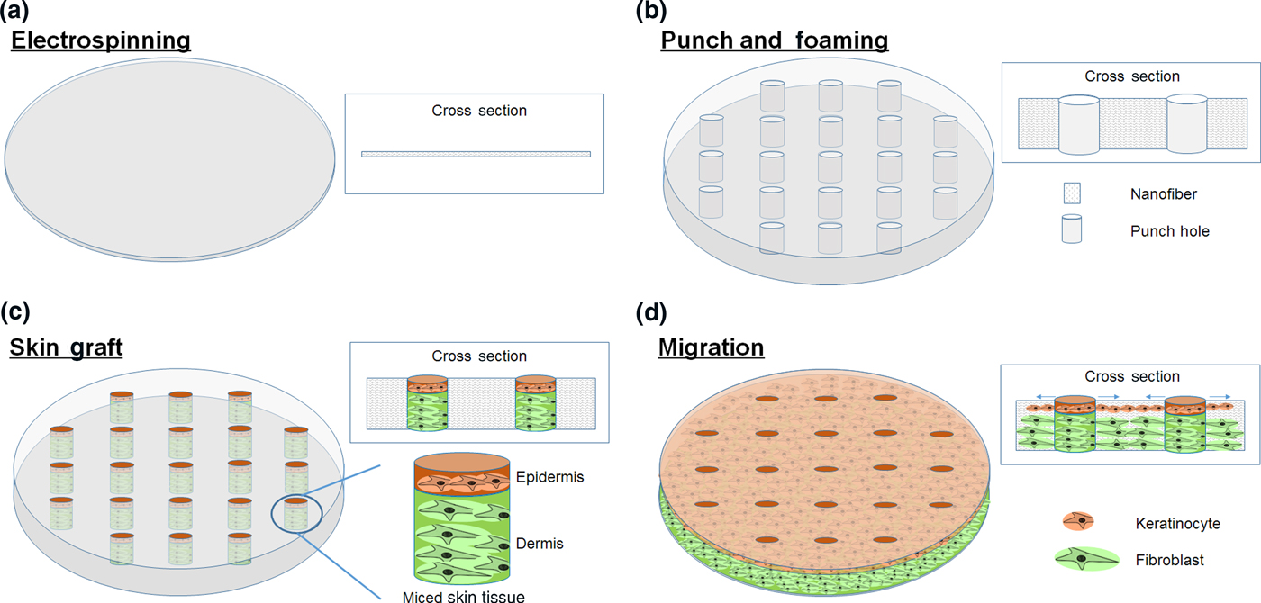
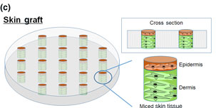
Our long-term goal is to use 3D expanded nanofiber scaffolds with arrayed holes together with minced autologous tissues (e.g., skin and bone) to form transplantable 3D tissue constructs for wound healing and bone regeneration. Compared with typical fibrous scaffolds (e.g., 2D nanofiber membranes and 3D expanded nanofiber scaffolds), it is expected that scaffolds with arrayed holes can greatly enhance cell penetration throughout the whole scaffold as the cells can migrate from both holes and surrounding areas of scaffolds (Fig. 1). The aim of the punched holes was to combine minced tissues (e.g., skin) with 3D expanded scaffolds for creation of 3D skin tissue constructs for wound healing and skin regeneration. To meet this goal, we firstly fabricated 2D nanofiber membranes using traditional electrospinning.[Reference Ma, Xie, Jiang and Wu9, Reference Xie, Zhong, Ma, Shuler and Lim12] Comparing with poly(ε-caprolactone) (PCL), the blend with gelatin renders the fiber scaffolds better biocompatibility and faster degradation rate.[Reference Maji, Dasgupta, Pramanik and Bissoyi13] In order to prepare a highly hydrophilic 3D nanofibrous scaffold, we punched arrayed holes throughout 2D scaffolds and then transformed 2D membranes into 3D nanofiber scaffolds using a modified gas-foaming technique.[Reference Jiang, Carlson, Teusink, Wang, MacEwan and Xie10, Reference Jiang, Li, Wang, Wang, Carlson, Teusink, MacEwan, Gu and Xie11] To demonstrate the proof-of-concept, green fluorescent protein-labeled human dermal fibroblasts (GFP-HDF) cell spheroids were prepared and seeded into the arrayed holes of 3D nanofiber scaffolds. The cell adhesion and migration were examined in vitro. In terms of composition of scaffolds, we chose a blend of PCL (a synthetic biodegradable and biocompatible polymer) and gelatin (a natural polymer) both approved by Food and Drug Administration for certain clinical applications.[Reference Kweon, Yoo, Park, Kim, Lee, Lee, Oh, Akaike and Cho14, Reference Ungerleider and Christman15]
Schematic illustrating the fabrication of nanofiber skin grafts and the proposed mechanism for chronic wound healing. (a) Nanofiber membrane generated by electrospinning. (b) Punch arrayed holes throughout nanofiber membranes and expand the membrane in the third dimension using the modified gas-foaming method. (c) Seed minced skin tissues to the arrayed holes to form nanofiber skin grafts ready for implantation to the chronic wound. (d) After implantation, cells migrated from minced skin tissues to the surrounding areas to form 3D tissue constructs and heal the chronic wound.

Materials and methods
PCL pellets (M w = 80,000) and gelatin powder of porcine skin were purchased from Sigma-Aldrich (St. Louis, MO). Hexafluoro-2-propanol (HFIP) was purchased from Oakwood Chemical, Inc. (Estill, SC). Glutaraldehyde in ethanol was bought from Ladd Research (Williston, VT). The [3-(4,5-dimethylthiazol-2-yl)-2,5-diphenyltetrazolium bromide] (MTT), Dulbecco's modified eagle medium, and phosphate buffer saline were obtained from Sigma-Aldrich (St. Louis, MO). Fetal bovine serum was received from Thermo Fisher Scientific (Waltham, MA) and GFP-HDF was gifted from Dr. Mark A. Carlson.
Nanofiber mats were produced utilizing an electrospinning setup following our previous studies.[Reference Jiang, Carlson, Teusink, Wang, MacEwan and Xie10] The parameters for electrospinning were as follows: 5% PCL and gelatin at a ratio of 1 : 1 (v/v) in HFIP, a flow rate of 0.4 mL/h, an applied voltage of 18 kV, a spinneret (a 22-gage needle), and a rotating drum located 20–23 cm away. The morphology of fiber samples was examined using scanning electron microscopy (SEM) (FEI, OR). In a sealed box, the PCL/gelatin nanofiber mats were crosslinked for 24 h with glutaraldehyde vapor at room temperature. To improve the hydrophilicity, plasma treatment (Harrick Plasma Inc., Ithaca, NY) at medium RF level was used to treat PCL/gelatin nanofiber mats for 1 min. The arrayed holes on the nanofiber mats were punched in liquid nitrogen. The nanofiber mats with arrayed holes were then expanded to form 3D scaffolds using a modified gas-foaming technique following our established protocols.[Reference Jiang, Carlson, Teusink, Wang, MacEwan and Xie10, Reference Jiang, Li, Wang, Wang, Carlson, Teusink, MacEwan, Gu and Xie11] Briefly, the nanofiber mats were immersed in freshly prepared 1% NaBH4 solutions at room temperature for 5–30 min. The expanded scaffolds were then gently rinsed three times with distilled water for 10 min each and finally freeze-dried overnight. The scaffolds were sterilized with ethylene oxide prior to cell culture. The morphology of the nanofiber scaffold was examined using SEM. One drop of distilled water was added to the surface of fibers and kept for at least 5 min. The water stability of PCL/gelatin nanofibers before and after crosslinking was examined using SEM.
A hanging drop method was used to generate GFP-HDF cell spheroids.[Reference Foty16, Reference Fennema, Rivron, Rouwkema, van Blitterswijk and de Boer17] Each drop contained 20 µL of cell suspension the concentration of which was 106 cells/mL. Cell spheroids of GFP-HDF were seeded to the surface of 2D PCL/gelatin scaffolds and the holes of 3D scaffolds. The samples were observed after 5 days of incubation. A fixative, which contains 2% glutaraldehyde and 2% paraformaldehyde in 0.1 M Sorensen's phosphate buffer (SPB), was used to fix the samples at 4 °C for overnight. The samples were washed three times using 0.1 M SPB and fixed using 1% osmium tetroxide solution for 30 min. The samples were washed with SPB and dehydrated in graded ethanol. Hexamethldisilazane solution was used to wash samples for 10 min and repeated for three times. The cell-seeded scaffolds were dried overnight. All the samples were fixed on a metallic stud with double-sided conductive tape and coated with platinum using a sputter coater. SEM images were acquired at an accelerating voltage of 15 kV.
Results and discussion
PCL/gelatin nanofibers were generated by electrospinning under different conditions (Supplementary Fig. S1). It appears that the fiber diameter gradually became uniform with decreasing the PCL/gelatin concentration from 10 to 5% (w/v). The smooth and uniform fibers were obtained when the flow rate was 0.4 mL/h and the PCL/gelatin concentration was 5% (w/v) [Supplementary Fig. S1(b) and S1(d)]. By controlling rotating speed of the drum collector, aligned and random fibers were obtained (Supplementary Fig. S2). The orientation of fibers was confirmed by a fast Fourier transform analysis (Supplementary Fig. S2). The chosen of PCL/gelatin as raw materials for nanofiber scaffold fabrication was because the combination of synthetic polymer and nature polymer can have an optimum mechanical property, biocompatibility, and degradation rate. The gelatin is water soluble, necessitating crosslinking to preserve the fiber morphology in an aqueous environment. The glutaraldehyde vapor was used to crosslink PCL/gelatin nanofibers for maintaining their structural integrity since glutaraldehyde has been widely applied to covalently cross-link functional groups in natural polymers, such as gelatin and collagen.[Reference Kim, Jun, Shin, Jang, Kim and Shin18] It is found that the fiber morphology of PCL/gelatin nanofibers was mainly preserved except for some minor swelling after water treatment (Supplementary Fig. S3). To examine the potential cytotoxicity of cross-linked PCL/gelatin nanofibers, relative growth rates (RGR) of GFP-HDF were quantified when incubating with extractions of various fiber samples (Supplementary Fig. S4). RGR of cross-linked and non-cross-linked PCL/gelatin nanofibers were higher than 80%, indicating a marginal cytotoxicity. To further reduce the cytotoxicity, a more benign cross-linking method could be used such as photo-crosslinker, genipin or natural phenolic compounds.[Reference Mazaki, Shiozaki, Yamane, Yoshida, Nakamura, Yoshida, Zhou, Kitajima, Tanaka, Ito, Ozaki and Matsukawa19–Reference Zhang, Do, Casey, Sulistio, Qiao, Lundin, Lillford and Kosaraju21]
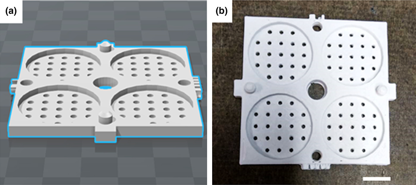
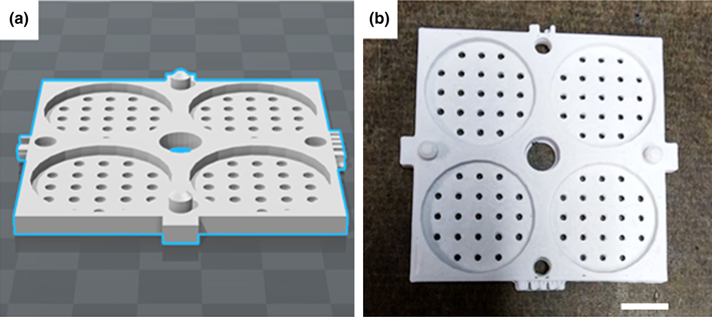
To form square arrayed holes throughout nanofiber membranes, we first immersed membranes into liquid nitrogen to make the materials brittle (below the glass transition temperature) and then created holes with a punch. Following our established protocols, we expanded nanofiber membranes with square arrayed holes in a mold generated by 3D printer using a modified gas foaming technique (Fig. 4).[Reference Jiang, Carlson, Teusink, Wang, MacEwan and Xie10, Reference Jiang, Li, Wang, Wang, Carlson, Teusink, MacEwan, Gu and Xie11] The nanofiber membrane was initially 0.4 mm thick and became about 4 mm thick after expansion. Figure 2 shows the morphology of a 3D nanofiber scaffold with arrayed holes. The hole was designed to fit the minced tissue and enhance cell infiltration/penetration to form 3D tissue constructs throughout the whole scaffold. As expected, the inner surface of punched holes displayed a layered structure [Fig. 2(c)]. The SEM image of cross sections indicated a layered and highly porous structure [Fig. 2(d)–2(f)].
Morphological characterizations of 3D PCL/gelatin (50 : 50) nanofiber scaffolds with arrayed holes. (a) Photograph of a 3D nanofiber scaffold with around 5 mm thick and arrayed holes. Scale bar = 5 mm. (b) The punched hole. Scale bar = 200 µm. (c) The corresponding highly magnified image of (b) showing layered structures. Scale bar = 50 µm. (d) Crosssections of the edge of 3D scaffolds. Scale bar = 100 µm. (e) Crosssections of the middle of 3D scaffolds. Scale bar = 100 µm. (f) The corresponding highly magnified image of (e) showing nanofiber layers after expansion. Scale bar = 10 µm The nanofibers were fabricated by electrospinning 5% PCL/gelatin (50 : 50) in HFIP. [Electrospinning parameters: flow rate = 0.4 mL/h, voltage = 18 kV, the distance between the collector (drum) and needle = 21 cm.]

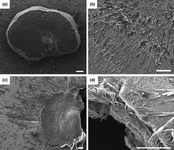
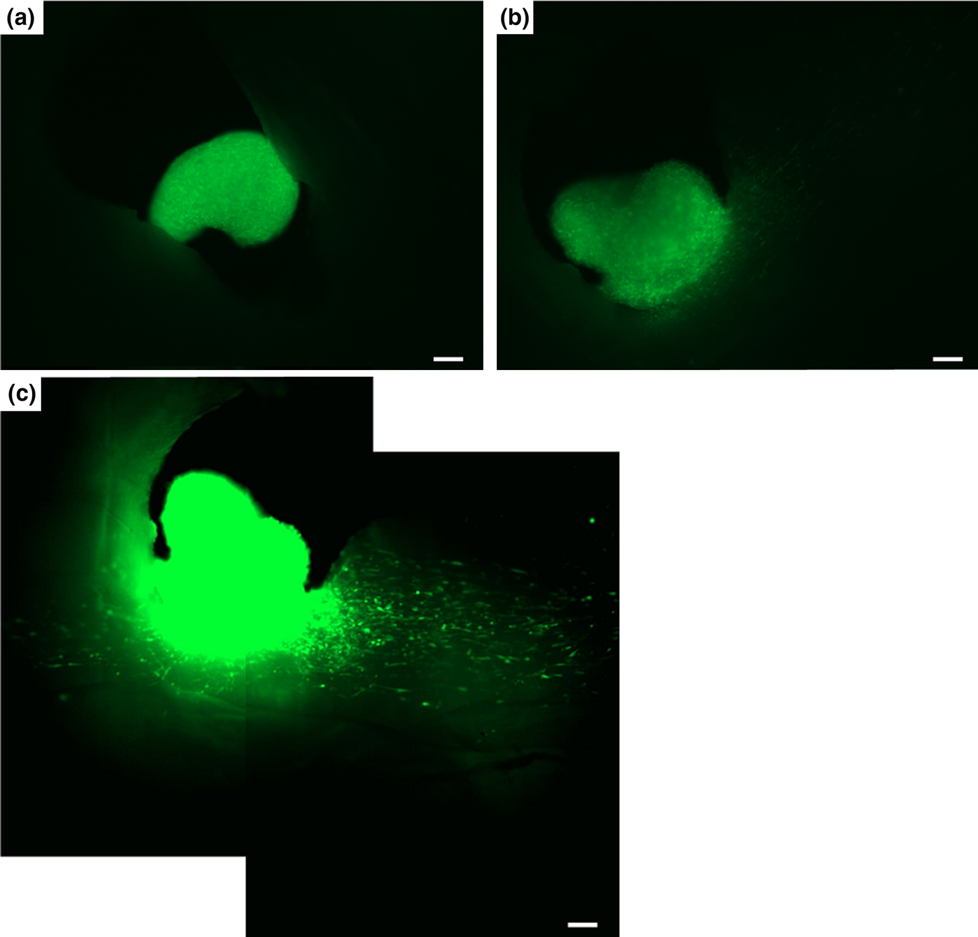
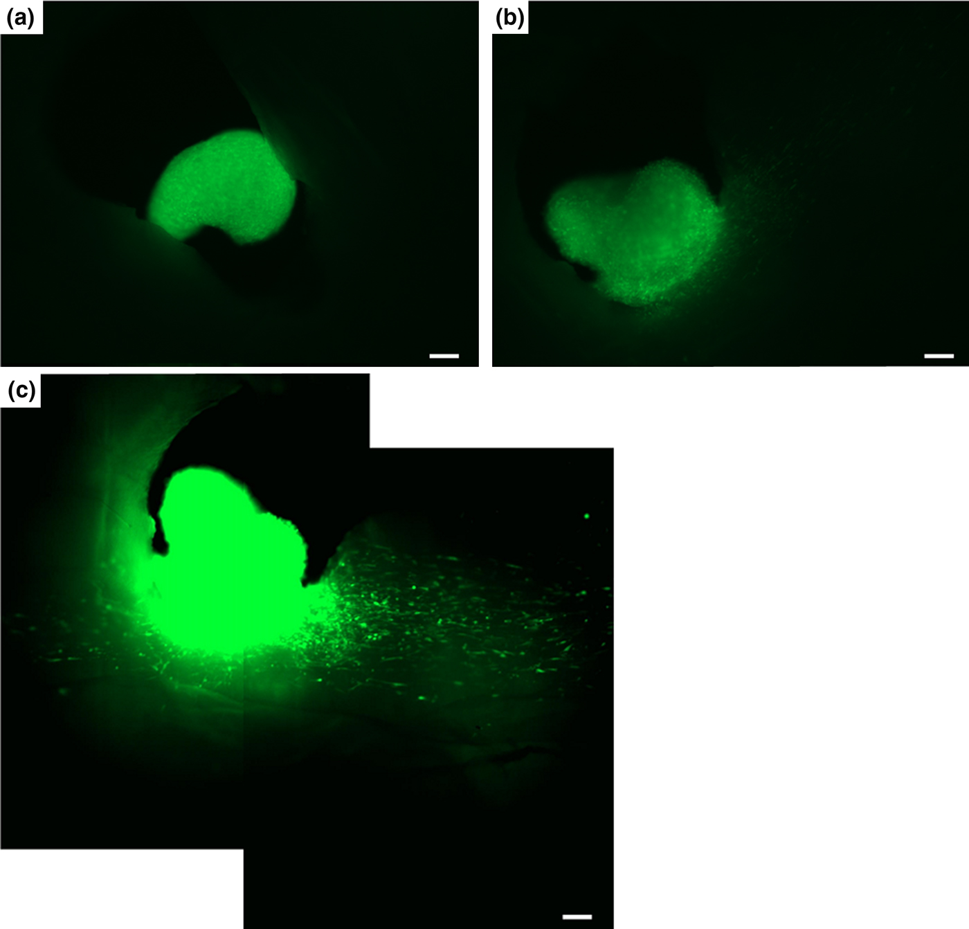
To demonstrate the proof-of-principle, we prepared GFP-HDF cell spheroids using a hanging drop method.[Reference Foty16] An average diameter of 442.35 ± 13.62 µm for cell spheroids was obtained after culture for 24 h. We then seeded cell spheroids to the 2D and 3D nanofiber scaffolds. After culture for 5 days, it is observed that fibroblasts migrated from the cell spheroids seeded to 2D nanofiber membranes adhered and proliferated on the surface only [Fig. 3(a) and 3(b)]. In contrast, fibroblasts that were migrated from cell spheroids seeded into the holes of 3D nanofiber scaffolds adhered to the surface and surrounding nanofiber walls of holes [Figs. 3(c) and 3(d) and 5]. Cells could further infiltrate into the scaffolds through the gaps between layers due to the layered and highly porous structures after expansion. The arrayed holes seeded with 3D cell spheroids showed a simultaneous cell infiltration and proliferation in three dimensions. It is expected that cells migrated from cell spheroids could infiltrate the surrounding space and form a 3D tissue construct. The plasma treated gelatin/PCL scaffolds showed better cell compatibility comparing with plasma-treated bare PCL scaffolds. All the cell spheroids came off from the PCL scaffolds during the culture. The cells migrated from the cell spheroids in 3D PCL/gelatin scaffolds and proliferated well on and in the scaffolds. We also vision that skin cells may migrate from the minced skin tissues seeded into the arrayed holes to the surrounding space and form 3D skin tissues for chronic wound closure. To provide a promising skin tissue construct, various methods of treatment have been suggested in the past, such as postage stamp grafting, mesh grafting, intermingled auto- and homograft transplantation, alternating strips of auto- and homograft transplantation, micro-skin grafting and the MEEK technique.[Reference Almodumeegh, Heidekrueger, Ninkovic, Rubenbauer, Hadjipanayi and Broer22] The presented 3D PCL/gelatin scaffolds can further decrease the need of large donor sites. It can also provide the physical protection and relieve the pain because its hydrophilicity can help out bathe the exposed nerve endings in wound bed.[Reference Doughty23] We reasonably foresee the application of this 3D scaffolds with arrayed holes as an ideal candidate for wound management including chronic wounds and acute wounds.
Cell spheroids-seeded 2D (a, b) and 3D (c, d) PCL/gelatin nanofiber scaffolds after incubation for 5 days. Scale bar = 100 µm.

(a) Mold design. (b) Mold generated by 3D printing. Scale bar = 10 mm.
GFP-HDF spheroid-seeded 3D PCL/gelatin nanofiber scaffolds after incubation for (a) 1, (b) 4, and (c) 5 days. Scale bar = 100 µm.
Utilizing this approach, these 3D nanofiber scaffolds together with cell spheroids or minced tissues may be able to create complex tissue architectures for wound healing, tissue regeneration, and engineering 3D in vitro tissue models.[Reference Chaisri, Chingsungnoen and Siri24, Reference Cesarz and Tamama25] The scaffolds can also combine with a variety of hydrogels or biologic matrices/cues to form 3D hybrid scaffolds with eliciting biologically functional molecules.[Reference Zanoni, Piccinini, Arienti, Zamagni, Santi, Polico, Bevilacqua and Tesei26–Reference Manning, Schwartz, Liu, Xie, Havlioglu, Sakiyama-Elbert, Silva, Xia, Gelberman and Thomopoulos28] In addition, these novel 3D scaffolds with cost-effective fabrication could likely lead to the drastic change in the utilization of electrospun nanofibers.
Conclusion
We have demonstrated a simple and reproducible approach to prepare 3D PCL/gelatin nanofiber scaffolds with arrayed holes. The crosslinking can significantly enhance the structural stability of nanofiber scaffolds in an aqueous environment. Seeded GFP-HDF spheroids displayed cell migration to the surface and surrounding nanofiber walls of punched holes. Combining 3D nanofiber scaffolds with arrayed holes and minced tissues could be a promising strategy for chronic wound healing, regenerative medicine, as well as engineering in vitro tissue models.
Supplementary material
The supplementary material for this article can be found at https://doi.org/10.1557/mrc.2017.49
Acknowledgments
This work was supported partially from startup funds from University of Nebraska Medical Center (UNMC) and National Institute of General Medical Science (NIGMS) grant 2P20 GM103480-06. The authors thank the McGoogan Library of Medicine at University of Nebraska Medical Center for use of the LulzBot TAZ 5 3D printer.