I. INTRODUCTION
Over the past few decades, the synthesis of inorganic nanomaterials with desired sizes and shapes has been intensively pursued due to their unique electronic, magnetic, optical, and catalytic properties with respect to multiple fundamental and practical technological applications. Reference Kim, Connor, Song, Kuykendall and Yang1–Reference Chen, Ko, Yen, Chen and Lin7 By means of further investigations on crystal growth kinetics and shape evolution, nanomaterials with different architectures have been successfully fabricated in the solution phase. Reference Sun and Xia8,Reference Lee, Habas, Kweskin, Butcher, Somorjai and Yang9 By means of the cooperative self-assembly of inorganic species and surfactants, where the surfactant surface aggregates as templates, the surfactant-assisted strategy is known to be a general and efficient method for the preparation of uniform inorganic nanomaterials with controllable sizes and shapes. Recently, various nano- and microstructures of metal oxides (particularly those of MnO2, SiO2, and V2O5) in the form of spheres, porous shapes, layers, tubes, wires, and rods have been successfully fabricated by using this approach. Reference Qiu, Huang, Dharmarathna, Benbow, Stafford and Suib10–Reference Nguyen and Do14
Manganese oxide (MnO2)—an important functional metal oxide—and its derivative compounds offer the following advantages: possibility of numerous attainable oxidation states of Mn, cost-effectiveness, variety of electrochemical behaviors, and environmental compatibility; because of these advantages, they have been the subject of intense investigations for a wide range of applications such as catalysts, water purification, water splitting, molecular absorption, and battery materials. Reference Huang, Lv, Yue, Attia and Yang15–Reference Kim, Ayalasomayajula, Gona and Choi17 In addition, they also provide reversible Faradaic (redox) reaction, appropriate electrode potential window, and high theoretical capacitance; therefore, they have been considered for use as a promising electrode material in electrochemical supercapacitors for yielding high power density, excellent reversibility, and long cycle. Reference Lee, Hall, Kim and Mallouk18,Reference Wei, Cui, Chen and Ivey19 More recently, manganese oxides with various crystal types, morphologies, and sizes have been systematically investigated with great enthusiasm for their unique properties that promote their performances in applications involving energy storage (e.g., supercapacitors). Reference Zhang, Liu, Lv, Tang and Ooi16,Reference Ghodbane, Pascal, Fraisse and Favier20,Reference Zhu, Shi, Xiao, Rui, Tan, Lu, Hng, Ma and Yan21 Among them, MnO2 structures with a spinel-type crystal phase of three-dimensional (3D) interconnected tunnels have been recently fabricated to obtain better capacitance performances in aqueous solutions as they can offer a larger number of electrolyte transport paths for electron transfer and proton/cation diffusion, thereby allowing enhanced charge transport efficiency through the electrodes during the charge/discharge process and leading to better supercapacitive performances. Reference Ghodbane, Pascal and Favier22 In addition, as we know, hierarchical microspheres assembled from small nanostructured particles have been reported to exhibit distinctive properties—different from those of the constituent nanoparticles—with various potential applications Reference Wang, Zhu and Tao23 ; therefore, the creation of such a shape of MnO2 is of great significance. Several types of morphologies of MnO2 materials have been previously reported including nanotubes, nanorods, nanowires, nanosheets, and nanoparticles Reference Qiu, Huang, Dharmarathna, Benbow, Stafford and Suib10,Reference Zhu, Shi, Xiao, Rui, Tan, Lu, Hng, Ma and Yan21,Reference Xia, Feng, Wang, Lai and Lu24–Reference Kai, Kobayashi, Yamada, Miyazaki, Abe, Uchimoto and Kageyama26 ; however, to the best of our knowledge, spinel-type MnO2 microspheres (MS-MnO2) constructed from small nanoparticles have not been investigated. In this paper, we report a simple and quick procedure for the one-pot synthesis of manganese oxides under a basic solvothermal condition in the presence of a cationic surfactant acting as a template in a 2-butanol/water solution for the direct synthesis of spinel-type MS-MnO2 comprising small MnO2 nanoparticles. The galvanostatic charge/discharge and cyclic voltammetry (CV) measurements in 1-M Na2SO4 aqueous electrolytes demonstrate that the MS-MnO2 display an improved specific capacitance (SC) of ∼190 F/g at 0.5 A/g. Interestingly, a superior SC retention of ∼100% and a stable Coulombic efficiency of ∼95% after 1000 cycles can be obtained, yielding excellent long-term charge/discharge cycling stability. In addition, by means of the solvothermal method, various morphologies of manganese oxides including schists, rods, fibers, and nanoparticles can be easily obtained by changing the ratio of 2-butanol and water and the nature of the precursors as well as cation surfactants. Therefore, the proposed method can provide a simplistic technique to fabricate MnO2 having novel architectures.
II. EXPERIMENTAL SECTION
A. Materials
Cetyltrimethyl ammonium bromide (CTAB), manganese sulfate (MnSO4·H2O), and (±)2-butanol used in the study were purchased from Sigma-Aldrich. Tetraethyl ammonium bromide (TEA; 98%) and potassium permanganate (KMnO4) were obtained from Alfa Aesar (UK) and SHOWA (Japan), respectively. All these reagents were used in the experiments without any further purification.
B. Preparation of MS-MnO2
In a typical experiment, the MnO2 precursor of the TEAMnO4 powder, which was presynthesized by using a modified Brock method by the cation-exchange process that involved the replacement of a hard potassium cation of the permanganate precursor with a soft organic TEA cation, Reference Brock, Sanabria, Suib, Urban, Thiyagarajan and Potter27 was dissolved in 2 mL of deionized water to yield a homogeneous solution; then, the solution was added to 10 mL of 2-butanol under stirring, where 2-butanol acted as a solvent as well as a reducing agent and TEA served as a template. The mixed solution was subsequently sealed in a Teflon-lined autoclave and heated to 120 °C at the rate of 5 °C/min and maintained at this temperature for 3 h. This aging process accompanied an increase in the pH, from the neutral pH of the initial sols up to pH of 11–12. After the solvothermal process, the precipitates (brownish-black color) were collected by centrifugation and washed in sequence with deionized water to remove the possible unreacted material. The final products of the MnO2 materials, denoted as MS-MnO2, were dried in vacuum desiccators at the ambient temperature.
C. Characterization
Powder x-ray diffraction patterns were recorded by an analytical X’Pert Pro MRD equipped with Cu Kα as the x-ray source (λ = 0.15418 nm). An operational voltage and current of 45 kV and 40 mA, respectively, were used and the data were collected stepwise in the 2θ range of 10–80° with a scan step of 0.033°/s. A Zeiss Ultra Plus field emission scanning electron microscope (FE-SEM; Germany) was used to examine the morphology and size of the as-prepared MS-MnO2. A JEM 2010 high-resolution transmission electron microscope (HRTEM; Japan) operated at 200 kV was used to examine the morphology and lattice structure of the MS-MnO2. The samples used in the HRTEM analysis were prepared by placing a drop of the MS-MnO2 aqueous suspension onto a copper grid covered with a carbon film and evaporating the drop in air. Thermal gravimetric analysis (TGA) curves were obtained using a Perkin-Elmer TGA-7lab instrument. In a typical analysis, approximately 10 mg of the as-synthesized MS-MnO2 sample was analyzed in a platinum pan at a heating rate of 5 °C/min under nitrogen atmosphere with a flow rate of 150 mL/min.
D. Electrochemical measurements
All the electrochemical measurements were carried out in a 1-M Na2SO4 aqueous electrolyte solution at room temperature by using the 672A electrochemical system (CH Instruments, Austin, TX) with a three-electrode cell that was composed of a MS-MnO2-based electrode as the working electrode, a Pt film as the counter electrode, and an Ag/AgCl electrode as the reference electrode. The working electrodes were fabricated using a mixture of 70 wt% MS-MnO2 powder with 25 wt% carbon black and 5 wt% poly(tetrafluoroethylene) (PTFE) binder; subsequently, a small amount of alcohol was added into the mixture to form a clay-like paste. The paste was then incorporated in nickel foam (1 × 1 cm), dried in a vacuum oven at room temperature, and followed by a compression treatment under a pressure of 6000 kg/cm2 for 5 min to ensure that the active material completely adhered to the nickel foam. The CV measurements were performed between 0 and 0.9 V (versus Ag/AgCl) for scan rates in the range of 2–100 mV/s. The charge/discharge properties were examined galvanostatically in the potential range of 0–0.9 V (versus Ag/AgCl) at current densities of 0.5 and 1 A/g. The SC of the fabricated electrodes was calculated according to the relation C (F/g) = IΔt/ΔVm, where I denotes the constant discharge current, Δt is the discharge time, m is the mass of the active materials (g), and ΔV is the total potential difference (V).
III. RESULTS AND DISCUSSION
Figures 1(a)–1(e) show the FE-SEM images of the typical samples of the MnO2 material prepared using our approach, revealing the morphological evolution with the reaction time. A series of impressive microspheres were observed at the reaction times in the range of 3–6 h [Figs. 1(b)–1(c)]; when the reaction time was above 12 h, a needle-like morphology of manganese oxides was generated [Fig. 1(d)]. Notably, the population of such shapes increased with an increase in the reaction time, whereas the population of the microspheres decreased concomitantly. After 48 h, nearly all the initial microspheres were converted into such needle-like structures, as shown in Fig. 1(e). These observations clearly show that the MnO2 composites with a needle-like architecture evolve from the initial microspheres. From the above observations, we can show that the optimal condition for the production of attractive MS-MnO2 can be obtained under a solvothermal condition at a heating temperature of 120 °C and a gel time of 3 h.
FE-SEM images of the as-synthesized MnO2 materials under solvothermal conditions and heating at 120 °C for (a) 1 h, (b) 3 h, (c) 6 h, (d) 12 h, and (e) 48 h.

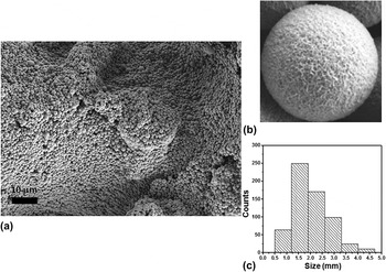
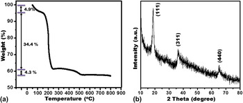
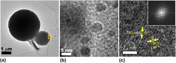
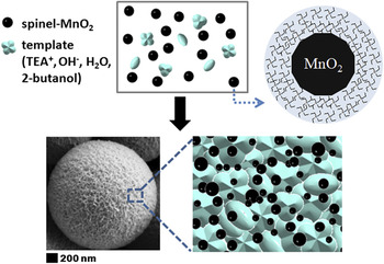
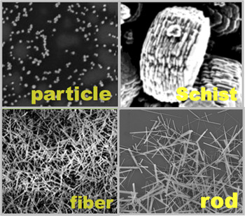
A large number of microspheres can be clearly seen in the FE-SEM image shown in Figs. 2(a) and 2(b), with the size distribution ranging from 0.5 to 4.5 µm [Fig. 2(c)]. The corresponding TGA profile, shown in Fig. 3(a), indicated that the thermal decomposition curve of MS-MnO2 involved three weight-loss steps: the first weight loss of 4.9% occurred below 145 °C, which was due to the evaporation of physically adsorbed water molecules in MS-MnO2, followed by a second weight loss of 34.4% between 145 and 230 °C, which was caused by the release of the TEA+ species according to the Hofmann degradation reaction; in the third step, a weight loss of 4.3% in the temperature range of 230–500 °C was demonstrated, which was associated with the combustion of remaining TEA and the phase transformation of MnO2 to Mn2O3 and Mn3O4. Reference Ghodbane, Pascal and Favier22,Reference Camblor, Corma and Valencia28 Importantly, a weight loss of 43.6% after calcination of MS-MnO2 was observed, and the weight loss of sample at temperatures 145–230 °C was significantly greater than that at any other heating temperature, which suggests that the TEA-based template played a significant role in the formation of the spinel MnO2 microspheres. To understand the crystalline structures of the as-prepared MS-MnO2 material, its corresponding XRD measurement was carried out, as shown in Fig. 3(b). All the peaks can be indexed as pure spinel-type MnO2 (JCPDS no. 44-0992), where it can be shown that the three main peaks at 2θ = 18.73°, 36.14°, and 65.09° can be correspondingly indexed to the (111), (311), and (440) reflections of the spinel-type MnO2; this indicates that the as-prepared products are phase-pure spinel-type MnO2 microspheres. To further examine the configuration of these microstructures, the HRTEM images were recorded. From Fig. 4(b), it is revealed that the MS-MnO2 material is composed of numerous spherical nanoparticles with an average diameter of 5 nm; interestingly, it is distinctly visible that these nanoparticles are surrounded by a TEA-based template. The corresponding clear lattice fringes and fast-Fourier-transformed diffraction pattern data of the individual small spherical nanoparticles of MS-MnO2, as shown in Fig. 4(c), indicate their efficient crystallization; additionally, two clear lattice fringes with basal distances of 2.02 Å and 1.86 Å can be observed, which are consistent with the (400) and (331) planes of the spinel-phase MnO2, respectively. These results indicate that the spinel-type MS-MnO2 microstructures are, in fact, built from these small nanoparticles that are spontaneously assembled through the assistance of the TEA-based template, finally forming a 3D network of spherical microstructures (Fig. 5). This is the first time that the spinel-type MnO2 with the shape of a microscopic sphere has been fabricated; further, we believe that it can offer additional electrolyte-transport paths for electron transfer and proton/cation diffusion due to its 3D interconnected tunnel crystal phase and unique microspherical architecture constructed from small nanoparticles. This architecture permits enhanced charge-transport efficiency through the electrodes during the charge/discharge process and leads to better supercapacitive performance. The considerable impact of the morphology on the electrochemical properties of MnO2 materials has been pointed out Reference Bach, Henry, Baffier and Livage29 ; therefore, much effort has been expended in the controllable synthesis of MnO2 materials with desired and uniform structures for energy storage applications. In this paper, we also employed the proposed one-pot solvothermal method for the successful synthesis of MnO2 with different architectures, including nanoparticles, fibers, rods, and schists, by changing the ratio of 2-butanol and water and the nature of the precursors as well as the cation surfactants, as shown in Fig. 6.
(a, b) FE-SEM image and (b) size distribution of the as-synthesized MS-MnO2 using the solvothermal method and heating at 120 °C for 3 h.

(a) TGA data and (b) XRD pattern of the as-synthesized MS-MnO2 using the solvothermal method and heating at 120 °C for 3 h.

(a) TEM image of the MS-MnO2 fabricated using the solvothermal process and heated at 120 °C for 3 h. (b) HRTEM image of the fringe region of an individual MS-MnO2 sample, indicating that MS-MnO2 was constructed from sphere-like nanoparticles surrounded by surfactants. (c) HRTEM image of an individual sphere-like nanoparticle of MS-MnO2 taken along the [011] axis and its corresponding Fourier-transformed diffraction pattern (inset), which can be indexed as the spinel-type structure of MnO2.

Schematic illustration of the solvothermal formation of the as-prepared spinel-type MS-MnO2.

FE-SEM images of the synthesized MnO2 materials through the developed solvothermal method and heating at 120 °C for 3 h under different experimental conditions and changing the ratio of 2-butanol and water, nature of the precursors, and cation surfactants. See also SI, Table 1.

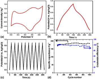
For testing the electrochemical performances of the spinel-type MS-MnO2 materials, galvanostatic charge/discharge measurements and CV measurements were carried out in 1-M Na2SO4 aqueous electrolytes by means of a conventional three-electrode system (Fig. 7). Broad redox peaks were obtained in the spinel-type MS-MnO2 which illustrated that the intercalation/deintercalation of protons and electrolyte cations of Na+ ions into the bulk material accompanied by a Faradaic reaction occurred in the spinel-type MS-MnO2 materials [Fig. 7(a)] Reference Brousse, Toupin, Dugas, Athouel, Crosnier and Belanger30 ; the Faradaic reaction can be a beneficial effect by the combination of surface-based double-layer capacitance and volume-based “pseudocapacitance,” leading to boost the energy current density of the 3D transport-pathway-based spinel-type MS-MnO2 electrode and resulting in improved capacitance performances. The SC values of the spinel-type MS-MnO2 were estimated through galvanostatic charge/discharge cycling at 0.5 A/g in 1-M Na2SO4 aqueous solution, as shown in Fig. 7(b). It can be clearly observed that the spinel-type MS-MnO2 exhibits a good SC value (190 F/g); significantly, the obtained SC values are competitive with those of other reported spinel-type MnO2 materials, including the slightly truncated nanoparticles (21 F/g at 20 mV/s), Reference Devaraj and Munichandraiah31 interconnected nanofibers (241 F/g at 5 mV/s), Reference Ghodbane, Pascal and Favier22 and particles with polyhedral shapes (53 F/g at 10 mA/cm2). Reference Xue, Chen, Zhang and Yan32 The cycling stability can be an area of concern for the practical applications of supercapacitor electrodes; therefore, an endurance test at a constant current density of 1 A/g for 1000 cycles was conducted for the as-synthesized spinel-type MnO2 electrode [Figs. 7(c) and 7(d)]. Almost 100% of the initial capacitance was retained and the corresponding Coulombic efficiency remained significantly high at ∼95% in the entire cycling process. Such results indicate that the proposed spinel-type MS-MnO2 material with 3D transport tunnels has the potential application as a supercapacitor electrode that can be used in long charge/discharge cycling without any significant microstructural modification or phase changes in the crystals of the MS-MnO2 material and without any dissolution of the active material of the electrode during the charge/discharge processes.
(a) Representative CV curves (third curve) of the as-prepared spinel-type MS-MnO2 material at a scan rate of 5 mV/s in 1-M Na2SO4 aqueous solution using a three-electrode system with Ag/AgCl as the reference electrode and Pt wire as the counter electrode. (b) Galvanostatic charge/discharge cycling tests recorded at a constant current density of 0.5 A/g for MS-MnO2 in 1-M Na2SO4 aqueous electrolyte. (c) Galvanostatic charge/discharge curves and (d) relationship of the SC and Coulombic efficiency with the number of cycles for the spinel-type MS-MnO2 electrode measured at a current density of 1 A/g in 1-M Na2SO4 aqueous electrolyte.

IV. CONCLUSION
In this paper, we have described the one-pot synthesis of a 3D network of spinel-type MS-MnO2 with the size distributions in the range of 0.5–4.5 µm; these microspheres are built from small MnO2 nanoparticles with average sizes of 5 nm assembled using the cationic surfactant as the template. Such spinel-type MnO2 microspheres have been fabricated for the first time by means of a solvothermal method in a 2-butanol/water solution under a basic condition. CV and galvanostatic charge/discharge measurements were carried out to characterize the fabricated MS-MnO2 material in the form of a supercapacitor electrode; its SC value was calculated to be ∼190 F/g. Furthermore, approximately 100% of the SC retention and approximately 95% of the Coulombic efficiency were obtained after 1000 charge/discharge cycles, exhibiting its excellent long-term stability. Significantly, the proposed solvothermal method can enable the morphological tuning of MnO2, which has been successfully used to obtain architectures of schists, rods, fibers, and nanoparticles. Therefore, this method can possibly offer an attractive technique toward synthesizing other metal oxides having the form of novel morphologies that can widen the range of its applications in biosensors, catalysis, fuel cells, and electronic nanodevices.
Supplementary Material
Supplementary materials can be viewed in this issue of the Journal of Materials Research by visiting http://journals.cambridge.org/jmr.
ACKNOWLEDGMENTS
We gratefully acknowledge the financial support from National Science Council of Taiwan (Grant Nos. NSC-101-2113-M-005-014-MY3 and NSC 101-2628-M-007-006). We also appreciate Dr. Lih J. Chen at National Tsing Hua University, Taiwan, for providing technical assistance in HRTEM measurements.