Introduction
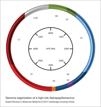
Human papillomaviruses (HPVs) are a heterogeneous group of nonenveloped, double-stranded, circular DNA viruses of 8000 nucleotides in size that infect the basal layer of the epithelium of skin (cutaneous HPV types) and mucous tissues (mucosal HPV types) (Ref. Reference zur Hausen1), (Fig. 1). Today, more than 150 HPV genotypes have been completely sequenced with the distinct HPV genotypes to have different life cycle features and oncogenic potential (Refs Reference Chen, de Freitas and Burk2, Reference Tommasino3, Reference Bernard4). A phylogenetic tree construction based on the nucleotide sequence of the L1 gene classified the HPV types into five genera – alpha, beta, gamma, mu and nu (Refs Reference Bernard4, Reference Bernard, Calleja-Macias and Dunn5, Reference Doorbar6). The alpha genus (Alphapapillomavirus) consists of 62 HPV types that infect the mucosal epithelium (Refs Reference Chen, de Freitas and Burk2, Reference Bernard4, Reference de Villiers7) The Alphapapillomaviruses are further classified into high-risk (HR) types and low-risk (LR) types, according to their potential to cause anogenital cancer (Refs Reference zur Hausen1, Reference Bernard4, Reference Bernard, Calleja-Macias and Dunn5).
Genome organisation of a HR Alphapapillomavirus. The viral genome is a small (8 kb in size), circular DNA that is divided into three regions: (i) the long control region (LCR), (ii) the early region and (iii) the late region. The LCR harbours binding sites for the E1 and E2 regulatory proteins as well as for cellular transcription factors. The early region encodes for the E1, E2, E4, E5, E6 and E7 genes and the late region encodes for the L1 and L2 genes.

HR-HPV infection frequently results in cervical cancer development (Ref. Reference Tommasino3). Cervical cancer is the third most common malignancy in women and the second most frequent cause of cancer-related death worldwide (Refs Reference Jemal8, Reference Forman9). Epidemiological analyses revealed that the most frequently identified HR-HPV genotype is HPV16, which is followed in prevalence by HPV18 and approximately 70% of cervical cancer cases are caused by these two genotypes (Refs Reference de Sanjose10, Reference Li11). Different triage approaches for HR-HPV-positive women have been estimated, including cytology (Ref. Reference Ronco12), p16/Ki67 immunostaining (Ref. Reference Ikenberg13), the detection of methylation of viral and/or human genes (Refs Reference Snellenberg14, Reference Brentnall15) and the identification of HR HPV DNA/RNA (Refs Reference Castle16, Reference Cox17).
Cancer of the uterine cervix is associated with persistent HR-HPV infection and increased expression of HPV E6/E7 oncogenes (Refs Reference McLaughlin-Drubin and Munger18, Reference Howie, Katzenellenbogen and Galloway19, Reference Moody and Laimins20, Reference Doorbar21). Persistent infection with HR-HPV genotypes triggers the integration of HPV DNA into the host genome that eventually leads to the accumulation of chromosomal damages and genome destabilisation in the infected cells (Refs Reference Hudelist22, Reference Cricca23, Reference Li24, Reference Boulet25, Reference Akagi26, Reference Tsakogiannis27) (Figs 2 and 3). The frequency of viral integration increases with the severity of cervical intraepithelial neoplasia (CIN) of stages I, II, III and the integrated form of the HPV DNA is found in the vast majority of cervical cancer cases (Refs Reference Doorbar21, Reference Akagi26, Reference Tsakogiannis27, Reference Wentzensen, Vinokurova and von Knebel Doeberitz28, Reference Xu29, Reference Tsakogiannis30). Over the last decades several studies have focused on the analysis of the physical status of HPV DNA considering its causal association with cervical cancer development. The identification of integrated viral genomes in clinical samples is regarded as a valuable prognostic tool that facilitates the accurate prediction of cervical cancer development (Refs Reference Hudelist22, Reference Cricca23, Reference Li24, Reference Boulet25, Reference Tsakogiannis27, Reference Tsakogiannis30, Reference Theelen31). In the present review, we describe molecular approaches for HPV-DNA detection and genotyping for intra-lesion analyses and review molecular assays for the determination of HPV-DNA physical status. Finally, we discuss the criteria for selecting the most appropriate regions of viral DNA to be incorporated in HPV genotyping and in the determination of HPV-DNA form in clinical samples.
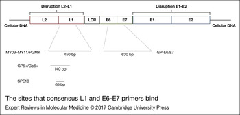
The sites that consensus L1 and E6–E7 primers bind. Upon integration, the L1 gene can be disrupted, while the E6 and E7 genes always remain intact. Thus, PCR with primers that target the L1 gene might lead to false-negative results when HPV-DNA integration occurs through disruption of the L1 gene.

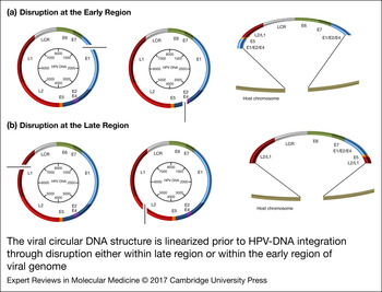
The viral circular DNA structure is linearised prior to HPV-DNA integration through disruption either within late region or within the early region of viral genome. The most frequent sites of disruption are located within the E2 and E1 genes (a), while disruption events have been also reported within the L1 and L2 genes (b). Despite the site of viral DNA disruption the LCR and E6, E7 oncogenes remain intact and integrated into the host DNA.

Food and Drug Administration (FDA) approved methods for HPV genotyping
Cytology-based tests have reduced the cervical cancer incidents and mortality rates, but they have low specificity as well as significant variability in the diagnosis of precancerous lesions. The strong causal relationship between HPV and cervical disease stimulated the development of molecular techniques that are based on the identification of HR-HPV DNA/RNA (Ref. Reference Abreu32). These assays are the gold standard for identifying the presence of viral infection and they permit early diagnosis and sufficient treatment of precancerous cases (Refs Reference Castle16, Reference Cox17, Reference Schiffman and Wentzensen33). The American Society for Colposcopy and Cervical Pathology (ASCCP; 2006, 2009 and 2012) proposes that women with a negative cytopathological test, but with a positive HPV test should be subjected to HPV genotyping screening. It is regarded that the identification of HR HPV16 and HPV18 in clinical samples provides valuable information concerning the triage of colposcopy of HR-HPV-positive women who have greater risk to develop more severe dysplasias (Refs Reference Castle16, Reference Cox17). The molecular techniques for HPV-DNA detection have been divided into three categories, including nucleic acid-hybridisation, signal amplification and target amplification methods (Refs Reference Castle34, Reference Cuzick35). Several of these assays have been commercialised and used in HPV diagnosis (Ref. Reference Abreu32).
Over the last years, the US FDA has approved a total of five HPV assays. In 2003, FDA first assigned the Hybrid Capture 2 (HC2) for HPV detection in cervical scrapings and formalin-fixed, paraffin-embedded tissues (FFPE). This method is a signal-amplification assay based on the use of labelled RNA probes that are hybridised with the target HPV-DNA. The method detects 13 HR HPV genotypes (16, 18, 31, 33, 35, 39, 45, 51, 52, 56, 58, 59 and 68) or 5 LR HPV genotypes (6, 11, 42, 43 and 44). Briefly, DNA is isolated from the exfoliated cervical cells and is denatured to produce single-stranded DNA targets. Subsequently, the RNA probes are hybridised to their specific HPV-DNA targets and the DNA–RNA hybrids are transferred to a microwell plate harbouring captured antibodies, specific to DNA–RNA hybrids immobilising them. Finally, DNA–RNA hybrids react with antibodies conjugated with alkaline phosphatase. With the presence of chemiluminescence substrate, emitted light indicates the presence of a positive result (Ref. Reference Bozzetti36). Although the assay is easy to perform, it is not able to carry out single HPV genotyping, since it is designed to distinguish HR and LR HPV groups. The assay presents high sensitivity (87–96%) for detecting high-grade CIN, but relatively low specificity (20–85%) (Refs Reference Castle16, Reference Ratnam37, Reference Luu38, Reference Meijer39) (Table 1). Finally, the assay can be used for semiquantitative viral loads of HPV (Ref. Reference Castle40). The cost of HC2 is estimated roughly at 40–60$ per test (Refs Reference Gravitt41, Reference Fotiadis42).
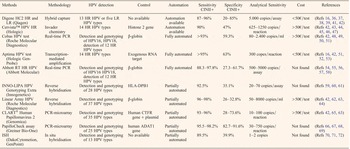
Brief description of FDA and non-FDA approved commercialised HPV tests.

The detected HPV genotypes, the availability of automation, the clinical sensitivity – specificity, the analytical sensitivity and the molecular approach of each individual method are reviewed and the cost of HPV tests is mentioned (Ref. Reference Fotiadis42)
Two more tests, the Cervista™ HPV HR and Genfind™ DNA Extraction Kit, and the Cervista™ HPV16/18 were approved by FDA in 2009 for identifying HPV DNA in cervical scrapings. The assay is a signal-amplification method for the detection of nucleic-acid sequences and is based on two simultaneous isothermal and signal amplification reactions to detect the specific nucleotide sequences. Briefly, a cleavage enzyme cleaves a type-specific probe when it overlaps an invader oligonucleotide at the target DNA recognition site. Subsequently, the 5′ region of the cleaved probe binds to universal hairpin fluorescence resonance energy transfer oligonucleotide, thus creating a second substrate for the cleavage enzyme. Finally, cleavage of the resonance energy transfer oligonucleotide between the fluorophore and quencher molecule lead to production of a fluorescence signal. Moreover the assay detects the human histone 2 as internal control. The Cervista™ HPV HR and Genfind™ DNA Extraction Kit detects 14 HR HPV types (the 13 HR HPV type that detects HC2 plus genotype 66) (Refs Reference Day43, Reference Einstein44). The Cervista™ HPV16/18 detects only HPV16 and HPV18 genotypes and may be used in conjunction or as a follow up of Cervista™ HPV HR test. The clinical sensitivity and specificity of Cervista™ HPV HR for the detection of CINII+ is 90 and 47% respectively and 95 and 87% for CINIII+, as well (Refs Reference Belinson45, Reference Tao46) (Table 1). Comparative analyses between HC2 and Cervista™ HPV HR test revealed that both tests have minor differences in clinical sensitivity and specificity for the detection of CINIII and cancer (Refs Reference Belinson45, Reference Tao46). In contrast to HC2 the Cervista™ HPV HR test contains an internal control that eliminates the false-negative results, while the automated integrated DNA extraction procedure reduces the steps, the time of the assay and it eliminates the specimen contamination prior to HPV test. Nevertheless, the assay has a high false-positive rate in cytology normal samples (Ref. Reference Boehmer47).
In 2011, FDA assigned the Cobas HPV test that is a highly automated real-time PCR assay, which is based on simultaneous detection and individual genotyping of the viral genotypes HPV16 and HPV18 and pooled detection of 12 more HR-HPV genotypes (HPV31, 33, 35, 39, 45, 51, 52, 56, 58, 59, 66 and 68) in cervical scrapings. The preparation of specimens is automated and is performed in the Cobas × 480 instrument. Briefly, after the lysis and digestion of cells the extracted DNA binds to magnetic glass particles. Subsequently, the extracted DNA is subjected to real-time PCR (Ref. Reference Rao48). The real-time PCR methodology uses a set of 16 primers to amplify a fragment of the L1 gene from the respective HPV genotypes and Taqman probes labelled with spectrally unique fluorescent dyes. The Taqman probes have a reporter dye at their 5′ end and a quencher dye at their 3′ end. When the target sequences are present the oligonucleotide probes are hybridised to the target sequence and subsequently they are cleaved by the 5′–3′ exonuclease activity of the DNA polymerase during PCR, thus generating a fluorescent signal. Moreover, the assay enables the detection of the β-globin gene as an internal control. The results are analysed in four different channels (channel 1, pool detection of the 12 HR HPV; channel 2, detection of HPV16; channel 3, detection of HPV18, channel 4, detection of β-globin) (Refs Reference Rao48, Reference Heideman49, Reference Cui50). Cobas HPV test has high clinical sensitivity for the detection of CINII+ and CINIII+ (>93%), while the specificity of the assay is 59.3 and 56.8% for the detection of CINII+ and CINIII+, respectively (Refs Reference Rao48, Reference Castle51). Finally, comparative analyses between the Cobas HPV test and the HC2 revealed that the Cobas HPV test achieves higher sensitivity and specificity in detecting HR-HPV genotypes and lower rate of cross-reactivity with LR-HPV genotypes than HC2 (Ref. Reference Cui50), (Table 1). The Cobas HPV test is regarded as a robust test with significant clinical value, since it is able to specifically identify the HPV16 and HPV18 genotypes in the tested samples (Ref. Reference Rao48).
In 2011, the FDA also approved the APTIMA HPV test. The Aptima HPV test is a highly automated method that is based on transcription-mediated amplification and probe hybridisation for the concurrent detection of E6/E7 mRNA of 14 HR genotypes (HPV16, 18, 31, 33, 35, 39, 45, 51, 52, 56, 58, 59, 66 and 68) in cervical scrapings (Ref. Reference Castle52). The APTIMA HPV test GT is provided as a separate test in HPV-positive samples for the identification of HPV16 and HPV18/45 (HPV18 and 45 are detected together) (Ref. Reference Castle52). Briefly, the lysis of cells is followed by selective capture of target E6/E7 mRNA to magnetic particles. Subsequently, the target E6/E7 mRNA is subjected to transcription-mediated amplification using a reverse transcriptase and T7 RNA polymerase. In particular, the reverse transcriptase enables the synthesis of a complementary DNA copy that harbours a promoter sequence for T7 RNA polymerase. The T7 RNA polymerase in turn initiates transcription of the DNA template producing multiple copies of RNA amplicons. Each newly synthesised RNA amplicon further stimulates the cycle of cDNA and RNA synthesis, thus facilitating the exponential amplification of target E6/E7 mRNA. Finally, the monitoring of results is performed through specific single-stranded nucleic acid probes with chemiluminescent labels that bind to the target RNA amplicons, while the emitted light is measured with a Luminometer (Ref. Reference Haedicke and Iftner53). Moreover, the assay contains an internal control (RNA noninfectious target) that is detected by probes with different light-emission kinetics. Aptima HPV test has similar sensitivity with HC2 (>95%) for the detection of CINII+ and CINIII+, but the AptimaHPV test demonstrates significantly higher specificity (42.2–94.5% for CINII+ and CINIII+) than the HC2 (Refs Reference Castle16, Reference Haedicke and Iftner53), (Table 1). In addition, comparative analysis between the Aptima HPV test and the Cobas test revealed that both assays have comparable risk stratification for patient management through HPV genotyping (Refs Reference Castle51, Reference Castle52). In addition, both HPV tests achieve similar sensitivity for detection of CINII+ and CINIII+ (>93%), but the Aptima HPV test is more specific in detection of CIN II+ and CINIII+ than the Cobas HPV test (63.1 versus 59.3%, 60.5 versus 56.8%) (Refs Reference Castle51, Reference Castle52) (Table 1).
PCR-based methods for HPV genotyping
Although several methods are available for HPV genotyping, research and clinical laboratories mainly use PCR-based assays to detect and identify the HPV DNA. PCR is regarded as an attractive, nonlabour methodology that requires nonsophisticated equipment and thus it can be easily applied in small laboratories (Ref. Reference Abreu32). Moreover, PCR-based assays for HPV genotyping can be easily automated and with high reproducibility, as well as specificity and enable the identification of multiple HPV genotypes with relatively low cost (Refs Reference Sotlar73, Reference Tsakogiannis74). Briefly, PCR requires a set of oligonucleotide primers to target a specific nucleic-acid region and a thermostable Taq DNA polymerase that recognises the primers and initiates DNA elongation. The assay is performed through repeated steps of DNA denaturation at 95°C followed by annealing of primers at lower temperature and DNA replication at 72°C. A high sensitivity that ranges from 1 to 10 copies per PCR reaction is accomplished by the majority of PCR-based methods, while signal amplification assays (HC2, Cervista) require 10–100-fold higher viral load (Refs Reference Sotlar73, Reference Tsakogiannis74). Although PCR reaches high detection rates of HPV DNA, comparative analyses between the HCII and conventional PCR (GP5–GP6, MY09–MY11, PGMY) revealed that HCII is more sensitive and specific than PCR for the detection of high-grade dysplasia (89 and 73% versus 85 and 62%), while PCR is more sensitive but less specific than HCII for the detection of low-grade dysplasia (70 and 56% versus 45 and 63%) (Ref. Reference Luu38). The PCR-based assays may either use either consensus or type-specific primer sets.
Consensus primer systems
The PCR-mediated methods with consensus primer sets target mainly conserved regions of the L1 open reading frame (GP5–GP6, MY09–MY11, PGMY and SPF10) (Refs Reference de-Roda75, Reference Gravitt76, Reference Karlsen77, Reference Kleter78), while there have been designed consensus primer sets that target the E6 and E7 oncogenes, as well (Refs Reference Sotlar73, Reference Sasagawa79, Reference Lin80) (Fig. 2). The consensus primer systems can amplify the most relevant HPV genotypes in a single reaction, while they have a considerable drawback in regard to their sensitivity to detect specific HPV genotypes (Refs Reference Abreu32, Reference Gravitt76, Reference Depuydt81). In particular, consensus primer systems could influence the specificity of the assay as the insertion of nucleotides at position of degeneracy is a random event and the accumulation of variations among separate syntheses could disturb a balanced distribution of all primers (Refs Reference Gravitt76, Reference Depuydt81). The identification of specific HPV genotypes is performed through postamplification analyses, including hybridisation with type-specific probes (Ref. Reference Shen-Gunther and Rebeles82), restriction fragment length polymorphism analysis (RFLP) (Refs Reference Maver83, Reference Naqvi, Wajid and Mitra84), direct sequence analysis (Ref. Reference Militello85) and a second PCR assay with HPV-type-specific primer sets (nested PCR) (Refs Reference Sotlar73, Reference Romero-Pastrana86).
Hybridisation
The hybridisation with type-specific probes is conducted through reverse hybridisation methods that are regarded as considerable and appealing tools for concurrent hybridisation of a PCR product to multiple HPV-type-specific oligonucleotide probes. Several reverse hybridisation protocols have been developed using biotinylated consensus primers (SPF10 – INNO-LiPA; PGMY09/11 – Linear Array; GP5+⁄ 6+ – beads; E1 consensus primers – PapilloCheck) and multiple probes that are immobilised on a solid support such as membrane strips, filters, microarrays and microsphere beads. Briefly after PCR the biotinylated amplicons are denatured under alkaline conditions and are hybridised with the immobilised probes. Subsequenlty, streptavidine – peroxidase conjugate is added to bind to the biotinylated hybrids and with the presence of a chromogenic substrate colour precipitates are formed which can be visually interpreted (Refs Reference Ngou59, Reference Pista, Verdasca and Oliveira65, Reference Gravitt87, Reference Szarewski88). Several of these methods have been commercialised.
Strip format use the INNO-LiPA HPV Genotyping Extra (Innogenetics, Gent, Belgium) and the Linear Array HPV Genotyping Test (Roche Diagnostics, Indianapolis, IN) (Refs Reference Alberizzi60, Reference van Hamont61, Reference Gravitt62). Comparative analysis between Linear Array and INNO-LIPA HPV test revealed that both provide similar sensitivity and specificity (Table 1) (Ref. Reference van Hamont61). However, the INNO-LiPA HPV test has higher sensitivity than HC2 for diagnosis of CINII+ but lower specificity (Table 1), (Ref. Reference Ngou59). In addition, Linear array and HC2 are found to have similar sensitivity, but Linear array is less specific than HC2 for the detection of high-grade dysplasias (Ref. Reference Gravitt62), (Table 1).
Microarray-based format use the CLART® Human Papillomavirus 2 (Genomica, Madrid, Spain), and the PapilloCheck assay (Greiner Bio-One, Germany) (Refs Reference Pista, Verdasca and Oliveira65, Reference Ejegod, Rebolj and Bonde63, Reference Crosbie66, Reference Heard67, Reference Vieira and Almeida68, Reference Didelot69). Comparative analysis between CLART and HC2 revealed that both have comparable clinical sensitivity, but CLART has higher clinical specificity for the detection of CINII+ than HC2 (Table 1) (Ref. Reference Ejegod, Rebolj and Bonde63). Moreover, comparative analysis between the PapilloCheck assay and HC2 revealed that PapilloCheck is more specific than HC2 with a small letdown in sensitivity (Ref. Reference Crosbie66).
Luminex bead-based format contains a flow-cytometry-based method using the xMAP platform (Luminex Corporation, Austin, TX). The assay use oligonucleotide probes that are immobilised on Luminex colour-coded microsphere beads. The microspheres are dyed with different ratios of two spectrally distinct fluorochromes creating an assay that is able to analyse up to 100 different probes in a single reaction. The microspheres are evaluated by two lasers on a Luminex analyser. The first laser stimulates the fluorochromes in the microsphere bead and enables the identification of the microsphere bead with the type-specific probe. The second laser facilitates the quantification of PCR product through stimulation of the reporter flouorochrome that is linked to the hybridised PCR amplicon (Refs Reference Schmitt89, Reference Zubach90). Several bead-based protocols have been designed that are able to detect 46 HPV genotypes (Ref. Reference Zubach90), 15 HPV genotypes (Ref. Reference Oh91), 22 HPV genotypes (Ref. Reference Crosbie66), 26 HPV genotypes (Ref. Reference Jiang92) and 18 HPV genotypes (Ref. Reference Seme93). The clinical sensitivity and specificity of the assay for detection of high-grade CINs has not been assessed. Although the assay is regarded as a sensitive and reproducible technique it is less sensitive for detecting multiple HPV genotypes compared with Linear Array (Ref. Reference Zubach90).
PCR–RFLP
The PCR–RFLP analysis is based on sequence-specific enzymatic digestion of PCR amplicons producing different size and number of DNA fragments (Refs Reference Maver83, Reference Naqvi, Wajid and Mitra84). The pattern of generated bands is specific to each individual type-specific amplicon, enabling the discrimination of different HPV genotypes with significant analytical sensitivity and specificity (Ref. Reference Chen94). The most commonly used restriction enzymes are BamHI, Dd6eI, HaeIII, HinfI, PstI and RsaI HpyCH4V (Refs Reference Chen94, Reference Naqvi, Wajid and Mitra95, Reference Gimenes96). The PCR–RFLP is suitable for identification of single and/or multiple HPV genotypes. Although the assay is simple, low-cost and nonlabour it is regarded as time consuming and less appropriate for routine diagnosis compared with hybridisation assays (Refs Reference Maver83, Reference Chen94, Reference Milutin Gasperov97, Reference Coser98). Finally, it has been proposed that hybridisation and RFLP assays have different sensitivity in the detection of various HPV genotypes, while both present 100% agreement only for identification of HPV types 16, 18, 34 and 59 (Ref. Reference Milutin Gasperov97).
Sequencing
The direct sequencing of amplicons (Sanger technique) is a more sufficient method for HPV genotyping, since it facilitates the identification of a broader spectrum of HPV genotypes with high resolution and it eliminates the chance of cross-reaction among similar HPV genotypes encountered in hybridisation assays (Ref. Reference Carvalho Nde99). It is significant to underline that direct sequencing is reliable in cases with single HPV infection. However, in mixed HPV-infected cases, the assay is regarded as less sufficient, since mixed chromatograms are produced, reducing the resolution of the assay (Refs Reference Milutin Gasperov97, Reference Carvalho Nde99, Reference Tardif100). In cases of multiple HPV infections, type-specific primers are used in order to perform HPV-DNA-type-specific sequencing (Refs Reference Lee101, Reference Wang102). Although sequencing is regarded as the ‘gold standard’ for HPV genotyping, it is time consuming, costly and it is not applicable for routine diagnosis.
Nested PCR
In nested PCR, the products of the first round of PCR with consensus primers are subjected to a second PCR (nested PCR) using either consensus primers in order to detect HPV DNA or HPV-type-specific primer sets that enable the discrimination of different HPV genotypes (Refs Reference Sotlar73, Reference Husnjak103). A considerable asset of nested PCR is the high sensitivity for the detection of viral DNA, which is higher than that of conventional PCR (Refs Reference Sotlar73, Reference Husnjak103). Nested PCR methodology is effective for the detection of multiple HPV infections in a single specimen (Ref. Reference Sotlar73). On the other hand, the assay is regarded as time-consuming, cumbersome and costly, since two sequential PCR assays are required to be performed. Moreover, there is an increased risk of cross-contamination between the studied specimens and this is the major issue that eliminates the diagnostic value of the assay (Ref. Reference Giovannelli104).
Type-specific primer systems
In recent years, there have been considerable debates as to whether the consensus primer systems are effective in HPV genotyping or as to whether type-specific primer sets are required in order to upgrade the sensitivity of the PCR-based assays. In general, it is thought that type-specific primer systems are more reliable screening tools in HPV diagnosis than consensus primers (Refs Reference Abreu32, Reference Sotlar73, Reference Depuydt81, Reference Lin105). In particular, type-specific primers allow for the identification of individual HPV genotypes with high sensitivity and specificity, while their ability to be arranged in primer mixtures facilitates the identification of multiple HPV genotypes simultaneously (Refs Reference Sotlar73, Reference Tsakogiannis74, Reference Lin80, Reference Brestovac106, Reference Nishiwaki107, Reference Souho and Bennani108)
The design of type-specific primer sets is based on nucleotide polymorphisms that are encountered mainly into the E6 and E7 oncogenes (Refs Reference Sotlar73, Reference Depuydt81, Reference Souho and Bennani108). Recent studies have described the design of type-specific primer sets that target the L1 gene of viral DNA (Refs Reference Tsakogiannis74, Reference Lin105, Reference Ozaki109). The selection of the L1 gene is based on the fact that it is a highly conserved region among the different HPV genotypes and evolves slower compared with the E6 and E7 oncogenes (Refs Reference García-Vallve, Alonso and Bravo110, Reference Shah, Doorbar and Goldstein111). As a result, the random insertion of nucleotide variations into the L1 gene is an exceptional event which significantly eliminates mismatches between type-specific primers and their template. Therefore the use of L1 type-specific primer sets upgrades the efficiency of HPV genotyping (Ref. Reference Tsakogiannis74). Recently, a novel PCR-based assay was established using type-specific primer sets that target highly conserved regions of the L1 gene (Refs Reference Tsakogiannis74, Reference Tsakogiannis112). The method combines the features of type-specific primer systems and the Multiplex PCR methodology for simultaneous detection and identification of HPV genotypes 16, 18, 45, 35, 66, 33, 51, 58 and 31. The different length of the type-specific amplicons permits the discrimination and identification of the HPV genotypes through a simple gel electrophoresis. The assay incorporates the amplification of the human b-actin gene as an endogenous control (Ref. Reference Tsakogiannis74). The analysis revealed that the L1 type-specific primer sets identify with high specificity (100%) the different HPV genotypes, while the assay is able to identify 10 copies of each individual HPV genotype per test. The application of the assay in clinical samples shows that the established method is a simple, rapid, nonlabourious assay that can be easily applied in small laboratories, in order to identify single and multiple HPV infections, as well.
Integration of the viral genome into the host chromosome is one of the major aspects that contribute to false-negative results in HPV genotyping methods using either consensus or type-specific primers that target the L1 gene (Fig. 2). The integration of viral genome can be conducted through disruption of the E1, E2, L2 or L1 genes (Figs 2 and 3). Therefore, PCR primers targeting L1 can be considered as less reliable than primers targeting the E6 or E7 oncogenes that always remain intact (Ref. Reference Tsakogiannis113) (Figs 2 and 3). To this end, three possible explanations can be provided, supporting the reliability of primer systems that target the L1 gene. First, a false-negative result using L1 primer systems can only be obtained in cases that harbour the pure integrated viral form and particularly the viral insertion has to be conducted through exclusive disruption of the L1 gene. Second, in regard to the integration of HPV16 genome into the host chromosome, it has been proposed that cervical samples may contain different HPV16 integrants that are inserted into the host chromosome through disruption of different viral genes (Refs Reference Akagi26, Reference Tsakogiannis30, Reference Tsakogiannis113). As a consequence, HPV integrants that are inserted into the host DNA through disruption of the L1 gene may coexist with HPV integrants that are inserted through disruption of the L1, E1 or E2 genes, respectively. Third, it has been proposed that disruption of the L1 and L2 genes may influence the formation and transmission of virions and thus cells that contain disrupted L1 and L2 genes may be selected against in a clone selection procedure. As a consequence, cells that harbour disruption events of the respective genes are expected to be eliminated (Ref. Reference Li114). Taking all these facts into consideration, it is concluded that false-negative results can be obtained through L1 primer systems, but their presence could be a rare event.
Real-time PCR-based methods for HPV genotyping
Real-time PCR is an appealing technique that has revolutionised the diagnosis of several infectious diseases (Ref. Reference Espy115). The methodology presents lower contamination risk as well as higher speed, sensitivity and reproducibility than conventional PCR (Refs Reference Cuzick54, Reference Hwang and Lee55, Reference Roberts64). Over the last few years, several real-time PCR-based assays have been established and have considerably contributed in the field of HPV diagnosis (Ref. Reference Abreu32). The technology of real-time PCR requires thermal cyclers supplied with fluorescence detection systems in order to monitor the fluorescent signal during the amplification, and thus eliminates the postamplification procedures in order to discriminate the different HPV genotypes. The identification of distinct HPV genotypes can be performed either through sequence-specific fluorescent-labelled probes (Refs Reference Cuzick54, Reference Depuydt116, Reference Micalessi117) or through SYBR Green I melting curve analysis (Refs Reference Tsakogiannis112, Reference Micalessi, Boulet and Bogers118).
Fluorescent-labelled probes
The fluorescent-labelled probes bind with high efficiency to specific target-sequences, and their ability to be arranged in multiplex reactions enables the amplification of different sequence targets in a single reaction (Refs Reference Depuydt116, Reference Micalessi117). The real-time PCR assays for HPV genotyping use either consensus or types specific primer systems. Consensus primer sets use the FDA approved test Cobas 4800 HPV, while similar features have been described in the Abbott Real-Time High Risk HPV test (Refs Reference Cuzick54, Reference Poljak and Oštrbenk56, Reference Park57, Reference Chung, Hahm and Lee58) (Table 1).
In addition, a recent study described the development of a Taqman-based qPCR assay using type-specific primer sets that target the E6, E7 genes. The assay enables the identification of 17 HPV genotypes (HPV 6, 16, 18, 31, 33, 35, 39, 45, 51, 53, 52, 56, 58, 59, 66, 67, 68) and β-globin gene in seven multiplex reactions (Ref. Reference Micalessi117). The analytical sensitivity of the assay is ≤100 copies per reaction for all the HPV types and the analytical specificity of each primer pair is 100%. Although the probed-based real-time PCR assays have high specificity in detecting multiple HPV genotypes in a single reaction, expensive reagents are required, while the design of the assay is considered rather laborious (Ref. Reference Wong and Medrano119).
SYBR Green I melting curve analysis
SYBR Green I is a fluorescent DNA-binding dye that binds nonspecifically to double-stranded DNA (dsDNA). After the reaction has ended, the amplification products are subjected to SYBR Green I melting curve analysis and the amplicons are detected by their melting temperature values (T m) in the melting curve diagram. The feature of Real-time PCR instruments to represent the specificity of amplicons through their T m values facilitates the development of HPV genotyping assays that are based on SYBR Green I melting curve analysis. These assays use either consensus or type-specific primer sets (Refs Reference Tsakogiannis112, Reference Micalessi, Boulet and Bogers118). For example, the SPF10 Real-time PCR assay is based on the pooled detection of a wide spectrum of HPV genotypes through the use of the SPF10 consensus primers (Ref. Reference Micalessi, Boulet and Bogers118). The presence of HPV DNA is performed through SYBR green I melting curve analysis, while the HPV identification is performed through a reverse line probe assay (INNO-LiPA HPV Genotyping Extra assay) (Ref. Reference Micalessi, Boulet and Bogers118). Although the SPF10 Real-time PCR with INNO-LiPA genotyping is sensitive and facilitates the determination of HPV-positive or -negative samples, it requires a second step in order to identify the particular HPV genotypes. However, a recent study combined the reliability of L1-type-specific primer sets and the benefits of Real-time PCR methodology in order to develop a new HPV genotyping assay that is based on simultaneous detection and identification of the HPV genotypes 16, 18, 31, 35, 51 and 66 (Ref. Reference Tsakogiannis112). The method facilitates the detection of the respective HPV genotypes in three Duplex Real-time PCR assays and it enables the quantification of type-specific viral load in each individual sample. The confirmation of products identity is carried out though SYBR green I melting curve analysis and the specific HPV genotypes are identified by their T m values in the melting curve diagram. The method is designed to eliminate postamplification manipulations that are required to identify the specific HPV genotypes and thus it reduces the time and cost of the assay. The analytical sensitivity of the assay is 10 copies for all the HPV types per reaction, while the assay presents 100% specificity for all L1-type-specific primer sets. The new established assay can be regarded as a rapid, nonlabour and reliable HPV genotyping assay that can be easily used in small laboratories (Ref. Reference Tsakogiannis112).
In situ DNA hybridisation (ISH)
ISH is a signal amplification assay that detects HPV DNA (HPV-ISH) in cells or tissue specimens (FFPE). The assay is designed to detect viral DNA within the nuclei of infected cells, while the validation of results is conducted microscopically (Ref. Reference Guo70). HPV-ISH enables concurrent detection and identification of HPV DNA into specific lesions preserving the morphological context with DNA signals, and thus the detection of viral genome is related with histopathology (Ref. Reference Sheng71). Moreover, HPV-ISH can be used in order to obtain considerable information regarding the cervical origin of dysplasia and metastatic lesions (Ref. Reference Sheng71). Although ISH is considered to be a cost effective technique that can be applied in cytological laboratories, it presents significant drawbacks that are based on sample preparation and data analysis. In particular, the assay requires several steps for the processing of samples (FPPE tissues should be deparaffinised and rehydrated in alcohol prior to ISH) and assay completion in order to be used in routine diagnosis. In addition, the collection and evaluation of outcomes are subjective, since they are based on colorimetric and morphological criteria. Moreover, it is significant to underline that the method presents high analytical specificity (100%) but low sensitivity, which is the major concern for laboratories that use ISH for HPV-DNA detection (Ref. Reference Dabic120).
Improved signal-detecting methods have a 10–100 fold increase the analytical sensitivity of HPV-ISH (Refs Reference Guo70, Reference Kelesidis121). In particular, a commercially available assay the DakoCytomation GenPoint is based on CARD (catalysed reporter deposition). The assay uses biotinylated probes to identify HPV genotypes 16, 18, 31, 33, 35, 39, 45, 51, 52, 56, 58, 59 and 68 (GenPoint HPV Probe Cocktail, Dako). Briefly, after the concurrent denaturation of DNA and the double-stranded biotinylated probes, the hybridisation process is performed. Subsequently, streptavidin–horseradish peroxidase (HRP) conjugate is applied to a DNA–DNA hybridisation mixture and incubated with biotinylated tyramide. The hybrids are detected by incubation with the chromogenic substance diaminobenzidine (Ref. Reference Larque122). In a previous study, it has been estimated that the sensitivity and specificity of ISH (Dako) for detection of CINII+ is 89.5 and 39.9%, respectively (Ref. Reference Kelesidis121). Moreover, ISH (Dako) and PCR–RFLP revealed comparable results with concordance between these two methodologies of 83% for the detection CINII+ and 91% for the detection of CINIII+ (Ref. Reference Kelesidis121) (Table 1).
In addition, a second commercially available method, the Ventana INFORM HPV-ISH assay (Ventana Medical Systems, Tucson, AZ, USA) with INFORM HPV III Family 16 probe uses a cocktail of HPV genomic probes to identify the HR-HPV genotypes 16, 18, 31, 33, 35, 39, 45, 51, 52, 56, 58 and 66. The assay is based on a stacked antibody approach and it can be used in cytological and histological specimens (Ref. Reference Guo70). Briefly, the primary antibody recognises a dinitrophenyl hapten, and signal amplification is conducted through antibody stacks consisting of a secondary antibody and a biotinylated tertiary antibody. Finally, a streptavidin–alkaline phosphatase conjugate is added and with the presence of chromogenic substrate colour precipitates are formed (Refs Reference Guo70, Reference Sheng71). A previous study revealed that both Ventana HPVIII ISH assay and DakoCytomation GenPoint have 100% specificity, while their sensitivity is 69.2 and 55.6%, respectively, in CINs (Ref. Reference Kong72). Moreover, it has also been suggested that INFORM HPVIII presents comparable results with PCR-based assay with reverse hybridisation format for detection of HPV DNA in all CIN groups (Ref. Reference Guo70).
A novel chromogenic RNA ISH technology (RNAscope HR) was developed to detect E6/E7 mRNA of HR-HPV genotypes on tissue (FFPE) specimens (Refs Reference Wang123, Reference Evans124). RNAscope enables the identification of E6/E7 mRNA in FFPE samples in which mRNA is typically rather degraded, providing significant information regarding the diagnosis of cervical dysplasia induced by HR-HPV genotypes with actively expressed oncogenic E6/E7 mRNAs. RNAscope HR detects E6/E7 mRNA for 18 HR-HPV genotypes (16, 18, 26, 31, 33, 35, 39, 45, 51, 52, 53, 56, 58, 59, 66, 68, 73 and 82) (Ref. Reference Evans124). Briefly, the test uses 10 pairs of RNA-specific probes for each HPV genotype to bind specifically with the respective E6 or E7 sequence. Signal amplification is performed through sequential hybridisation of amplifier sequences that bind to the pre-amplifiers and label-probes conjugated with HRP that bind the amplifiers. The visualisation of results is performed through a standard bright-field microscope (Ref. Reference Wang123). Moreover, it has been suggested that in the future, RNAscope could be applied for routine HPV screening, since it can be supported by the Ventana DISCOVERY XT/ULTRA automated ISH platform. RNA ISH is more sensitive than DNA-ISH, while it presents more abundant and strong staining within a lesion. In addition, it has been estimated that RNAscope may be used to distinguish normal tissues and CINs from their mimickers (Ref. Reference Evans124). However, the clinical sensitivity and specificity of the assay has not been validated.
PCR in situ hybridisation (PISH)
PISH is a methodology that uses the PCR assay for HPV-DNA detection on tissues followed by ISH. The assay is designed to combine the sensitivity of PCR with the tissue localisation of ISH, and thus the morphologic change of the cell and the HPV-positive signals can be observed concurrently (Refs Reference Nuovo125, Reference Xiao126). Briefly, the assay is conducted through PCR on FFPE tissue slides and subsequently the slides are washed, dehydraded in alcohol and dried. The PCR products that are produced on the slide are hybridised with DNA probes. PISH presents higher detection rates compared with ISH and comparable results with Immunocytochemical assays (Refs Reference Xiao126, Reference Venuti and Paolini127). Although the assay combines the features of PCR and ISH to detect an HPV infection in topographical relationship with neoplasia, it is extremely laborious to be applied for routine diagnosis and requires specialised and skilled laboratories (Ref. Reference Venuti and Paolini127). Moreover, limited studies have investigated the efficacy of PISH in detection HPV DNA in cervical samples.
Integration of HPV DNA
Following the initial infection of the cells of the basal epithelium, the HPV DNA is established as an extra-chromosomal circular episome (Fig. 3). The infection is followed by primary restricted number of DNA replication cycles and thus the virus is maintained as a low copy number episome (Refs Reference Doorbar21, Reference McKinney, Hussmann and McBride128). In differentiated epithelial cells of the upper layers, the expression of viral oncogenes E6 and E7 promote the extensive chromosomal instability and the accumulation of DNA damages (Ref. Reference Akagi26). These chromosomal alterations promote, in turn, cellular repair mechanisms that enable the integration of HPV DNA into the host cell (Refs Reference Wentzensen, Vinokurova and von Knebel Doeberitz28, Reference Ziegert129, Reference Jeon, Allen-Hoffmann and Lambert130). The integration of the HPV genome results in formation of viral–cellular fusion transcripts with extended lifetime that facilitate the elevated production of the E6 and E7 oncoproteins (Ref. Reference Jeon, Allen-Hoffmann and Lambert130). Subsequently, viral integration further deregulates the cellular cell cycle that eventually results in the accumulation of additional chromosomal damages and the development of cancer (Refs Reference Moody and Laimins20, Reference Peter131).
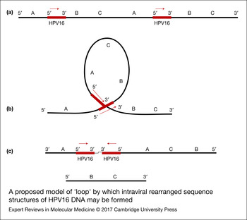
Although HPV integration contributes to the deregulated expression of the E6 and E7 genes, the integration of HPV DNA induces structural damages at the locus of viral insertion, as well. The structural changes contain extensive genomic amplifications and rearrangements (Refs Reference Akagi26, Reference Xu29, Reference Peter131). In particular, it has been proposed that the DNA replication of HPV16 integrants and recombination events lead to the construction of viral – host DNA concatamers through a model of ‘loop’. This procedure results in intrachromosomal amplifications and rearrangements that finally lead to structural and functional changes in neighbouring genes (Ref. Reference Akagi26). In addition to structural changes of the host DNA, intraviral sequence changes of HPV16 integrants are also detected. These changes contain the conjunction of different fragments of HPV16 DNA with inverted orientation. These sequence forms were primarily described in the Caski cell line (Refs Reference Akagi26, Reference Tsakogiannis27), while a more recent analysis revealed their presence in precancerous and cervical cancer cases, as well (Ref. Reference Tsakogiannis27). One possible explanation in order to explain the formation of HPV16 intraviral sequence structures is that the HPV16 integrants, which are located within the viral – host DNA concatamers might initiate the formation of additional DNA ‘loops’ and subsequently replication and recombination events among the different viral integrants might enable the conjuction of different viral genes with inverted orientation. This mechanism could further promote the inversion and deletion of adjacent sequences of the host chromosome (Fig. 4).
A proposed model of ‘loop’ by which intraviral rearranged sequence structures of HPV16 DNA may be formed. (a) HPV16 DNA – host DNA concatamers. (b) Circular structures might be formed within the region of viral – host DNA concatamers facilitating the replication of HPV16 integrants, while subsequent recombination events might generate the intraviral rearranged sequences of HPV16 DNA. (c) This putative mechanism enables the conjunction of noncontiguous viral fragments with inverted orientation as well as facilitates the inversion and deletion of adjacent sequences of the host chromosome.

Today, more than 250 viral integration sites have been recorded in all chromosomes of the host cell (Refs Reference Wentzensen, Vinokurova and von Knebel Doeberitz28, Reference Matovina132, Reference Chandrani133, Reference Hu134). The sites of viral integration are randomly dispensed across the host genome and often take place in transcriptionally active regions (myc oncogene) (Ref. Reference Ferber135) or in chromosomal unstable portions, which are called common fragile sites (Refs Reference Thorland136, Reference Thorland137). In regard to HPV16 genome, recent analyses revealed that there are preferential sites of viral insertion. In particular, the HPV16 DNA was found to be integrated in chromosomal spots that have considerable homology with viral DNA (Refs Reference Tsakogiannis113, Reference Hu134, Reference Schmitz138). The viral portion that presents homology with chromosomal spots is encountered between E5 and L2 genes (nucleotide positions 4100–4240) (Ref. Reference Schmitz138). In addition, the HPV16 is also found to be inserted in repetitive elements throughout the host chromosome, several of which are located close to cancer-related genes (Ref. Reference Li114).
In general, it is thought that HPV integration is strongly associated with more severe dysplasias and cervical cancer development, while viral integrations have been recorded in low-grade lesions, as well (Refs Reference Cricca23, Reference Tsakogiannis30, Reference Tsakogiannis113, Reference Andersson139, Reference Huang140). The presence of viral integration events in clinical samples is regarded as a valuable biomarker that facilitates the prediction of cervical malignancy progression and subsequently the development of cervical cancer (Refs Reference Boulet25, Reference Tsakogiannis30, Reference Han141, Reference Das142). Although cervical cancer is related to the integration of HPV DNA into the host chromosomes, cancer can be generated from infected epithelial cells that contain exclusively the viral episomal DNA form. In regard to HPV16 DNA, HPV16-positive cervical cancers were found to contain exclusively the episomal viral DNA form (Refs Reference Pett and Coleman143, Reference Klaes144, Reference Vinokurova145, Reference Chaiwongkot146). In contrast, in HPV18-related cervical cancers the viral genome is exclusively found integrated into the host chromosome (Refs Reference Fehrmann and Laimins147, Reference Badaracco148, Reference Woodman149, Reference Cullen150, Reference Pirami, Giache and Becciolini151). Absence of viral integration events in cervical cancer cases has lead to the conclusion that different mechanisms might contribute to cell transformation. Such mechanisms include the methtylation status of HPV DNA (Refs Reference Chaiwongkot146, Reference Bhat152, Reference Jiménez-Wences153) or mutations into the viral genome that change the protein functions of the E2 and E6 proteins and subsequently augmented the viral capacity to transform the epithelium. Such mutations have been described at position 3240 (E2, C3410T) (Refs Reference Tsakogiannis154, Reference Graham and Herrington155) as well as at position 350 (E6, T350G) (Refs Reference Tsakogiannis156, Reference Grodzki157, Reference Andersson158).
Preferential sites of HPV-DNA disruption
Upon integration the circular episomal HPV DNA is converted into a linear structure and inserted into the host chromosome. The disruption of viral genome can take place within the early region of viral genome (E1 and E2 genes) (Refs Reference Xu29, Reference Tsakogiannis113, Reference Hu134, Reference Arias-Pulido159, Reference Cricca160), or within the late region (L1 and L2 genes) (Refs Reference Xu29, Reference Li114, Reference Wang161) (Fig. 3). Although disruption events modify the initial state of HPV DNA (episome), the Long Control Region (LCR), and E6 and E7 oncogenes remain intact and expressed as viral-host fusion transcripts in all HPV-positive specimens (Refs Reference Akagi26, Reference Xu29, Reference Khoury162) (Fig. 3). The disruption of E2 gene is considered as the key event that contributes to the extended oncogenicity of the virus. The E2 gene encodes a multifunctional protein that is involved in replication and transcriptional regulation of the viral DNA. The E2 transcriptional factor binds to 12 bp motifs into the LCR and regulates the expression of the E6 and E7 oncogenes (Ref. Reference McBride163). Since the E2 transcription factor suppresses the expression of the viral oncogenes, disruption of the E2 ORF constraint this transcriptional control and consequently results in the overexpression of E6 and E7 genes (Refs Reference Wentzensen, Vinokurova and von Knebel Doeberitz28, Reference Xu29, Reference Thorland136, Reference Kraus164). Moreover, disruption of the E1 and/or E2 genes influences the HPV-DNA replication, as well. The E2 protein binds with high fidelity to the origin of viral replication and simultaneously interacts with the E1 protein. The E2 protein recruits E1 monomers to the viral origin of replication and promote their assembly into an active double E1 hexameric helicase (Refs Reference D'Abramo and Archambault165, Reference Lehoux, Gagnon and Archambault166). As a result, loss of function of either E2 or E1 proteins results in the prevention of HPV-DNA replication control and DNA expression that finally augments the oncogenic capacity of the integrated viral genome.
In recent years, a growing interest has emerged for the identification of the most frequent sites of disruption within the HPV16 genome in order to establish specific molecular assays necessary to determine the physical status of viral DNA. The distribution of preferential sites of HPV-DNA disruption varied among different studies. In particular, four recent analyses provide considerable variability in the rate of disruption of E1, E2, L1 and L2 genes among different studied groups (Refs Reference Xu29, Reference Li114, Reference Hu134, Reference Wang161). The variability of the preferential sites of HPV16 DNA disruption should be further investigated in order to reveal if the disruption of particular viral genes is associated with different studied populations or if it is related with differences in the assays that have been used. These insights will provide valuable information regarding the selection of the most appropriate genomic region specific to each individual population in order to further analyse the physical status of viral DNA and at the same time will provide significant data in order to select the most suitable method to study the physical status of HPV DNA in each studied population.
Although significant variability has been recorded in the distribution of the most frequently disrupted genes, considerable variability has also been described in the distribution of the most prevalent disrupted sites within the E1 and E2 genes. In general, it is accepted that the most frequent site of disruption within the E2 ORF is located in the portion that encompasses the hinge region of the E2 protein (Refs Reference Xu29, Reference Li114, Reference Arias-Pulido159, Reference Cricca160, Reference Kalantari167). In contrast, the preferential sites of E1 gene disruption varied among different studies. In particular disruptions within E1 gene have been found to be more frequently located either at the 5′ end (Refs Reference Tsakogiannis113, Reference Kalantari167), or at the 3′ end of the E1 gene (Ref. Reference Wang161). Moreover, significant association between the grade of cervical malignancy and the disruption of E1 and E2 genes was observed. In particular, in high-grade CIN s and cervical cancer cases disruption of both E1 and E2 genes occurred, while in low-grade lesions only the disruption of the E1 gene was involved (χ2 test, P < 0.005) (Refs Reference Tsakogiannis113, Reference Li114, Reference Kalantari167). The high prevalence of E2 gene disruption in cervical cancer cases enabled several studies to evaluate the physical status of HPV DNA through the study of E2 gene (Refs Reference Cricca23, Reference Boulet25, Reference Li114, Reference Han141, Reference Das142, Reference Briolat168). However, considering the strong implication of E1 gene disruption in low-grade lesions it is noteworthy to point out that the study of E1 gene is mandatory in order to determine viral integration events especially in cases of mild dysplasia (Ref. Reference Tsakogiannis30). Consequently, the selection of viral region in order to determine the physical status of HPV DNA should be specific to each individual HPV-positive patient considering the preferential sites of viral disruption and the grade of cervical malignancy.
Assays for the identification of HPV-DNA physical status
Over the last years there have been established molecular methods that facilitate the identification of HPV integrated genomes in the examined cervical samples. The assays are considered as valuable prognostic tools, since they provide considerable insights about the physical status of HPV DNA, the distribution of preferential sites of viral disruption, the locus of viral insertion, as well as the catastrophic effects of viral integration in the host chromosome (Refs Reference Boulet25, Reference Tsakogiannis30, Reference Li114, Reference Hu134, Reference Han141). The presence of viral integrants indicates the extensive accumulation of chromosomal damages in the infected cells, which eventually lead to cervical cancer development (Refs Reference Akagi26, Reference Xu29, Reference Peter131). Therefore, we suggest that future incorporation of molecular approaches for the detection of viral integrants could further stratify prognosis by selecting patients with the highest risk for development CINIII+ and cervical cancer. Moreover, insights derived from these molecular approaches will provide doctors not only the opportunity to better evaluate patient prognosis but also to select the most suitable therapeutic strategy for the effective treatment of cervical disease. These methods include: (i) hybridisation assays (FISH), (ii) PCR-mediated methods (DIPS, RS-PCR, APOT, qReal-time PCR) and (iii) whole genome sequence analysis [next generation sequencing (NGS)].
Fluorescent in situ hybridisation (FISH)
The FISH assay is performed in cervical smears in order to detect chromosomal abnormalities in regions (3q26, 8q24, 5p15, 20q13, centromere 7) that are frequently detected in HPV-associated cancers as well as to identify the presence of HPV episomal or integrated HPV DNA (Refs Reference Houldsworth169, Reference Olthof170, Reference Gimenes171). The method enables the localisation of specific sequences through a complementary polynucleotide sequence (probe) that is linked to a fluorescent reagent. The probe hybridised with the specific target – sequence and the observation of signal is performed through ultraviolet (UV) light (Refs Reference Houldsworth169, Reference Olthof170). The episomal form of HPV DNA is detected by a uniform signal in the nucleus of cell, while the integrated viral genomes are identified when a punctuate signal is observed (Ref. Reference De Marchi Triglia172). Although the assay enables the identification of integrated and episomal viral genomes, it presents a significant limitation in regard to the detection of HPV episomal DNA form. In particular, the episomal DNA can be lost during FISH procedure, or the increased prevalence of integrated viral genomes might constraint the detection of episomal DNA form (Refs Reference Theelen31, Reference Hopman173). In addition, FISH analysis might present a considerable false-negative rate for the presence of HPV DNA, as well. The low copy number of viral genomes or loss of HPV DNA because of deregulated cell proliferation may prevent the detection of viral DNA (Ref. Reference Sokolova174).
PCR-based assays
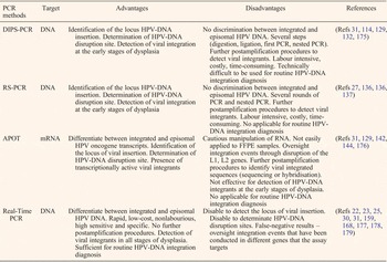
The Detection of Integrated Papillomavirus Sequences (DIPS) method is an adaptor ligation-based PCR assay that has been designed specifically to identify integrated viral sequences of HPV DNA (Ref. Reference Luft175). The assay allows the amplification and identification of host DNA sequences flanking the integrated viral fragments (Ref. Reference Luft175). In particular, the method is based on the digestion of genomic DNA through the restriction enzyme Sau3AI that is followed by ligation to a Sau3AI-specific double-stranded adaptor through T4 DNA ligase. Subsequently, a first round of linear PCR is performed using HPV-specific primer sets and is followed by a second round of nested PCR. The nested PCR is conducted through a nested primer specific for HPV DNA in conjunction with an adaptor-specific primer (AP). The amplicons are further subjected to sequencing and the resulting sequences encompass the disrupted site of viral DNA and the locus of viral insertion, as well (Refs Reference Theelen31, Reference Li114, Reference Ziegert129, Reference Matovina132, Reference Luft175) (Table 2).
Comparison of PCR-based assays for HPV integration analysis

The restriction site PCR (RS-PCR) is a PCR-based assay that retrieves unknown sequences that are flanking a known sequence (Ref. Reference Sarkar, Turner and Bolander180). The principle of the assay lies mainly in the use of restriction site oligonucleotide (RSO) Primers that have been constructed specifically to identify a restriction enzyme recognition site (BamHI, EcoRI, Sau 3AI, TaqI) (Refs Reference Thorland136, Reference Sarkar, Turner and Bolander180). In particular, the RSO primers are designed in order to harbour a T7 promoter sequence, a random sequence of 10-bases in length and the sequence of a restriction enzyme recognition site (Refs Reference Thorland136, Reference Thorland137). The method is performed through a first round of PCR that utilises HPV site-specific primers in conjunction with an individual RSO primer. Subsequently, a second round of nested PCR is conducted by using as template an aliquot of the first RS-PCR, a nested primer specific for HPV DNA and the same RSO (Refs Reference Tsakogiannis27, Reference Thorland136, Reference Thorland137). The final product is subjected to sequencing in order to determine HPV-DNA sequences adjoining cellular sequences (Table 2). Although DIPS-PCR and RS-PCR assays facilitate the identification of the locus of viral insertion and the site of viral disruption with high specificity, they are not able to discriminate episomal or mixed HPV-DNA forms (Table 2).
An alternative method that can identify with high sensitivity the episomal, mixed and integrated viral form in a specimen is the assay of papillomavirus oncogene transcripts (APOT). The method is based on the detection and analysis of HPV E6, E7 transcripts that are derived from episomal or integrated viral genomes (Refs Reference Theelen31, Reference Das142, Reference Klaes144, Reference Lu176). The APOT is based on a first step of reverse transcription of the total RNA using an adaptor linked oligo(dT)17-primer. Subsequently, the first-strand cDNAs are subjected to PCR using a forward primer specific for HPV DNA and the sequence of the adaptor as reverse primer. In addition, a second nested PCR is performed by using as template an aliquot of the first PCR, a nested primer specific for HPV DNA as forward primer and the adaptor linked oligo(dT)17 as reverse primer (Ref. Reference Klaes144). The analysis of the amplicons can be performed either through hybridisation with HPV E7 and E4-specific probes or through sequencing in order to determine viral transcripts that are derived from episomal and integrated HPV genomes (Refs Reference Theelen31, Reference Ziegert129, Reference Das142, Reference Klaes144, Reference Lu176) (Table 2).
The assay provides significant insights considering the presence of integrated and transcriptionally active viral genomes in HPV-positive patients as well as it presents considerable data about the locus of viral insertion into the host DNA (Ref. Reference Lu176). The principle of the assay lies mainly in the detection of HPV E6, E7 transcripts. As a consequence, it enables the detection of viral integrants that have been inserted into the host chromosome through disruption of the E1 and/or E2 genes and thus it might oversight integration events that have been conducted through disruption of the L1 and/or L2 genes, respectively. In addition, the assay is based on the cautious manipulation of RNA, which is an unstable molecule compared with DNA. As a result, the time and storage conditions of cervical samples might influence the RNA stability and subsequently the outcomes of the assay (Table 2). Finally, it is considered that DIPS-PCR, RS-PCR and FISH DNA assays are more reliable techniques for the detection of integrated HPV genomes at the early stages of cervical lesions compared with APOT, because of the fact that transcripts of integrated HPV genomes are produced at an advanced stage of cervical dysplasia (Ref. Reference Theelen31), (Table 2).
Quantitative real-time PCR
The increased popularity of quantitative real-time PCR assay facilitates the development of a nonlabour assay that permits the rapid quantification of viral copy numbers and the physical state of HPV DNA (episomal, integrated or mixed) with high specificity and sensitivity (Refs Reference Hudelist22, Reference Cricca23, Reference Boulet25, Reference Tsakogiannis30, Reference Theelen31, Reference Arias-Pulido159, Reference Briolat168, Reference Saunier177, Reference Jiang178, Reference Peitsaro, Johansson and Syrjänen179). The assay is able to identify the integration of viral DNA even at the early stages of cervical malignancy (Ref. Reference Tsakogiannis30), (Table 2). The main principle of the methodology lies mainly in the quantification of the E2 (the most prevalent site of viral disruption) and E6 genes (always remain intact and integrated into the host DNA) through real-time PCR assay, while the determination of the viral physical status is determined through E2/E6 ratio analysis. The episomal DNA is determined when there are equivalent amounts of E2 and E6 genes, while disruption events into the E2 gene indicate the mixed or integrated viral form (Refs Reference Hudelist22, Reference Cricca23, Reference Boulet25, Reference Tsakogiannis30, Reference Theelen31, Reference Arias-Pulido159, Reference Briolat168, Reference Saunier177, Reference Jiang178, Reference Peitsaro, Johansson and Syrjänen179).
Although the methodology provides significant sensitivity in identifying viral integration events through E2/E6 ratio analysis, it presents a considerable limitation in regard to integration events that have been occurred through disruption for example the E1 gene. The method may present high false-negative rates, particularly in low-grade CINs in which implication of the E1 gene disruption has been observed (Refs Reference Tsakogiannis113, Reference Cricca160). To this end, a recent analysis proposed the simultaneous analysis of the E1/E6 and E2/E6 ratio values in each cervical sample in order to determine with accuracy the physical status of HPV16 DNA (Ref. Reference Tsakogiannis30). Interestingly, low-grade CINs that were characterised as episomal DNA forms through E2/E6 ratio values, revealed integration events through E1/E6 ratio values. Moreover the simultaneous analysis of E1/E6 and E2/E6 ratio values enables the determination of multiple HPV16 integration events as different HPV16 integrants (integrants with E1 gene disruption and integrants with E2 gene disruption) were found to coexist in the same cervical sample (Ref. Reference Tsakogiannis30).
Although the assay is considered as a reliable method that facilitates the detection of integrated viral genomes with no further postamplification procedures, it is not able to locate the position of viral insertion into the host DNA. Moreover, it is important to underline that the accuracy of the assay's outcome is strongly associated with the selected sequences that target the respective primer sets (Ref. Reference Tsakogiannis30), (Table 2). Two main requirements emerged from this conclusion. First, extensive mapping analysis has to be performed in each studied group in order to determine the most frequent sites of disruption of HPV DNA. Second, nucleotide analysis of the E6 gene is required to be conducted in order to determine the most conserved portion of the viral gene among the different HPV16 isolates that are circulating in the studied population (Refs Reference Tsakogiannis30, Reference Tsakogiannis113).
Next generation sequencing
The NGS technology has upgraded the traditional sequencing process by allowing massively parallel sequencing reactions (Refs Reference Chen181, Reference Datta182, Reference Soper183). The capacity of NGS systems to perform whole genome sequencing of an individual sample in a short time as well as to examine multiple individuals concurrently has introduced the NGS in the field of HPV-DNA analysis. In particular, the NGS technology provides a wide range of data specific to each individual patient considering the HPV genotype, the presence of multiple viral infections, the viral copy number, the physical status of viral DNA, the sites of viral disruption, the locus of viral insertion as well as the presence of chromosomal damages induced by HPV integrants, regardless the grade of cervical lesion (Refs Reference Akagi26, Reference Xu29, Reference Militello85, Reference Hu134, Reference Wang161, Reference Flores-Miramontes184, Reference Fonseca185). The NGS technology offers crucial information for prognosis of cervical disease that makes the assay desirable for routine clinical use. However, data analysis and the increased cost of the assay remain the major issue for most clinical laboratories. The NGS technology requires expensive reagents and sophisticated equipment, while the collection of outcomes demands individuals with specialised knowledge in bioinformatics (Ref. Reference Liu186).
Nowadays several NGS platforms have been developed including 454 pyrosequencing (Roche), Ion Torrent (Life Technologies), SOLID (Applied BioSystems), Illumina, Heliscope (Helicos Biosciences) and PacBio (Pacific Biosciences). With a such rapidly advancing field, several NGS platforms have already become obsolete (e.g. 454 pyrosequencing, SOLID), others are in still use such as Illumina (being the predominant one) and Ion Torrent, while new ones are emerging or are highly anticipated (Pacific Biosciences) (Refs Reference Liu186, Reference Bahassi and Stambrook187, Reference Buermans and den Dunnen188, Reference van Dijk189, Reference Goodwin, McPherson and McCombie190). The NGS platforms are divided into two categories according to the preparation of their sequence libraries that is required in order to generate the appropriate templates to detect the signal during the extension of one base at the DNA template (Ref. Reference Buermans and den Dunnen188). In particular, the 454 pyrosequencing, Ion Torrent, SOLID and Illumina platforms are regarded as less sensitive and thus clonal amplification of the initial template is conducted in order to augment the signal, while the Heliscope and PacBio platforms are more sensitive and no previous amplification procedures are required (Ref. Reference Buermans and den Dunnen188). The NGS platform that has been mainly used in order to investigate with high efficiency the HPV-DNA insertion sites into the host chromosome as well as to describe the catastrophic effects of viral integration into the host DNA is the Illumina Hiseq platform (Refs Reference Akagi26, Reference Xu29, Reference Hu134, Reference Wang161). The Ilumina Hiseq platform is based on sequencing by synthesis (SBS) (Refs Reference Liu186, Reference Buermans and den Dunnen188). In particular, the sequence library with fixed adaptors is denatured to single strands and it is inserted into a flow cell. Subsequently bridge amplification is performed in order to generate template clusters (Refs Reference Liu186, Reference Mardis191). Finally the library is merged into single strands and the sequencing starts when the four different fluorescently tagged nucleotides (ddATP, ddGTP, ddCTP, ddTTP) synthesise the new strand according to the sequence at the template. The signal is detected through a charge-coupled device (Refs Reference Liu186, Reference Buermans and den Dunnen188). The Illumina platform is widely used in clinical laboratories as it provides the highest throughput and lowest cost compared with the other NGS platforms and recently read lengths have improved significantly from 2 × 100 bp to 3 × 100 bp (Refs Reference Liu186, Reference Buermans and den Dunnen188, Reference Goodwin, McPherson and McCombie190).
Conclusion
The strong causal relationship between HPV and cervical cancer development motivated several studies to develop HPV-DNA tests. An HPV test enables the diagnosis of patients who are identified as negative for CIN by cytological tests and consequently can stratify the risk by detecting individuals with augmented risk for development of cervical dysplasia and cancer. The HPV genotyping and specifically the identification of HPV16 and HPV18 genotypes increases the stratification of risk by identifying women who have higher risk for development of CINII+, CINIII+. Although the specific identification of HPV genotypes allows a more effective evaluation of cervical carcinogenesis risk, the molecular assays that define the presence of viral integrants as well as the assays that determine the locus of viral insertion into the host chromosome should be incorporated in HPV diagnosis in order to expand the predictive value of current HPV screening approaches. Therefore, we recommend that the incorporation of HPV-DNA physical status analysis in routine clinical diagnosis can further stratify the risk by detecting women who have the maximum risk for the development of CINIII+ and cervical cancer. The selection of patients with the highest risk for development of cancer at the pretreatment stage would allow the selection of the most suitable therapeutic strategy for the effective treatment of cervical disease. Moreover, it is important to mention that the selection of the most appropriate molecular assay should be performed taking into consideration particular attributes of studied samples, including the grade of cervical lesion and disruption events within the viral genome in order to establish personalised prognosis, diagnosis and treatment of HPV-positive patients. It is substantial to consider that the molecular approach of HPV diagnosis is a valuable and powerful tool that could be extensively used for the effective management of HPV infection.
Acknowledgements
This work was supported by a research grant of the Postgraduate Programme “Toxicology” code 5050, of the University of Thessaly, School of Health Sciences, Department of Biochemistry & Biotechnology. The authors would like also to thank Dr G. Amoutzias for invaluable language support and for helpful scientific discussions.
Conflict of interest
All authors declare that they have no conflicting or dual interests.