Introduction
The subfamily Fimbriariinae Wolffhügel, 1898 (Cestoda: Hymenolepididae) comprises members of five genera parasitising waterfowl and shorebirds: Fimbriaria Froelich, 1802; Fimbriarioides Fuhrmann, Reference Fuhrmann1932; Profimbriaria Wolffhügel, Reference Wolffhügel1936; Fimbriariella Wolffhügel, Reference Wolffhügel1936; and Fimbriasacculus Alexander & McLaughlin, 1996. The genus Fimbriarioides, which is discussed in this paper, contains five species: F. intermedia (Fuhrmann, Reference Fuhrmann1913); F. lintoni Webster, Reference Webster1943; F. haematopodis Webster, Reference Webster1943; F. tadornae Maksimova, Reference Maksimova1976; and F. spasskyi Maksimova, 1989. The former three species are probably transmitted in marine ecosystems, judging from the fact that they were described from sea ducks and shorebirds (Fuhrmann Reference Fuhrmann1913; Webster Reference Webster1943). Fimbriarioides intermedia, the type species of the genus, is usually recorded in definitive hosts represented by different eider species (Belopolskaja Reference Belopolskaja1952; Cooper Reference Cooper1921; Kornyushin et al. Reference Kornyushin, Iskova, Smogorzshevskaia and Ardamatskaia1996; Kuklina et al. 2019; Kulachkova Reference Kulachkova1958; Schiller Reference Schiller1955a; Skirnisson Reference Skirnisson2016). It is presumed that its intermediate host is the barnacle Semibalanus balanoides L., 1767, but this assumption is based on the similarity of rostellar hooks of adults from birds and from metacestodes (Belopolskaja Reference Belopolskaja1953; Regel Reference Regel2024) and has not been confirmed by experimental infection or genetic analysis.
We collected abundant material on metacestodes from barnacles at the eastern Murman coast (Barents Sea) and sexual adults of F. intermedia from eiders in the Barents Sea and the White Sea. Molecular-genetic analysis of this material made it possible to elucidate the life cycle of this species. Besides, we discovered another Fimbriarioides species, F. lintoni, which has been known only from the first description (Webster Reference Webster1943). It was found in an unrecognised host, Velvet Scoter Melanitta fusca. In this paper, we provide emended diagnoses of F. intermedia and F. lintoni with an emphasis on their differentiating characters, ascertain their position in the molecular phylogenetic tree of the Fimbriariinae, and characterise the ways of their transmission in high latitudes.
Materials and methods
Collection and processing of material
The material was collected from multiple localities in the Barents Sea, the White Sea, and the Sea of Okhotsk in 2019–2023. Adult cestodes Fimbriarioides spp. were extracted from the small intestine of Common Eider Somateria mollissima (Linnaeus, 1758) (taken near Srednii Island in the White Sea in 2019), King Eider Somateria spectabilis (Linnaeus, 1758), and Velvet Scoter Melanitta fusca (Linnaeus, 1758) (taken in the Khaipudyr Bay of the Barents Sea in 2022) (Table 1). The common eider from the White Sea was taken as bycatch; the ducks from the Barents Sea were provided by local hunters. The ducks were shot in accordance with the hunting regulations for local people. Strobilae were cleared of chyme, fixed, and stored in 96% ethanol for subsequent molecular and morphological analysis. For morphological studies, fixed worms (whole individuals and fragments of vouchers – see below) were stained with Ehrlich’s haematoxylin and Blazhin’s carmine. After dehydration in an ascending alcohol series, stained worms were cleared in clove oil and embedded in Canada balsam on slides, which were deposited in the Collection of Helminths in the Zoological Institute of the Russian Academy of Sciences (hereafter, ZISP), ‘Cestodes’ section.
List of samples used in this study and their GenBank accession numbers

Table 1. Long description
The table contains 9 columns: Sample I D, Species, Host species, Place, Region, Coordinates, and three sub-columns under GenBank accession numbers: 28 S, 16 S, and cox 1.
Key data points include:
* Samples 01-22-P1 through 01-22-P4 are Fimbriarioides lintoni collected from Melanitta fusca in Khaipudyr Bay, Pechora Sea.
* Samples 03-22-P1 through 03-22-P3 are Fimbriarioides intermedia collected from Somateria spectabilis in Khaipudyr Bay, Pechora Sea.
* Samples S M-2019-1 through S M-2019-4 are F. intermedia from Somateria mollissima at Srednii island, White Sea.
* A large group of samples (Ya 29 to Ya 116) are F. intermedia collected from the host Semibalanus balanoides in Yarnyshnaya Bay, Barents Sea, at coordinates 69.6.181 N 36.3.704 E.
* The final sample, N 22, is F. intermedia from S. balanoides in Nayakhanskaya Bay, Sea of Okhotsk.
* Accession numbers vary by sample, with many 16 S sequences provided (e.g., P V 8 2 9 6 1 1, P X 6 7 8 7 2 7) and fewer cox 1 sequences (e.g., P X 6 7 5 3 1 1, P X 6 7 5 3 1 7).
Additionally, in order to count the number of longitudinal muscle bundles, we made several serial cross-sections (0.1–0.3 mm thick) in the anterior part (immediately after the pseudoscolex) of the stained strobilae of F. intermedia (from both hosts) and F. lintoni.
For molecular analysis, we chose four voucher strobilae (vouchers) from each duck and cut out fragments, 2–5 mm long, located immediately after the pseudoscolex. The remaining fragments of the vouchers were used to make slides, which were deposited in ZISP (see above). Photographs were made with the use of a DMLS microscope (Leica Microsystems, Germany) equipped with digital cameras of the same manufacturer. We also examined slides from the Cestode section of ZISP.
Barnacles Semibalanus balanoides (Linnaeus, 1758) were collected together with the substrate in the littoral zones of the Yarnyshnaya Bay of the Barents Sea and the Nayakhanskaya Bay of the Sea of Okhotsk (Table 1). In the laboratory, the barnacles were cut off the substrate and dissected as suggested by Regel (Reference Regel2024). The body extracted from the capitulum was squeezed between two slides and viewed in transmitted light under a stereomicroscope. Live metacestodes were photographed using an Olympus CH40 microscope (Olympus Optical Co. Ltd., Tokyo, Japan) equipped with an Olympus XC-30 digital camera (Olympus Optical Co. Ltd.) at the ‘Kartesh’ White Sea Biological Station of the Zoological Institute RAS. The metacestodes found during examination were freed from the capsule and the exocyst, fixed in 96% ethanol, and stored until further treatment. To measure cysticercoids and their rostellar hooks, 1–4 specimens from each infected barnacle were embedded in polyvinyl-based clearing medium as described by Regel (Reference Regel2024).
All measurements given in the text and the tables are in micrometres, unless otherwise indicated. Prevalence, intensity, and mean abundance were calculated in accordance with Bush et al. (Reference Bush, Lafferty, Lotz and Shostak1997).
DNA isolation, amplification and sequencing
Molecular analysis was performed by sequencing fragments of the nuclear 28S rRNA and mitochondrial cox1 and 16S rRNA genes. Genomic DNA was extracted from 96%-ethanol-fixed fragments of strobilae with the use of cetyltrimethylammonium bromide detergent (CTAB) according to the published protocols (Galaktionov et al. Reference Galaktionov, Solovyeva, Miroliubov, Romanovich and Skírnisson2024; Winnepenninckx et al. Reference Winnepenninckx, Backeljau and De Wachter1993). Prior to extractions, the fragments were washed three times in 1× phosphate buffer for 15 min.
One metacestode was taken from each infected barnacle for DNA extraction. Before DNA extraction, the metacestode was washed thrice in phosphate buffer, placed into lysis buffer containing 100 mM Tris-HCl, pH 8, 20 mM EDTA, pH 8, and 100 μg/mL proteinase K, and incubated for 20 min at 60 °C and then for 10 min at 90 °C.
Polymerase chain reaction (PCR) and purification of its products were performed according to the published protocol (Galaktionov et al. Reference Galaktionov, Solovyeva, Blakeslee and Skírnisson2023). Nucleotide sequences of the primers and their annealing temperatures are given in Table 2. Primers for amplification of the 28S rRNA gene fragment were designed using Primer3 software (Untergasser et al. Reference Untergasser, Cutcutache, Koressaar, Ye, Faircloth, Remm and Rozen2012) based on the nucleotide sequences of this gene in Fimbriaria spp. (OP906285.1, OP906284.1, AF28916.1) available in GenBank. Sanger sequencing was performed in the St. Petersburg State University Research Park, Centre for Molecular and Cell Technologies. All nucleotide sequences obtained in this study were deposited in National Center for Biotechnology Information (NCBI) GenBank (Table 1).
A list of primers used in this study

Table 2. Long description
The table contains five columns.
1. Gene: 28S r R N A. Primer I D: 28S f 3. Nucleotide sequence: 5 prime dash C G G T G T G T G C T T G T C A T G T G dash 3 prime. Temperature: 58 degrees Celsius. Reference: Current study.
2. Gene: 28S r R N A. Primer I D: 28S r 3. Nucleotide sequence: 5 prime dash G G T A G C T C G T C T A C C A C A G C dash 3 prime. Temperature: 58 degrees Celsius. Reference: Current study.
3. Gene: 16S r R N A. Primer I D: Cest 3 1 6 S f. Nucleotide sequence: 5 prime dash G T G Y D A A G G T A G S A T A A T dash 3 prime. Temperature: 55 degrees Celsius. Reference: Chan et al. 2022.
4. Gene: 16S r R N A. Primer I D: Cest 3 1 6 S r. Nucleotide sequence: 5 prime dash C C G G T Y T Y A A C T C A R C T C A T dash 3 prime. Temperature: 55 degrees Celsius. Reference: Chan et al. 2022.
5. Gene: cox 1. Primer I D: J B 3. Nucleotide sequence: 5 prime dash T T T T T T G G G C A T C C T G A G G T T T A T dash 3 prime. Temperature: 53 degrees Celsius. Reference: Bowles et al. 1992.
6. Gene: cox 1. Primer I D: J B 4 dash 5. Nucleotide sequence: 5 prime dash T A A A G A A A G A A C A T A A T G A A A A T G dash 3 prime. Temperature: 53 degrees Celsius. Reference: Bowles et al. 1992.
Alignment and phylogenetic analysis
Alignment, trimming, and basic analyses of the newly generated sequences, as well as the 28S rRNA, cox1, and 16S rRNA gene partial sequences retrieved from GenBank for other cestodes, were performed with BioEdit (Hall Reference Hall1999) and MEGA 11 (Kumar et al. Reference Kumar, Stecher, Li, Knyaz and Tamura2018). Phylogenetic relationships were reconstructed using Bayesian inference (BI) in MrBayes v. 3.2.6 (Ronquist et al. Reference Ronquist, Teslenko, van der Mark, Ayres, Darling, Hohna, Larget, Liu, Suchard and Huelsenbeck2012) and maximum likelihood (ML) in MEGA 11. The most suitable evolutionary models were determined by the corrected Akaike information criterion in MEGA 11. Phylogenetic trees were constructed with the use of sequences of 28S for Fimbriaria fasciolaris (OP906285.1), F. teresae (OP906284.1), Fimbriaria sp. (AF286916.1); cox1 for F. fasciolaris (OP896837.1), F. teresae (OP903372.1), Fimbriaria sp. (EU665472.1); 16S for Fimbriaria sp. (EU665472.1) as well as one mitochondrial genome of F. fasciolaris MH551238.1 (Chang et al. Reference Chang, Gao, Wang and Li2018; Greben et al. Reference Greben, Kornyushin, du Preez, Melnychuk, Yevstafeva and Syrota2023; Littlewood et al. Reference Littlewood, Waeschenbach and Nikolov2008).
The Hasegawa–Kishino–Yano model with estimates of gamma-distributed among-site rate variation (HKY+G) was chosen as the one with the best fit for all analysed genes. Genetic divergences between the taxa were calculated as uncorrected p-distances with 1000 bootstrap iterations for each gene region using MEGA 11. Haplotype network reconstruction and Tajima’s D neutrality test were performed with PopArt 1.7 (Leigh and Bryant Reference Leigh and Bryant2015).
Results
Using molecular markers, we showed species-level differences between specimens identified as F. intermedia and F. lintoni based on morphological characters. This is the first record of the latter species in the Palaearctic. It was found in an unrecognised host, Velvet Scoter M. fusca. Using molecular methods, we also proved that metacestodes from barnacles S. balanoides belonged to F. intermedia (see Molecular results).
Description
Family Hymenolepididae Fuhrmann, 1907, subfamily Fimbriariinae Wolffhügel, 1898.
Fimbriarioides intermedia (Fuhrmann, Reference Fuhrmann1913) Fuhrmann, Reference Fuhrmann1932 emend.
[syn. Fimbriaria intermedia Fuhrmann, Reference Fuhrmann1913; Fimbriaria intermedia Fuhrmann, Reference Fuhrmann1914; Fimbriaria intermedia Cooper, Reference Cooper1921; Fimbriarioides intermedia Fuhrmann, Reference Fuhrmann1932; Fimbriarioides intermedia Schiller, Reference Schiller1955a; Cysticercoid-a Belopolskaja, Reference Belopolskaja1953 (metacestode); Cysticercoid-a Ouspenskaia, Reference Ouspenskaia1960 (metacestode)]
Type host (definitive): Common Eider Somateria mollissima (Linnaeus, 1758)
Other hosts (definitive): King Eider Somateria spectabilis (Linnaeus, 1758); Long-tailed Duck Clangula hyemalis (Linnaeus, 1758). Records of F. intermedia from Anser albifrons, Anas platyrhynchos dom., Cereopsis novae-hollandiae, Aythya fuligula, A. nyroca, Netta rufina, Melanitta nigra, and M. fusca in Spasskaja (Reference Spasskaja1966) and McDonald (Reference McDonald1969) are questionable since no descriptions of the worms are given in these works. Unfortunately, the voucher specimens of Spasskaja (Reference Spasskaja1966) have been lost, and we do not know what happened to the materials of McDonald (Reference McDonald1969).
Site in definitive host: intestine
Type locality: Iceland
Other localities (in definitive host – eiders): Arctic Canada (Cooper Reference Cooper1921); Alaska, USA (Schiller Reference Schiller1955a,Reference Schillerb); Black Sea (Kornyushin et al. Reference Kornyushin, Iskova, Smogorzshevskaia and Ardamatskaia1996); Barents Sea (Eastern Murman) (Belopolskaja Reference Belopolskaja1952; Galaktionov et al. Reference Galaktionov, Kuklin, Ishkulov, Galkin, Marasaev, Marasaeva and Prokofev1997; Kuklina and Kuklin Reference Kuklina and Kuklin2019); White Sea (Kulachkova Reference Kulachkova1958, Reference Kulachkova1979); Sea of Okhotsk (Atrashkevitch et al. Reference Atrashkevitch, Orlovskaya, Regel, Galaktionov and Dobrovolskij2008)
Representative slides: vouchers from S. mollissima nos. 1657–1659 and from S. spectabilis nos. 1660–1663 deposited in ZISP; slides of M.M. Belopolskaja nos. 168–169 deposited in ZISP. Slide of V.V. Kuklin no. 1646 deposited in ZISP.
Intermediate host: Semibalanus balanoides (L., 1767) (Crustacea: Cirripedia: Balanidae) and Balanus crenatus Bruguière, 1789 (questionable – see Results for details) (natural)
Site in intermediate host: body cavity (mostly on surface of mid-intestine)
Localities (in intermediate host): White Sea, Barents Sea, Pechora Sea, Sea of Okhotsk
Representative DNA sequences: cox1 (PX675312âPX675317), 28S (PV829576âPV829579), 16S rRNA (PV829610; PV829614âPV829624; PX678717âPX678730) (according to Table 1).
The subsequent description is based on specimens of F. intermedia from two species of eiders: Somateria mollissima and S. spectabilis (ZISP nos. 1657–1663).
A total of 23 specimens of F. intermedia were recovered from S. spectabilis; the poor preservation of the strobilae fragments from Somateria mollissima prevented an accurate determination of intensity. Strobilae of average size (Table 3; Figures 1b,c, and 3a). Scolex small (Figure 1a), rostellar hooks (similar in shape to diorchoid type) with long handle and well-visible, slightly bifid guard. Blade, about 1/3 of hook length, bent back parallel to root process. Pseudoscolex sickle-shaped, segmented, weakly plicate. Common genital primordia well-visible in its posterior third or even posterior half. Externally, no distinct border between pseudoscolex and strobila; internally, border marked by start of differentiation of genital primordium. In sections of anterior part of strobila inner muscle layer numbering more than 150 muscle bundles (Figure 1b). Excretory system consisting of 4 pairs and 1–3 unpaired excretory canals in each lateral field of strobila. Paired excretory canals joined by metameric transverse anastomoses. External segmentation indistinct, external borders of segments not corresponding to internal segmentation represented by complex of female and male reproductive organs. Each proglottid usually containing complex of reproductive organs (3 testes, ovary, vitellarium, and copulatory apparatus) corresponding to morphogenetic stage. Rarely, proglottids lacked one, two, or all three testes as well as cirrus sac.
A comparison of the measurements of Fimbriarioides intermedia and F. lintoni

Table 3. Long description
The table compares measurements for two species across seven columns. The first five columns pertain to Fimbriarioides intermedia from various sources (Fuhrmann 1913, Cooper 1921, Belopolskaja 1952, Schiller 1955a, and current study data N equals 10). The final two columns pertain to Fimbriarioides lintoni (Webster 1943 and current study data N equals 4).
Key measurements include:
* Strobila length: Ranges from 10 to 60 mm for F. intermedia and 60 to 130 mm for F. lintoni.
* Scolex width: 82 to 150 micrometers for F. intermedia and 90 to 123 micrometers for F. lintoni.
* Rostellar hooks length: 22 to 25 micrometers for F. intermedia and 18.5 to 20 micrometers for F. lintoni.
* Longitudinal muscle bundles: 140 to 170 for F. intermedia and 80 to 90 for F. lintoni.
* Testes width: 110 to 315 micrometers (mean 202.2 plus or minus 6.8) for F. intermedia and 260 to 460 micrometers (mean 352.5 plus or minus 6.1) for F. lintoni.
* Ovary width: 480 to 1150 micrometers for F. intermedia and 740 to 1600 micrometers for F. lintoni.
* Embryonal hooks length: 14.3 to 15.3 micrometers for F. intermedia and 12 to 16 micrometers for F. lintoni.
Other rows include measurements for suckers, rostellum, neck width, pseudoscolex, excretory ducts, seminal vesicles, cirrus pouch, vitelline gland, and oncosphere dimensions. Dashes indicate missing data for specific sources.
Microphotographs of Fimbriarioides intermedia from Somateria mollissima (SM-2019: a, e, g, h) and S. spectabilis (3-22 P: b, c, d, f). (a) Scolex. (b) Transverse section of the anterior part of the strobila. (c) Hermaphroditic proglottids (ventral view). (d) Copulatory apparatus isolated from the uterine segment (ventral view). (e) Cirrus (dorsal view). (f) Uterine proglottids (fragments of the uterus with developing chains of eggs are marked with arrows). (g) String (chain) with mature eggs (in glycerine). (h) Fragment of this chain of eggs. Scale: (a, d, e, h) – 50 μm; (b, c, f, g) – 300 μm. Abbreviations: apt-mt-pt, aporal-medial-poral testes; cs, cirrus sac; ov, ovary; pec, pairs of excretory canals; sph, vaginal sphincter; sr, seminal receptacle; vc, copulative part of vagina; vcp, conductive part of vagina; vg, vitelline gland (vitellarium).

Figure 1. Long description
The figure consists of eight panels labeled a through h.
* Panel a: A microphotograph of the scolex, showing the anterior attachment organ with visible hooks.
* Panel b: A transverse section of the anterior strobila, labeled with aporal site on the left and poral site on the right.
* Panel c: Ventral view of hermaphroditic proglottids. Labels from left to right include c s (cirrus sac), v c p (conductive part of vagina), I p e c (first pair of excretory canals), p t (poral testes), s r (seminal receptacle), v g (vitelline gland), o v (ovary), m t (medial testes), II p e c, a p t (aporal testes), III p e c, and I V p e c.
* Panel d: Ventral view of the isolated copulatory apparatus. Labels point to the v c (copulative part of vagina), s p h (vaginal sphincter), and v c p (conductive part of vagina).
* Panel e: Dorsal view of the cirrus, showing a dense arrangement of small hooks.
* Panel f: Uterine proglottids. Arrows point to developing chains of eggs. Labels include s r (seminal receptacle) and Roman numerals I through I V p e c marking the pairs of excretory canals.
* Panel g: A long, winding chain of mature eggs photographed in glycerine.
* Panel h: A high-magnification fragment of the egg chain, showing the detailed structure of individual mature eggs.
Vertical scale bars are present in each panel. Panels a, d, e, and h represent 50 micrometers. Panels b, c, f, and g represent 300 micrometers.
Three testes strongly elongated transversally, arranged in line, as well as developing copulatory apparatus differentiating at a distance of about 1 mm behind pseudoscolex. At this stage, all primordia (those of testes and that of copulatory apparatus) located between first and third pairs of excretory canals, this localisation persisting for a further 2–3 mm. Further still, developing cirrus sac and vagina gradually shifting into lateral field, with genital atrium formed. Genital pores unilateral (dextral). Cirrus conical, its basal dome-shaped part armed with hooked spines, with blades bent towards base, arranged in chessboard fashion; apical part unarmed (Figure 1e). Cirrus sac cigar-shaped, muscular (Figures 1c and 3a), not intersecting first pair of excretory canals. Broad vas deferens connecting rounded external seminal vesicle lying anteriorly of poral testis with internal seminal vesicle occupying almost entire sac volume (Figures 1c and 3a). Fully developed testes in non-relaxed strobilae weakly lobed, those in relaxed strobilae rounded.
Female genital primordium located posteriorly, between poral and middle testes, starting to differentiate when external seminal vesicle starts to fill up. Ovary reticulate, elongated transversely, its aporal part reaching or (rarely) intersecting third pair of excretory canals. Vitellarium multi-lobed, stellate (Figures 1c and 3a). Vagina located ventrally of cirrus sac. Copulative part of vagina funnel-shaped, with broad distal cup-shaped part densely covered inside with spine-like structures, and narrow tubular proximal part with sphincter at the border with conductive part. Long convoluted conductive duct passing into large ovoid seminal receptacle adjoining the poral testis anteriorly (Figure 1d,e,f). Non-metameric reticulate uterus forming in hermaphroditic part of strobila at start of testes resorption. Maturing uterus breaking up into fragments containing up to 10–30 oncospheres. Mature eggs released in strings (Figure 1g, h).
Microphotographs of Fimbriarioides lintoni from Melanitta fusca (01-22 P). (a) Rostellar hooks. (b) Poral side of the proglottids with male organs (dorsal view). (c) Cirrus armament (dorsal view). (d) Cirrus and copulatory part of vagina (ventral view). (e) Hermaphroditic proglottids (ventral view). (f) Uterine proglottids (ventral view). (g) Mature eggs. Scale: (a) – 20 μm; (b, e, f) – 300 μm; (c, d, g) – 50 μm. Abbreviations: apt-mt-pt, aporal-medial-poral testes; cs, cirrus sac; esv-isv, external and internal seminal vesicles; mw, muscular wall; ov, ovary; pec, pairs of excretory canals; rfg, resorbed female gonads; sr, seminal receptacle; vg, vitelline gland (vitellarium); ur, uterus reticulate.

Figure 2. Long description
A collection of seven microphotographs labeled a through g.
Panel a shows rostellar hooks with a 20 mu m scale bar.
Panel b displays the dorsal view of the poral side of proglottids. Labels include s r for seminal receptacle, e s v for external seminal vesicle, and p t for poral testes. Double-headed arrows indicate the i s v (internal seminal vesicle), c s (cirrus sac), and m w (muscular wall). Scale bar is 300 mu m.
Panel c shows the cirrus armament in dorsal view with a 50 mu m scale bar.
Panel d shows the ventral view of the cirrus and copulatory part of the vagina, with arrows pointing to v c. Scale bar is 50 mu m.
Panel e presents hermaphroditic proglottids in ventral view. Labels from left to right include p t (poral testes), e s v (external seminal vesicle), s r (seminal receptacle), v g (vitelline gland), m t (medial testes), o v (ovary), and a p t (aporal testes). Roman numerals I p e c through I V p e c indicate pairs of excretory canals. Scale bar is 300 mu m.
Panel f shows uterine proglottids in ventral view. Arrows point to s r (seminal receptacle), r f g (resorbed female gonads), and u r (uterus reticulate). Scale bar is 300 mu m.
Panel g shows mature eggs with a 50 mu m scale bar.
Metacestodes of Fimbriarioides intermedia (Figure 4).
In barnacles S. balanoides from the Yarnyshnaya Bay (Table 1), we found metacestodes corresponding in morphological characters to the larvae of F. intermedia described by Regel (Reference Regel2024). Prevalence of metacestodes of F. intermedia in barnacles S. balanoides was 69.1 ± 4.2% (N = 123); infection intensity was 3–46 (15.2 ± 1.3); mean abundance was 10.6 ± 0.9. A detailed description of metacestodes of F. intermedia is given in Regel (Reference Regel2024). Here we provide only some details characteristic of our material.
Metacestodes localised in the body cavity of barnacles, mostly on mid-intestine (Figure 4a). We provide measurements of cysticercoids that were extracted from the capsule and, as a rule, from the exocyst and then fixed in alcohol (Figure 4b). Endocyst size 177.5–225 (196.2 ± 3.1) × 142–192 (171.5 ± 2.6) (N = 34) (Figure 4c). Exocysts retained in 4 specimens; their size 280–375 (312.4 ± 5.7) × 260–350 (307.2 ± 1.2). Length of caudal appendage 1.5–2.45 (1.9 ± 0.1, N = 15) mm (Figure 4b). Width of scolex 94–156 (117.6 ± 2.9, N = 28). Diameter of suckers 45–63. Length of rostellar hooks 22.5–26 (24.6 ± 0.1, N = 37) (Figure 4d, e).
Fimbriarioides lintoni Webster, Reference Webster1943
[syn. Fimbriaria falciformis Linton, Reference Linton1927, in part; Fimbriarioides falciformis Fuhrmann, Reference Fuhrmann1932, in part; Fimbriariella falciformis Wolffhügel, Reference Wolffhügel1936, in part.]
Type host (definitive): Melanitta perspicillata (L., 1758)
Other hosts (definitive): Melanitta deglandi (Bonaparte, 1850), Clangula hyemalis (L., 1758), and Fulica americana Gmelin, 1789 (by Webster Reference Webster1943); Melanitta fusca (L., 1758) (our data)
Site in definitive host: intestine
Type locality: Woods Hole, Massachusetts, USA
Other localities (in definitive host): Khaipudyr Bay of Pechora Sea, Russia
Representative slides: slides nos. 1664–1671 deposited in ZISP
Intermediate host: unknown, presumably barnacles Balanidae
Representative DNA sequences: cox1 (PX675311), 28S (PV829575), 16S rRNA (PV829611âPV829613) (according to Table 1).
We recovered 17 specimens of Fimbriarioides lintoni from Velvet Scoter Melanitta fusca. The following description is based on these specimens (Table 3; Figures 2 and 3; ZISP nos. 1664–1671).
Schematic drawing of the hermaphroditic proglottids (ventral view). (a) Fimbriarioides intermedia. (b) Fimbriarioides lintoni. Scale: (a, b) – 300 μm. Abbreviations : apt-mt-pt, aporal-medial-poral testes; cs, cirrus sac; esv-isv, external and internal seminal vesicles; ov, ovary; pec, pairs of excretory canals; sr, seminal receptacle; vc-vcp, copulative-conductive part of vagina; vd, vas deferens; vg, vitelline gland (vitellarium).

Figure 3. Long description
The schematic consists of two panels, a and b, each showing a ventral view of tapeworm proglottids with a 300 micro-meter scale bar below the central structures.
Panel a, Fimbriarioides intermedia:
Moving from left to right, the poral side features the v c and v c p leading to a c s containing an i s v. A v d connects to an e s v. Four vertical pairs of excretory canals, labeled Roman numeral 1 p e c through Roman numeral 4 p e c, divide the proglottid segments. Between the first and second p e c are the p t and s r. Centrally located is the dark, lobed v g. To the right of the second p e c are the m t and a p t. The o v is a large, granular structure spanning the lower portion of the segments.
Panel b, Fimbriarioides lintoni:
This panel shows a similar anatomical layout but with more densely packed and lobulated organs. On the left, the c s and i s v are more robust. The v d leads to a prominent, rounded e s v. The p t and s r are larger and more distinct between the first and second p e c. The v g appears as a dense, dark cluster at the center. The m t and a p t extend toward the third p e c. The o v is highly branched and fills a significant portion of the space between the excretory canals. The four pairs of excretory canals, Roman numeral 1 p e c to Roman numeral 4 p e c, are spaced similarly to panel a.
Microphotographs of the metacestodes of Fimbriarioides intermedia from the body cavity of the barnacles Semibalanus balanoides. (a) Multiple metacestodes on the surface of the middle gut (alive). (b) General view of cysticercoid, extracted from the capsule and exocyst (voucher slide Ya 67, polyvinyl). (c) Cysticercoid endocyst. (d, e) The crowns of rostellar hooks (voucher slides Ya 79 and Ya116). Scale: (a) – 500 μm; (b, c) – 100 μm; (d, e) – 30 μm.

Figure 4. Long description
A five-panel microphotograph series.
Panel a, top left. A wide-field color micrograph shows numerous translucent, spherical metacestodes embedded on the purple-tinted surface of a host middle gut. A vertical scale bar on the right represents 500 micrometers.
Panel b, top center inset. A circular black and white micrograph shows a single cysticercoid with a rounded head and several elongated, coiled appendages extending downward. A vertical scale bar represents 100 micrometers.
Panel c, bottom left. A high-resolution micrograph of a cysticercoid endocyst. It is a large, circular structure with thick, concentric outer layers and a central cavity containing a cluster of small, dark hooks. A vertical scale bar represents 100 micrometers.
Panel d, top right. A close-up view of the rostellar hooks arranged in a circular crown. The hooks are curved and sharp, radiating from a central point. A vertical scale bar represents 30 micrometers.
Panel e, bottom right. A further magnified view of a subset of rostellar hooks, showing their distinct curved shape and attachment points. A vertical scale bar represents 30 micrometers.
Phylogenetic relationships among Fimbriarioides spp. and other cestodes inferred from 28S rRNA gene fragments. The tree was reconstructed using maximum likelihood (ML) and Bayesian inference (BI) methods. ML bootstrap support values (based on 1000 replicates) are shown first, followed by posterior probabilities from the BI analysis.

Figure 5. Long description
A horizontal phylogenetic tree with branches extending from left to right. Support values are presented as M L bootstrap forward slash B I posterior probabilities at each node.
At the top, a green shaded box contains the Fimbriarioides intermedia clade. This clade includes four isolates: 03-22-P1 and N22 (66/100 support), Ya75 (88/100 support), and S M-2019-3 (81/97 support). Below this, a yellow shaded box highlights Fimbriarioides lintoni (isolate 01-22-P1), which is sister to the F. intermedia clade with 80/98 support.
Moving down the tree, the next branches include:
* O P 906285.1 Fimbriaria fasciolaris (47/81 support).
* A F 286916.1 Fimbriaria sp. O L B M 1 and O P 906284.1 Fimbriaria teresae (100/100 support).
* G U 166246.1 Microsomacanthus crenatus (73/73 support at the basal node).
A separate major lower clade (72/77 support) contains:
* Kontrimavichusia sp. h A M-2023 (two isolates, O R 992639.1 and O R 992642.1) with 99/99 support.
* A sub-clade (45/63 support) containing Neoskrjabinolepis singularis and N. fertilis (89/100 support) and Rodentolepis straminea and R. nana (99/100 support).
The most basal branch at the bottom is G U 166276.1 Monocercus arionis.
Phylogenetic relationships among Fimbriarioides spp. and other cestodes inferred from cox1 gene fragments. The tree was reconstructed using maximum likelihood (ML) and Bayesian inference (BI) methods. ML bootstrap support values (based on 1000 replicates) are shown first, followed by posterior probabilities from the BI analysis. Asterisk indicates bootstrap support values.

Figure 6. Long description
The phylogenetic tree branches from a common root on the left.
At the top, a green shaded box contains the Fimbriarioides intermedia clade. This clade includes nodes for Ya79 and Ya81 (78/100 support), Ya31 (83/100), Ya82 (100/100), S M-2019-3 (94/83), and N22 (88/99).
Below this, a yellow shaded box highlights Fimbriarioides lintoni (01-22-P2), which branches off a node with 72/94 support shared with the green clade.
Moving down the tree, the next branches include:
- Fimbriaria teresae (O P 903372.1) branching from a 62/65 support node.
- A clade containing Fimbriaria sp. (E U 665472.1) and Fimbriaria fasciolaris (O P 896837.1) with 88/98 support.
- A clade containing Hymenolepis hibernia (L C 063179.1) and Hymenolepis diminuta (L C 063184.1) with 76/98 support.
- A bottom clade branching from an 89/94 support node, containing Echinococcus granulosus (M Z 345697.1) and a sub-clade of Versteria mustelae (M W 476515.1) and Huinaylepis elegans (M H 663464.1) with 66* support.
Support values are presented as M L bootstrap values forward slash B I posterior probabilities.
Phylogenetic relationships among Fimbriarioides spp. and other cestodes inferred from mitochondrial 16S rRNA gene fragments. The tree was reconstructed using maximum likelihood (ML) and Bayesian inference (BI) methods. ML bootstrap support values (based on 1000 replicates) are shown first, followed by posterior probabilities from the BI analysis. Asterisk indicates bootstrap support values.

Figure 7. Long description
The tree originates from a root on the left. Moving right, the first major split separates a clade containing Dilepis undula and Monocercus arionis at the bottom with a bootstrap value of 94 asterisk. Above this, several outgroups are listed including Cloacotaenia megalops, Clistobothrium montaukensis, and a clade of Mesocestoides species with support values of 99 forward slash 100.
The main upper branch leads to the Fimbriariidae family. A yellow shaded box highlights the Fimbriarioides lintoni clade, containing specimens 01-22-P1, 01-22-P2, and 01-22-P4, supported by values of 93 forward slash 78 and 69 forward slash 82. Above this is a single branch for Fimbriaria species E U 6 6 5 4 7 2 dot 1.
The top section features a large green shaded box for Fimbriarioides intermedia. This clade is supported by a value of 85 forward slash 99 at the base. It contains 13 specimens: 03-22-P3, S M-2019-1, Ya112, Ya81, 03-22-P2, Ya75, Ya82, Ya84, S M-2019-3, S M-2019-4, Ya79, Ya45, and N22. Internal nodes within this green clade show support values of 54 asterisk and 64 forward slash 88.
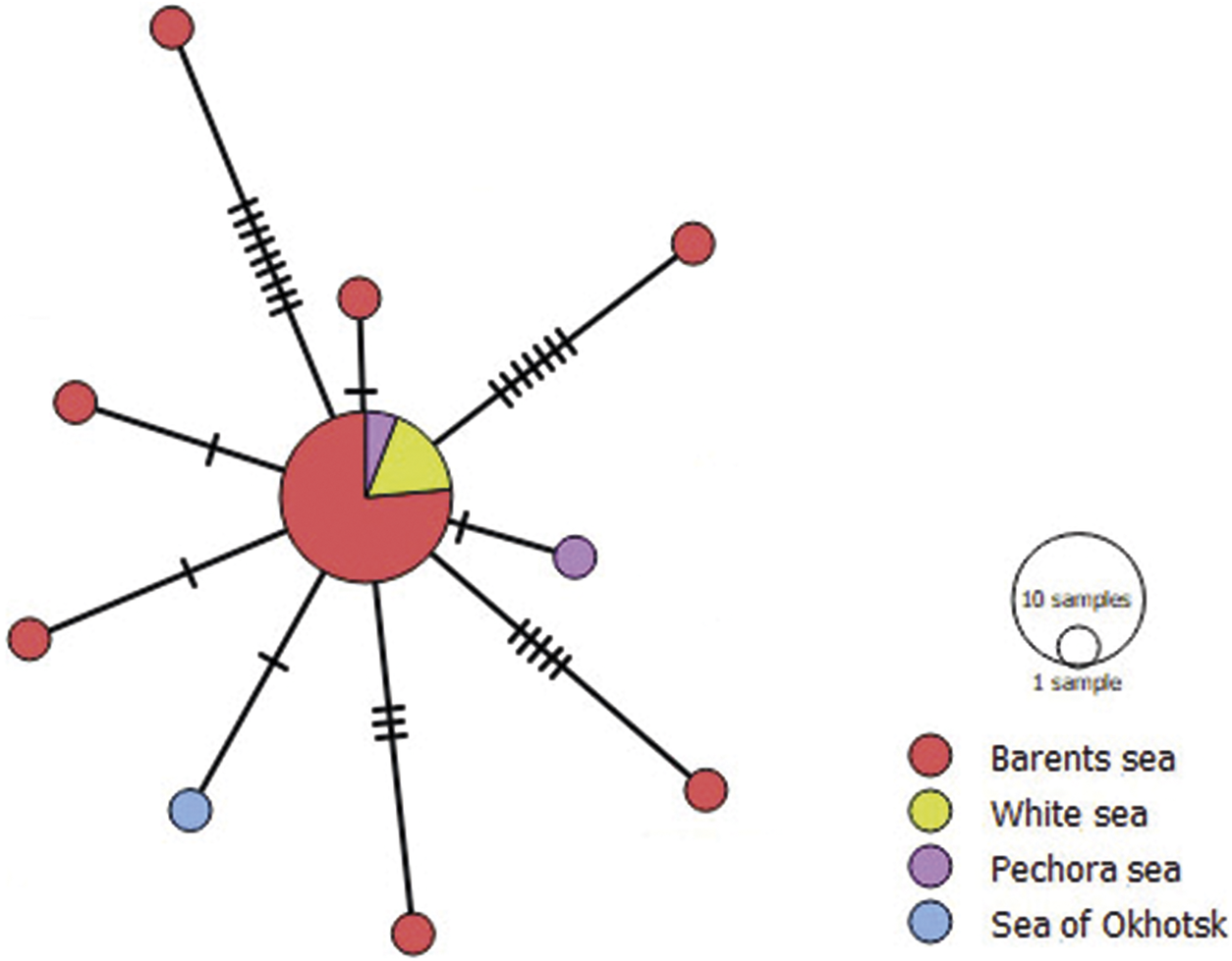
A minimum-spanning haplotype network for Fimbriarioides intermedia based on partial 16S gene sequences. The colours in the haplotype network indicate sampling regions, and the circle size is proportional to the sample size. Hatch marks represent nucleotide substitutions.

Figure 8. Long description
The diagram features a large central circular node connected to nine smaller peripheral nodes by black lines. The central node is a pie chart dominated by red (Barents Sea), with smaller slices of yellow (White Sea) and purple (Pechora Sea).
Radiating from this center:
* To the Northwest, a line with nine hatch marks leads to a red node.
* To the North, a line with one hatch mark leads to a red node.
* To the Northeast, a line with seven hatch marks leads to a red node.
* To the East, a line with one hatch mark leads to a purple node.
* To the Southeast, a line with five hatch marks leads to a red node.
* To the South, a line with three hatch marks leads to a red node.
* To the Southwest, a line with one hatch mark leads to a blue node (Sea of Okhotsk).
* To the West, two separate lines with one hatch mark each lead to individual red nodes.
A legend on the right indicates circle size represents sample size, with a large circle for 10 samples and a small circle for 1 sample. Color coding identifies:
* Red: Barents sea
* Yellow: White sea
* Purple: Pechora sea
* Blue: Sea of Okhotsk
Strobilae relatively large. Scolex small, rostellar hooks similar in shape to diorchoid type but with well-developed root process (Figure 2a). Pseudoscolex large, segmented, slightly plicate. Genital primordia identified starting approximately from mid-length of pseudoscolex. Border of strobila indistinct. In sections of anterior part of strobila inner layer of longitudinal muscles numbering not more than 90 bundles. Excretory system represented by at least 11 excretory canals, including 8 paired canals (4 pairs) and at least 3 unpaired canals. Excretory canals connected by transverse and diagonal anastomoses. First pair near bottom of cirrus sac, second pair between middle and aporal testes, third pair sinistral to aporal testis, fourth pair near aporal edge of ovary (Figures 2e and 3b). Unpaired canals occupying lateral fields of strobila: 1–2 canals in poral lateral field and 2–3 strongly convoluted canals in aporal lateral field.
External segmentation not corresponding to internal strobilation. Genital pores unilateral, dextral (Figure 2b). Cirrus conical, its basal part armed with hooked spines with bent blade arranged in chessboard fashion (Figures 2c and 3b). Apical part of cirrus unarmed. Cirrus sac cigar-shaped, elongated, with thick muscular wall. Internal seminal vesicle occupying almost ⅘ of sac volume. External seminal vesicle pear-shaped, located anteriorly of poral testis (Figures 2b, 2e, and 3b). Male gonads represented by three strongly lobed testes arranged in line. Poral testis often encroaching into lateral field of strobila (Figures 2e and 3b). Middle and aporal testes in male proglottids overlapping, and their borders therefore poorly discernible; can be mistaken for one large testis.
Ovary reticulate, large, transversely elongated, often encroaching into lateral fields of strobila. Vitellarium multi-lobed, stellate. Vagina located ventrally of cirrus. Copulative part of vagina funnel-shaped, with dense walls (Figure 1d,e), covered with characteristic spine-like structures. Proximal part narrow, relatively short. Conductive part of vagina long, tubular, thin-walled. Elongated seminal receptacle located between poral and middle testes, reaching vitellarium in hermaphroditic proglottids (Figure 3b). Large (spherical) seminal receptacles retained in all proglottids of uterine part of strobila (Figure 2f). Uterus reticulate, non-metameric. Fragments with mature eggs expelled. Two specimens retained traces of expulsion: terminal end of strobila forked and filled with mature eggs, usually arranged in strings but sometimes single.
Molecular results
We obtained 5 sequences of the partial 28S rRNA gene (444 bp) and 23 sequences of mitochondrial genes: cox1 (7 sequences, 379 bp) and 16S rRNA (16 sequences, 296 bp) for the specimens identified as Fimbriarioides intermedia and F. lintoni based on morphological characters (Table 1).
The phylogenetic reconstruction based on 28S rRNA gene fragments (Figure 5) and the analysis of genetic distances indicate that the sequences collected in our study belong to two species: F. lintoni ex M. fusca (1-22-P1) and F. intermedia ex S. mollissima (SM-2019-3), S. spectabilis (3-22-P1), and S. balanoides (N22, Ya75). Sequences SM-2019-3 and Ya75 branch from the clade made up by sequences 3-22-P1 and N22. However, the values of p-distances between all sequences of F. intermedia (Figure 5 and Supplementary Materials, Table S1) were 0.006 ± 0.002, which is less than the mean interspecies distance of 0.017 ± 0.005 between sequences of F. intermedia and F. lintoni (Supplementary Materials, Table S1). The values of mean interspecies distance between sequences of F. intermedia, F. lintoni, and the sequences of other species of the Fimbriariinae were greater than the intraspecies distance (Supplementary Materials, Table S1).
The phylogenetic reconstruction based on the cox1 gene fragment (Figure 6) had a similar topology to the dendrogram based on 28S rRNA gene fragment. The sequences of F. intermedia and F. lintoni formed sister clades and were separated from Fimbriaria spp. Mean intergroup distance between sequences of F. intermedia (Ya79; Ya81; Ya31; Ya32; SM-2019-3; N22) was 0.07 ± 0.008. Intergroup distance between F. intermedia and F. lintoni was 0.107 ± 0.013, which exceeded the values of intragroup distance. This result indicates that F. intermedia and F. lintoni are independent species. The values of p-distances between sequences of F. intermedia, F. lintoni, and the sequences of other species of the Fimbriariinae are given in Supplementary Materials, Table S2.
The phylogenetic reconstruction based on the 16S mitochondrial rRNA gene (Figure 7) showed a greater divergence between F. intermedia and F. lintoni. The sequences of the larvae (Ya112; Ya81; Ya75; Ya82; Ya84; Ya79; Ya45; N22) and the sequences of the strobilae of F. intermedia ex Somateria mollissima (SM-2019-1; SM-2019-3; SM-2019-4) and S. spectabilis (3-22-P3; 3-22-P2) clustered together into the same clade. The value of mean intergroup distance between all sequences of F. intermedia was 0.008 ± 0.002. Mean intergroup distance between sequences of F. lintoni ex Melanitta fusca (01-22-P1; 01-22-P2; 01-22-P4) was 0.002 ± 0.002. Mean intergroup distance between sequences of F. lintoni and F. intermedia (Figure 7 and Supplementary Table S3) was 0.044 ± 0.011, exceeding the values of intraspecies distances considerably.
Fimbriaria sp. (EU665472.1) was closer to F. intermedia than to F. lintoni. However, the values of interspecies distances for these three species were greater than the values of intraspecies distances (Figure 7 and Supplementary Table S3).
The analysis of 27 sequences of the 16S rRNA gene of F. intermedia revealed 10 haplotypes that formed a star-like network (Figure 8). The sequences of metacestodes from barnacles of the Yarnyshnaya Bay (Barents Sea) and those of adult worms ex Somateria mollissima (White Sea) and S. spectabilis (Pechora Sea) formed the dominant haplotype. Other isolates from the Barents Sea, the Sea of Okhotsk, and the Pechora Sea were separated from the central haplotype by 1–9 substitutions. Tajima’s D value for F. intermedia was –3.58817 (p = 0), which indicates a fast growth of the population in the recent past. Neutrality test for F. lintoni was not performed because of the small sample size.
Remarks
Adult cestodes
The first description of F. intermedia was made by Fuhrmann (Reference Fuhrmann1913, Reference Fuhrmann1914) based on strobilae without scolices from Common Eider in Iceland. Apparently, he attributed these parasites to the genus Fimbriaria because they were very similar in appearance to small specimens of Fimbriaria fasciolaris, having, in particular, a powerful pseudoscolex. Cooper (Reference Cooper1921) supplemented some morphometric parameters of F. intermedia based on materials from the Pacific subspecies of Common Eider (S. mollissima v-nigrum) from the Arctic coast of north-western Canada. Fuhrmann (Reference Fuhrmann1932) erected for F. intermedia (as the type species) a new genus Fimbriarioides and included in it one more species, F. falciformis (Linton, Reference Linton1927), described by Linton (Reference Linton1927) ex White-winged Scoter Melanitta deglandi, Surf Scoter M. perspicillata, Long-tailed Duck Clangula hyemalis, and American Coot Fulica americana (Woods Hole, Massachusetts, USA). Wolffhügel (Reference Wolffhügel1936) erected for F. falciformis a separate (monotypic) genus Fimbriariella and included it, alongside Fimbriaria, Fimbriarioides, and Profimbriaria, into the subfamily Fimbriariinae Wolffhügel, 1898.
Webster (Reference Webster1943), while studying the collection of cestodes ex Black Oystercatcher Haematopus bachmani from Alaska, re-examined Linton’s material (the type collection of F. falciformis from the US National Museum) and found not one but two species. Webster specified the description of Fimbriariella falciformis (Linton, Reference Linton1927), limiting the range of its hosts to M. deglandi, and described a new species, Fimbriarioides lintoni Webster, Reference Webster1943 (Syn. Fimbriaria falciformis Linton, Reference Linton1927, in part), based on Linton’s materials from Melanitta perspicillata, M. deglandi, Clangula hyemalis, and Fulica americana (Woods Hole, Massachusetts, USA). Further, he described one more species of this genus, Fimbriarioides haematopodis Webster, Reference Webster1943, based on material ex Haematopus bachmani (Sitka, Alaska, USA). In conclusion of his revision of the Fimbriariinae, Webster (Reference Webster1943) gave brief diagnoses of three genera: Fimbriarioides Fuhrmann, Reference Fuhrmann1932, Fimbriaria Froehlich, 1802, and Fimbriariella Wolffhügel, Reference Wolffhügel1936. The former genus comprised three species, while the latter two were at that time monotypic. Belopolskaja (Reference Belopolskaja1952) was the first to describe scolices of F. intermedia from two species of eiders at the White Sea.
Schiller (Reference Schiller1955a) found complete cestodes in the material from the Pacific subspecies of Common Eider (S. mollissima v-nigrum) and from King Eider of Alaska and provided parameters and drawings of scolex and rostellar hooks of F. intermedia. Having examined strobila sections, he found numerous (158) longitudinal bundles in the inner layer of the musculature, but noted that other morphological characters corresponded to the first description of this species (Schiller Reference Schiller1955a).
Our data are a considerable addition to the information about the morphology of F. intermedia and F. lintoni. Though these two species are very similar, they can be distinguished using the following meristic, metric, and morphological characters identified in our study:
-
- shape and size of strobilae: those of F. lintoni are broader and larger than those of F. intermedia;
-
- number of bundles in the inner layer of the musculature: 80–90 (F. lintoni) vs 140–170 (F. intermedia);
-
- length of rostellar hooks: 18.5–20 in F. lintoni vs 22–25 in F. intermedia;
-
- shape of mature testes: strongly lobed in F. lintoni vs smooth in F. intermedia;
-
- length of ovary: 145.9 ± 3.9, n = 83 in F. lintoni vs 55.6 ± 5.7, n = 33 in F. intermedia.
We also noted some differences in the structure of the excretory system in these two species. F. intermedia has a distinct metameric network of excretory canals, with commissures starting from the canals almost at right angles. F. lintoni has a less regular excretory system represented by a complex network of canals connected by transverse and diagonal anastomoses.
Metacestodes and life cycle
The first data about the life cycle of F. intermedia were obtained by Belopolskaja (Reference Belopolskaja1953). She reported the finding of metacestodes, which she called cysticercoid-a, in barnacles S. balanoides from Sem’ Ostrovov (Seven Islands) Nature Reserve (Barents Sea). Comparing their rostellar hooks with the scolex hooks in adult F. intermedia from eiders obtained in the same region, she hypothesised that cysticercoid-a was the larva of this species (Belopolskaja Reference Belopolskaja1953). Ouspenskaia (Reference Ouspenskaia1960) examined barnacles at the eastern Murman coast (Barents Sea) near Dalnie Zelentsy and noted that they were infected with cysticercoids-a and that their prevalence in barnacles ranged from 2% to 90%.
A detailed examination of metacestodes from barnacles S. balanoides in the material collected in 2020 and 2022 from the Barents Sea and the Sea of Okhotsk confirmed the morphological similarity of their rostellar hooks with those of adults of F. intermedia (Regel Reference Regel2024). The length of the hooks varied from 23 to 25 μm.
Intermediate hosts are known only for two Fimbriarioides spp. They are marine or brackish-water animals: S. balanoides and B. crenatus for F. intermedia (Regel Reference Regel2024) and Artemia salina for F. tadornae (Greben et al. Reference Greben, Kudlai, Kuzmin, Korol and Syrota2024; Maksimova Reference Maksimova1976). The final confirmation that the metacestodes from barnacles belong to F. intermedia was obtained in our molecular analysis: 28S, 16S, and cox1 gene sequences of the metacestodes either coincided with the corresponding sequences of the adult stages or demonstrated variation at the intraspecies level. Though it has not yet been confirmed molecularly that metacestodes from B. crenatus belong to F. intermedia, their complete morphological correspondence to metacestodes from S. balanoides is a convincing argument in favour of this assumption (Regel Reference Regel2024).
Discussion
Using new molecular and morphological data, we confirmed that F. intermedia and F. lintoni are independent species and verified their diagnoses. Fimbriarioides lintoni, previously known only from eastern North America (Webster Reference Webster1943), was recorded in the Palaearctic for the first time in our study. Moreover, we found this parasite in Velvet Scoter Melanitta fusca, which has not been noted as its definitive host before. We made a detailed morphological description of the strobilae of F. intermedia and F. lintoni and performed a differential diagnosis. The descriptions given in our study considerably facilitate the identification and differentiation of these two species.
Though there are few molecular data on representatives of the subfamily Fimbriariinae, we managed to obtain phylogenetic reconstructions based on 28S rDNA, cox1, and 16S mtDNA gene fragments. The phylogenetic trees based on the analysis of the 28S rDNA and cox1 dataset had a similar topology. Fimbriarioides lintoni and F. intermedia appeared as sister taxa in both trees, with the values of intraspecies and interspecies genetic distances confirming their species status and separating them from members of the genus Fimbriaria.
In the phylogenetic tree based on the analysis of the 16S mt rRNA dataset, the sequences obtained from strobilae of F. lintoni formed a separate clade, which was sister to Fimbriaria sp. and F. intermedia. This position of Fimbriaria sp., different from that in phylogenetic reconstructions based on 28S rDNA and cox1 sequences, may be associated with the limited resolving power of the 16S rRNA marker, which is more conservative than the other markers used in our study, as well as with the fact that this fragment is shorter and contains fewer polymorphic sites. At the same time, this marker is better suited for the analysis of the genetic structure of populations than cox1 precisely because it is more conservative. The tree topology is likely to change after the inclusion of other members of the Fimbriariinae in the analysis.
Our molecular data made it possible to confirm that metacestodes from barnacle S. balanoides belonged to F. intermedia, as suggested earlier based on the similarity of morphological characters of their metacestodes and strobilae (Belopolskaja Reference Belopolskaja1953; Regel Reference Regel2024). The intermediate hosts of F. intermedia, barnacles S. balanoides, are widespread and abundant in the littoral of temperate and Arctic seas except Siberian seas. Therefore, they are readily available to definitive hosts, especially the main one, the Common Eider, which usually feeds in the littoral during nesting and duckling growth (Bustnes and Tertitski Reference Bustnes, Tertitski, Anker-Nilssen, Bakken, Strom, Golovkin, Bianki and Tatarinkova2000; Cantin et al. Reference Cantin, Bédard and Milne1974; Korjakin Reference Korjakin and Karpovich1989; Pertsov and Flint Reference Pertsov and Flint1963).
Besides littoral S. balanoides, the role of intermediate hosts of F. intermedia may be played by sublittoral barnacles Balanus crenatus (Regel Reference Regel2024). The author noted a high prevalence of metacestodes of F. intermedia in individuals attached not to sediment such as stones and gravel but to filter-feeding bivalves Mytilus edulis. Apparently, dynamic water filtering by the mussels promotes the transfer of strings of eggs with water flows, increasing the chances of the parasite’s encounter with the epibiotic barnacle. Mussels are the major diet component of many adult marine ducks, including Common Eider and King Eider (Bianki et al. Reference Bianki, Boyko, Ninburg and Shklyarevich1979; Bustnes and Erikstad Reference Bustnes and Erikstad1988; Cantin et al. Reference Cantin, Bédard and Milne1974; Krasnov et al. Reference Krasnov, Shklyarevich and Goryaev2009, Reference Krasnov, Shklyarevich and Sukhotin2014; Larsen and Guillemette, Reference Larsen and Guillemette2000). The ducks feeding on mussels also ingest epibiotic barnacles, which increases the chances that metacestodes would get into the definitive host. Notably, mussels are second intermediate hosts of several trematodes successfully transmitted in the study area (Galaktionov et al. Reference Galaktionov, Solovyeva, Blakeslee and Skírnisson2023, Reference Galaktionov, Solovyeva, Miroliubov, Romanovich and Skírnisson2024). The accumulation of infective agents in barnacles attached to mussels may be an important factor intensifying transmission of F. intermedia.
Unfortunately, the intermediate host of F. lintoni is unknown. Only descriptions of its strobilae from different definitive hosts can be found in the literature (Webster Reference Webster1943). It cannot be ruled out that the intermediate hosts of F. lintoni are barnacles, as in the sister species F. intermedia. Velvet Scoter Melanitta fusca, which was registered as the definitive host of F. lintoni in our study, mainly feeds on crustaceans (Fox Reference Fox2003). However, these birds also appear to consume bivalves, which may serve as substrate for some barnacle species. For instance, the diet of Surf Scoter Melanitta perspicillata, White-winged Scoter M. deglandi, and American Coot Fulica americana (three of the four registered definitive hosts of F. lintoni) includes mussels and other bivalve molluscs (Linton Reference Linton1927). Thus, we may conjecture that barnacles attached to mussels play the same role in the life cycle of F. lintoni as they do in the life cycle of F. intermedia.
A star-like haplotype network for 16S rRNA gene sequences of F. intermedia indicates a recent expansion or a bottleneck effect. These events could take place after the last glaciation, when favourable conditions for the fast growth of the population were formed. This interpretation has been suggested for populations of several marine invertebrates in the North Atlantic (Jenkins et al. Reference Jenkins, Capon, Negin, Marais, Sorrell, Parkes and Horwitz2018; Maggs et al. Reference Maggs, Castilho, Foltz, Henzler, Jolly, Kelly, Olsen, Perez, Stam, Väinölä, Viard and Wares2008) and, speaking of parasites, for several trematodes with North Atlantic or circumpolar distribution, which, similarly to F. intermedia, have a star-like topology of the haplotype network (Galaktionov and Gonchar Reference Galaktionov and Gonchar2020; Galaktionov et al. Reference Galaktionov, Solovyeva, Blakeslee and Skírnisson2023, Reference Galaktionov, Solovyeva, Miroliubov, Romanovich and Skírnisson2024).
A small number of nucleotide substitutions between the haplotypes of F. intermedia is evidence of the continuity of its population in the northern Palaearctic, sustained by an intense gene flow between its parts. The fact that the only isolate from the Sea of Okhotsk examined in our study differed from the central haplotype by a single substitution may indicate a recent separation of the Atlantic and Pacific parts of the population or, which seems more realistic, the possibility of the present-day genetic exchange between them. Transarctic transfer of cestodes F. intermedia may be realised by their definitive hosts, sea ducks, which make long-distance migrations along the coasts of the northern Palaearctic seas (Hanssen et al., Reference Hanssen, Gabrielsen, Bustnes, Brathen, Skottene, Fenstad, Strøm, Bakken, Phillips and Moe2016; Isakov Reference Isakov and Salverda1967; Krechmar and Kondratyev Reference Krechmar and Kondratyev2006; Scott and Rose Reference Scott and Rose1996). Their intermediate hosts, boreal-arctic barnacle S. balanoides and B. crenatus, have an almost circumpolar distribution with a disruption in the Siberian seas (from the Kara Sea to the East Siberian Sea) (Jones et al. Reference Jones, Lister, Osborn, Harpham, Salmon and Morice2012; Sirenko Reference Sirenko2001, Reference Sirenko2013). The lifetime of the parasite in the definitive hosts appears to be sufficiently long for them to survive the transarctic flight and then infect the intermediate host. Considering our conjectures about the intermediate host of F. lintoni (see above), it is probable that this species, which is recorded in the Eastern and in the Western Hemisphere, has a similar scheme of geographic colonisation.
Supplementary material
The supplementary material for this article can be found at http://doi.org/10.1017/S0022149X26102375.
Acknowledgements
The authors are grateful to the White Sea Biological Station of the Zoological Institute of the Russian Academy of Sciences (ZIN RAS) and the Institute of Biological Problems of the North of the Far East Branch of the Russian Academy of Sciences (IBPN RAS) for providing fieldwork infrastructure. We thank the crew of the RV ‘Professor Vladimir Kuznetsov’, whose professional skills made it possible for us to work in the coastal waters of the SE Barents Sea (including the Pechora Sea). We also thank the members of our research group: Kirill Nikolaev, Daniil Fedorov, Alexandra Dyumina, and Georgy Kremnev for their help with sampling and primary treatment of the material. We wish to acknowledge the IEPUI, ‘Taxon’ Research Resource Center (http://www.ckp-rf.ru/ckp/3038/) of ZIN RAS. The authors acknowledge Saint Petersburg State University for partial financial support of Sanger sequencing performed at the Resource Center for the Development of Molecular and Cellular Technologies under research project no. 125022803066-3. We are grateful to Natalia Lentsman for her help with the manuscript preparation.
Financial support
This research was supported by the Russian Science Foundation (23-14-00329). Sampling at the Barents Sea, the White Sea, and the Sea of Okhotsk and work at the ‘Taxon’ Research Resource Center were partly financed by the State Academic Programs 125012800903-5 and 1021060307693-0.
Competing interests
The authors declare none.
Ethical standard
The authors assert that all procedures contributing to this work comply with the ethical standards of the relevant national and institutional guides on the care and use of laboratory animals.