Systemic lupus erythematosus (SLE) is a systemic autoimmune disease, characterised by auto-antibody production, complement activation and immune-complex deposition which eventually lead to tissue and organ damage (Ref. Reference Xu1). It has now been widely accepted that a combination of genetic and environmental factors might associate with the pathogenesis of SLE. Interferon (IFN) regulatory factors (IRFs) are the best characterised transcriptional regulators of type I IFNs and IFN-inducible genes, which are now recognised to play pivotal roles in the regulation of many facets of innate and adaptive immune responses (Ref. Reference Tamura2). IRF5, a member of the IRFs family of transcription factors, is known for its activation of Toll-like receptor (TLR)-mediated innate immune responses. Expressed in B cells, dendritic cells (DCs), monocytes and macrophages (Ref. Reference Savitsky3), IRF5 mediates antiviral response, induces IFN-dependent- and apoptosis-related gene expression and regulates the production of IFNα/β (Ref. Reference Fang4). The myeloid differentiation factor 88 (MyD88)-mediated activation of IRF5 involves the formation of a tertiary complex comprising of MyD88 tetramers, TNF receptor-associated factor 6 (TRAF6), IL-1 receptor-associated kinase 1 (IRAK1) and IRF5 (Ref. Reference Tamura2). Recently, findings suggested that the absence of IRF5 resulted in aberrant production of auto-antibodies and pro-inflammatory cytokines, which correlates with the pathogenesis of SLE. Furthermore, functional analysis revealed genetic association of IRF5 and SLE. The strong association of this transcription factor with autoimmune disease on multiple levels encourages us to systematically review what had been published recently on the crucial nature of IRF5 in relation to SLE gaining attention for its regulatory capability in autoimmune lupus. The information obtained may result in a better understanding of the pathogenesis and development of novel therapeutic strategies for this systemic autoimmune disease.
IRF5, a transcription factor
Murine Irf5 gene is primarily expressed as a full-length transcript in the B220+ mature B cells with only a single splice variant, detectable at low levels in the bone marrow (BM) (Refs Reference Shen5, Reference Paun6). This BM Mu Irf5 transcript includes a 288-nucleotide deletion in the coding region, with an impaired transcriptional activity of IRF5 (Ref. Reference Paun6). Both Tank binding Kinase 1 (TBK1) and MyD88 can activate the murine IRF5 to generate homodimers or activate transcription of type I IFN (Ref. Reference Paun6). Furthermore, IRF5 contributes to the expression of inflammatory cytokines such as TNFα, regulated on activation normal T cell expressed and secreted (RANTES) and IL-6 (Refs Reference Paun6, Reference Yanai7, Reference Takaoka8).
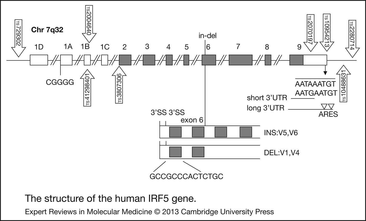
Human Irf5 gene is mapped to the chromosome 7q32 (Ref. Reference Kozyrev and Alarcon-Riquelme9) (Fig. 1). There are 9 exons in Irf5 gene. It has a CpG island, encompassing exon 1A and 1B (there are four alternative 5′UTR exons in IRF5 gene: 1A, 1B, 1C and 1D) (Ref. Reference Kozyrev and Alarcon-Riquelme9). Of the four exons, only exon 1A transcripts are highly expressed. Therefore, it is possible that methylation of the CpG island might result in IRF5 gene being epigenetically silenced. Indeed, methylated CpG island in promoter regions has been found in lung cancer cells (increased IRF5 hyper-methylation in CRL5800, CRL5807, CRL5872 and CRL5810 cell lines) (Refs Reference Li and Tainsky10, Reference Kondo and Issa11). Allele T of the single nucleotide polymorphism (SNP) rs2004640 creates the 5′ donor splice site, causing transcription of alternative exon 1B. SNP rs10954213 is located in the polyA site AATAAA. IRF5 gene has 2 alternative polyA sites, producing transcripts with either long or short 3′UTR (Refs Reference Kyogoku12, 13, Reference Kozyrev14). mRNA with long 3′UTR is less stable owing to the presence of the AU-rich elements (AREs) which target mRNA for rapid degradation (Ref. Reference Khabar15). Exon 6 has a 30-bp in-frame indel polymorphism, which is located in a proline-, glutamic acid-, serine-, threonine-rich domain, and known to affect protein stability and function in the IRF family of proteins. Exon 6 can be spliced at an alternative site with 48-bp (SV-16) downstream of the canonical splice junction. The 30-bp insertion/deletion (in/del-10) has distinct ability to initiate transcription of IRF5 target genes and differs from IRF5 protein isoforms in part (Refs Reference Barnes16, Reference Mancl17, Reference Barnes18).
The structure of the human interferon regulatory factor 5 (IRF5) gene. Proteins coding exons are displayed in grey and noncoding exons are shown in white. The relative positions of the single nucleotide polymorphisms (SNPs) and indel are displayed.

Correlation between IRF5 and IFNα in SLE
IRF5 is found to be up-regulated (Ref. Reference Feng19), and contributes to the heritability of serum IFNα activity in SLE and affects variance in IFNα activity (Ref. Reference Niewold20). Serum levels of IFNα were increased in SLE patients (Refs Reference Hooks21, Reference Baechler22), and associated with disease activity (Ref. Reference Kirou23), which makes it possible that high IFNα levels expressed in SLE patients might contribute to the increased IRF5 expression (Refs Reference Mancl17, Reference Barnes24). Minigene experiments demonstrated that IFNα could be a factor contributing to the enhanced IRF5 expression (Ref. Reference Feng19). IFNα is known to be a pleiotropic type I IFN with the potential to break self-tolerance by activating antigen-presenting cells following uptake of self-material (Ref. Reference Blanco25). High levels of IFNα were related to the presence of anti-double-stranded DNA (anti-dsDNA) and anti-RBP antibodies (Refs Reference Kirou23, Reference Weckerle26). Some patients who received recombinant human IFNα for the treatment of malignancy and viral hepatitis have developed de novo SLE, which typically resolves after withdrawal of the cytokine therapy (Refs Reference Rönnblom, Alm and Oberg27, Reference Niewold and Swedler28). Collectively, these findings suggest a potential role of IFNα in SLE susceptibility.
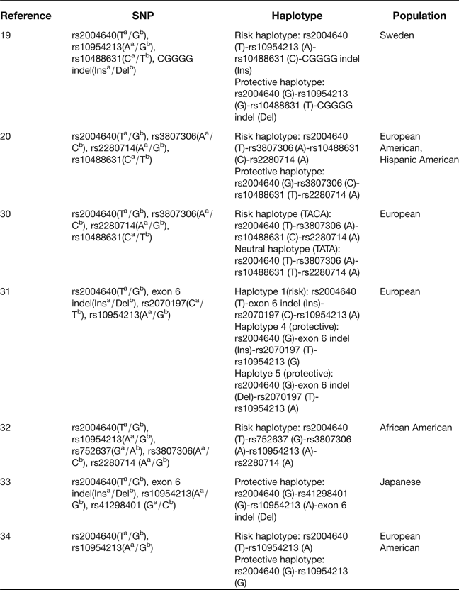
As noted above, high serum IFNα is a risk factor for SLE. Interestingly, the immune complexes (ICs) formed by auto-antibodies in SLE have been recognised to stimulate the IFNα synthesis by activating endosomal TLRs (Ref. Reference Lövgren29). IRF5 downstream of endosomal TLRs may exacerbate chronic endogenous stimulation provided by ICs. Therefore, genetic variation of IRF5 may affect IFNα production, depending on particular auto-antibodies (Ref. Reference Niewold30). In European ancestry patients with SLE, both anti-dsDNA and anti-Ro antibodies were associated with a risk haplotype (TACA), and anti-dsDNA correlated with a neutral haplotype (TATA), but anti-Ro antibodies were not significantly related to the TATA haplotype, which lacked the exon 6 insertion (Table 1) (Ref. Reference Niewold30). Furthermore, the haplotypes related to particular auto-antibodies can lead to higher serum IFNα in the presence of that particular auto-antibody, suggesting that auto-antibodies are necessary for dysregulation of serum IFNα ascribed to IRF5 risk variants. Although in SLE patients of European American and Hispanic American ancestry, risk haplotype of IRF5 was related to the increased serum IFNα activity in comparison to those with protective haplotype, and this effect was prominent in patients positive for either anti-RBP or anti-dsDNA auto-antibodies (Ref. Reference Niewold20) (Table 1). Collectively, these findings may support a model of disease pathogenesis where specific interaction of auto-antibodies with particular IRF5 variants contributes to abnormal IFNα synthesis, leading to up-regulated risk of SLE (Fig. 2) (Refs Reference Kozyrev and Alarcon-Riquelme9, Reference Niewold30). In addition, IRF5 haplotype correlated with anti-Ro antibodies in SLE patients is associated with the same auto-antibody in healthy individuals who carry this auto-antibody, suggesting that IRF5 plays a role in serologic autoimmunity in humans (Ref. Reference Cherian35).
Relationships among the proposed association of interferon regulatory factor 5 (IRF5) haplotypes, IFNα and Systemic lupus erythematosus (SLE)-specific auto-antibodies in the pathogenesis of SLE. (1) ?: IRF5 haplotypes may affect autoimmune susceptibility to generate particular auto-antibodies. (2) These auto-antibodies (anti-RBP, anti-Ro and anti-dsDNA) in cells may trigger endosomal Toll-like receptor 7/9 (TLR7/9). (3) Specific IRF5 related risk variants enhance downstream IFNα synthesis in the context of auto-antibodies, leading to high serum IFNα and SLE susceptibility.
Association of IRF5 haplotypes with SLE
aRisk allele.
bProtective allele.
Del, deletion; Ins, insertion; IRF5, interferon regulatory factor 5; SNP, single nucleotide polymorphism; SLE, systemic lupus erythematosus.
Genetic association between IRF5 and SLE
Genetic association studies on SNPs in IRF5 have suggested different haplotypes that confer either susceptibility to, or protection from SLE (Refs Reference Graham31, Reference Sigurdsson36, Reference Graham37). Three putative functional alleles (rs2004640, rs10954213 and exon 6 indel) were believed to be associated with SLE in European descent (Ref. Reference Graham31). When combined, these alleles and another SNP, rs2070197, haplotype 1 was significantly associated with the risk of SLE, and the risk haplotype 1 was predicted to express exon 1B isoform (because of rs2004640) (Ref. Reference Graham31) (Table 1). Haplotypes 4 and 5 indicated strong evidence for protection. Moreover, individuals who carried haplotype 1 in trans with either of the haplotypes (haplotyes 4 or 5) which lacked the expression of exon 1B isoform displayed a down-regulation of SLE susceptibility. Thus, the highest risk to SLE for a haplotype is predicted to be expressed at high level transcripts containing exon 1B. As for the haplotypes in SLE patients in Sweden, the risk haplotype contains the risk alleles of the four polymorphisms: 4xCGGGG indel Ins, rs2004640 T, rs10954213 A and rs10488631 C, whereas the corresponding protective haplotype contains the nonrisk alleles of the polymorphisms (Table 1) (Ref. Reference Feng19). With respect to the haplotype structure of IRF5 in African Americans, results of five variants (rs2004640, rs752637, rs3807306, rs10954213 and rs2280714) revealed a significant haplotype (TGAAA) determined by the minor A risk allele at rs3807306 (Table 1) (Ref. Reference Kelly32). Multivariate analysis demonstrated that rs3807306 was required for the association with SLE because all other variants, individually, and the haplotype, as a unit lost significance, when conditioned on rs3807306. In another study on SLE in Japanese population, a protective haplotype carrying rs2004640 G, rs41298401 G, a deletion in exon 6 and rs10954213 A, defined by the intron 1 SNP rs41298401, was identified (Ref. Reference Kawasaki33) (Table 1). Collectively, these findings suggest that IRF5 haplotypes were significantly associated with SLE in multiple ethnicities.
TLRs are able to recognise endogenous nucleic acids in the context of ICs detected in SLE patients, which promotes DCs maturation and IFNα synthesis, a process that involves IRF5 (Ref. Reference Yasuda38). A TATA-less promoter region, designated P1, was mapped in Epstein-Barr virus (EBV) BamHI-A rightward transcripts, where P1 had relatively high basal activity in B cells. After EBV infection of B cells, transcription from P1 was detected soon upon infection, but IRF5 negatively regulated P1 activity (Ref. Reference Chen39). In addition, transient expression of IRF5 decreased activation of the EBV oncoprotein 1 (LMP1) promoter, and transfection of either an IRF5 dominant-negative construct or IRF5 small interfering RNA in type II EBV cells (C1D12 and C2G6) led to up-regulation in endogenous levels of LMP1, suggesting that IRF5 can down-regulate the expression of LMP1 (Ref. Reference Ning, Huye and Pagano40). Furthermore, EBV infection of type III latently infected B-cell lines LCL, Mutu III and EREB2-5 induced IRF5 expression, and examination of IRF-5 transcripts revealed a novel splice variant, V12, which was induced by EBV infection, constitutively localised in nuclear, performed as a dominant negative form in IRF-5 (Ref. Reference Martin41). These data indicate that EBV is significantly related to IRF5. EBV infection is known to be associated with SLE susceptibility (Refs Reference James, Harley and Scofield42, Reference James43, Reference James44, Reference Lu45). Since B cells are the primary host cell type for EBV infection, it is likely that IRF5 affects B cell responses (B cell receptor crosslinking) to cause the disease. Compared with the protective haplotype (Table 1), peripheral blood mononuclear cells (PBMCs) in SLE patients from European American descents with the risk haplotype (both stimulated and un-stimulated with EBV) indicated three pathways with significance: the INF, TLR and B cell receptor (BCR) pathways (Ref. Reference Guthridge34). IFN-induced transmembrane protein 1 (IFITM1) had higher expression in cells with risk haplotype than cells without risk haplotype at steady states (Ref. Reference Guthridge34). Fos and MyD88 were both under-expressed in the un-stimulated cells with risk haplotype compared with the cells with protective haplotype (Ref. Reference Guthridge34). In addition, CD79A and CD79B were decreased 2.2-fold in the uninfected cells with risk haplotype, but this difference disappeared after EBV infection. Collectively, these data suggest that IRF5 haplotype may be a key determinant in not only the IFN, TLR pathways, but also the BCR pathways in SLE.
Recently, the A allele of rs10954213 was found to be related to the high level of IRF5 in SLE patients (Ref. Reference Kozyrev14). In mice, mutations disrupting AREs in the tumor necrosis factor α (TNFα) gene can result in up-regulation of circulating TNFα and hypersensitivity in the presence of stimulation with TLR4 ligand lipopolysaccharide (LPS) (Refs Reference Jacob, Lee and Strassmann46, Reference Kontoyiannis47). Therefore, it is possible that rs10954213 in the 3′UTR polyA site of IRF5 might be a functional mutation required for high levels of IRF5. IRF5 expression and alternative splicing were significantly increased in SLE patients compared with healthy controls, especially when the transcription from exon 1A and exon 1D was markedly up-regulated in the patient cells (Ref. Reference Feng19). It is known that increased serum levels of small nuclear ribonucleoproteins (snRNPs) and serine/arginine-rich (SR) proteins, which are components of the spliceosome, can be found in SLE patients (Refs Reference Lerner and Steitz48, Reference Neugebauer49). Following co-transfection with snRNPs or SR proteins, the transcription from exon 1A and alternative splicing of exon 5–7 were enhanced. In particular, co-transfection with U1 snRNP led to the strongest increase of IRF5 expression, implying its contribution to the enhanced alternative splicing (Ref. Reference Feng19). Furthermore, IRF5 transcript expression was higher in patients carrying the risk haplotype versus patients carrying the protective haplotype, and IRF5 protein expression was increased in myeloid DCs and monocytes from SLE patients, correlating with the risk haplotype (Ref. Reference Feng19). In addition, the functional effects of exon 6 in/del-10 and SV-16 within IRF5 were demonstrated in SLE, where the expression of either SV-16 or in/del-10 confers IRF5 the ability to impair apoptotic response, and is related to the down-regulation of IRF5 nuclear translocation in murine embryonic fibroblasts (MEFs) after treatment with DNA-damaging stimulus (Ref. Reference Wen50). MEFs that expressed IRF5-bearing SV-16 displayed up-regulated IL-6 production upon LPS stimulation. By contrast, MEFs containing in/del-10 produced IL-6 with no significant difference from the controls. Intriguingly, the absence or presence of both in/del-10 and SV-16 leads to abrogation of these effects. Together, these observations indicate that specific genetic architecture of IRF5 correlates with the up-regulated expression of IRF5 in SLE patients, and IRF5 variants carried on SLE risk haplotypes modulate IRF5 function.
IRF5 and the immune mechanisms of autoimmune lupus
It has been observed that compared with the nonlupus-prone strain of mice (C57BL/6), lupus-prone mice [NZB, (NZB/W)F1, NZM2410] produced higher levels of IRF5 mRNA in their splenic cells (Ref. Reference Shen5). In nonautoimmune mice, IRF5 deficiency reduced the expression of Blimp-1, IFNβ and p202 proteins, whereas the levels of Aim2 and FcγRIIB proteins were up-regulated. On the contrary, overexpression of IRF5 in RAW264.7 cells can rescue these defective effects whereas the levels of IRF5 mRNA and proteins were increased in Aim2−/− and FcγRIIB−/− mice (Ref. Reference Panchanathan51). In lupus-susceptible B6.Nba2 and B6.Sle123 congenic mice, the expression of IRF5 mRNA and proteins as well as IFNβ mRNA from their splenic cells were up-regulated compared with the age-matched B6 mice, which was positively related to the increased expression of Blimp-1 and p202 proteins, but negatively associated with the levels of Aim2 and FcγRIIB proteins (Ref. Reference Panchanathan51). These data demonstrated that IRF5 regulates the expression of lupus susceptibility genes, and implied that IRF5 may contribute to lupus susceptibility in part by down-regulating the expression of aim2 and fcγriib genes (Refs Reference Panchanathan51, Reference Panchanathan52, Reference Panchanathan53). Since the aberrant expression of IRF5 in human and murine cells is associated with SLE as discussed above, and IRF5 exhibits pathogenic potential by controlling immune response, advanced insight of regulatory mechanism of IRF5 in immune cells becomes critical to understand the pathogenesis and develops better therapeutic strategies for SLE (Ref. Reference Xu54).
The role of IRF5 in B cells
IRF5 plays an important role in B cell function. It promotes B cell maturation in part by modulating the expression of Blimp-1, a master regulator of plasma cell maturation (Ref. Reference Lien55). IRF5 deficiency in mice resulted in an altered isotype switching, leading to diminished IgG2a/c responses to TI and TD antigens and to virus infection (Ref. Reference Fang4). In responding to immunisation with NP-KLH, a TD antigen, IgG1 response was markedly lower in IRF5−/− mice compared with the control mice (Ref. Reference Fang4). On the contrary, the virus-specific antibody response of IRF5−/− mice to the major polyoma virus (PyV) capsomer protein VP1 demonstrated small difference in the levels of antiviral IgG1 in comparison with the control mice. Similarly, stimulation of IRF5−/− B cells with LPS and IL-4 led to upgraded levels of IgG1 germline transcripts as well as IgG1-expressing B cells (Ref. Reference Fang4). Adoptive transfer experiments indicated that the attenuated IgG2a/c response is an intrinsic property of IRF5−/− B cells.
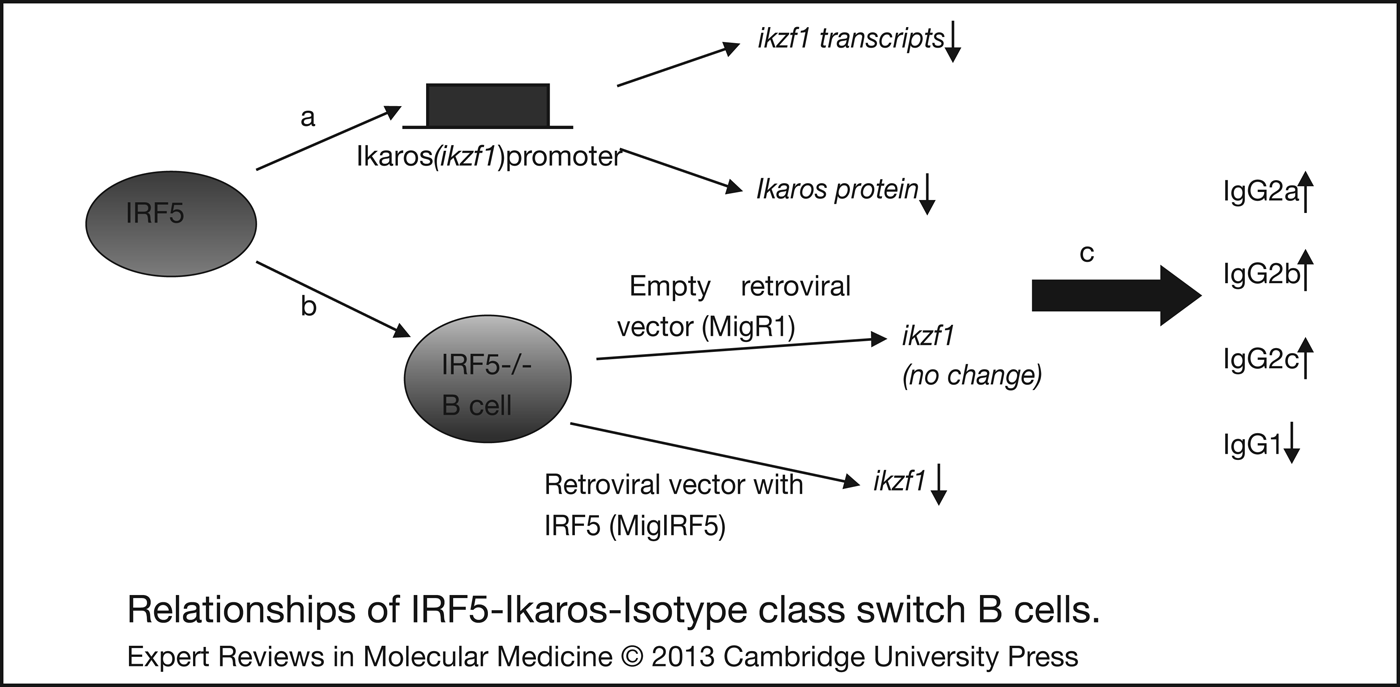
It is well-known that class switch recombination (CSR) can be induced in B cells in vitro by combination of multiple B cell activators, such as TLR ligands and cytokines (Ref. Reference Stavnezer56). Purified B cells from naive IRF5−/− mice induced less preferential switching to IgG2a/c than that of wild-type mice cultured with LPS alone or co-cultured with IL-4, or IFNγ (Ref. Reference Tamura2). These findings were demonstrated by the fact that modulation of the IgG2a/c responses was mediated by the IRF5-Ikaros (ikzf1) axis. Evidence has indicated that IRF5 mediates induction of IgG2a in virtue of controlling Ikaros expression (Ref. Reference Kim57). The Ikaros family of transcription factors is able to silence their target genes through recruitment of transcriptional inhibitor complexes, for instance, histone deacetylase complexes. Ikzf1−/− mice cannot generate B cells (Ref. Reference Wang58), whereas Ikzf1L/L mice (‘L’ for Lac Z) up-regulated their production of IgG2a/c during LPS- and IFNγ-induced CSR in culture (Refs Reference Kirstetter59, Reference Sellars60). The levels of ikzf1 transcripts were elevated in IRF5−/− B cells undergoing CSR, indicating that the expression of ikzf1 was regulated by IRF5, which was confirmed by sequence analysis where there was an IRF-binding site in the promoter of the ikzf1 gene that specifically bound IRF5 in the presence of MyD88 (Ref. Reference Fang4). Together, these results identify the IRF5-Ikaros axis as a critical modulator of IgG2a/c class switching in B cells (Fig. 3).
Relationships of interferon regulatory factor 5 (IRF5)-Ikaros-Isotype class switch B cells. (a) IRF5 binds to ikzf1 promoter, results in Ikaros (ikzf1) transcripts and proteins down-regulation. (b) Transduction of IRF5 deficient B cells with empty retroviral vector (MigR1), or IRF5 (MigIRF5), leads to no changed ikzf1 or decreased ikzf1 transcripts, respectively. (c) IRF5 negatively regulates Ikaros (ikzf1), which may up-regulate the levels of IgG2a, IgG2b and IgG2c, but down-regulate the levels of IgG1.
IRF5 deficiency in pristane-induced lupus mice did not produce IgG2b, IgG2c, IgG3, IgM anti-U1A Abs, anti-Sm/RNP auto-antibodies, anti-nuclear antibodies (ANA) and anti-dsDNA auto-antibodies (Refs Reference Savitsky3, Reference Xu61, Reference Feng62). Similarly, the production of auto-antibodies, including ANA, anti-dsDNA antibody, anti-Sm antibody and anti-RNP antibody, was dramatically attenuated in MRL/lpr, FcγRIIB−/− Yaa and FcγRIIB−/− lupus mice deficient in IRF5 (Refs Reference Xu61, Reference Feng62, Reference Richez63, Reference Tada64). When the correlation between the absence of auto-antibodies and defective isotype switching was evaluated in IRF5−/− mice, the pristane-treated IRF5−/− mice produced decreased levels of IgG2b, IgG2c and IgG1 anti-U1A Abs (Ref. Reference Xu61). However, IgM levels were comparable between IRF5−/− and control mice (Ref. Reference Xu61). These results were later confirmed by the findings that IRF5−/− mice revealed significant decreases in both IgG2a/c and IgG2b levels (Refs Reference Savitsky3, Reference Feng62). Therefore, the down-regulation of IgG2a/c and IgG2b may be associated with significant decreases in specific lupus auto-antibodies. In the pristane-induced lupus mice, IRF5 deficiency indicated upgraded serum levels of total IgG1, implying that IRF5 deficiency does not decrease long-lived IgG1 expressing plasma cells (Ref. Reference Feng62). After class switching, auto-reactive B cells might undergo further selection and expansion, where IRF5−/− mice were lack of the generation of IgG1 auto-antibodies, implying that a mechanism other than class switching controls antigen specificity in these mice. Instead, IRF5 might be of importance for selection or expansion of auto-reactive clones from B cell repertoire (Refs Reference Xu61, Reference Feng62). However, the clear mechanism is not known to date, which needs further investigation.
When the role of type I IFN in inducing IgG2a secretion by IRF5+/+ and IRF5−/− B cells was investigated, it was found that exogenous type I IFN did not rescue the defect in CpG-B (a TLR9 ligand)-induced IgG2a secretion in the absence of IRF5, in spite of their response to type I IFN (Ref. Reference Savitsky3). Furthermore, there were no differences in the levels of type I IFN mRNA between CpG-B-stimulated IRF5+/+ and IRF5−/− B cells. Thus, these results suggest that IRF5 may regulate IgG2a synthesis via its ability to control type I IFN gene expression, which was verified by the absence of IgG2a production in type I IFN receptor-deficient B cells stimulated with CpG-B (Ref. Reference Savitsky3). Moreover, IRF5 was found to be necessary for IL-6 production in B cells (Ref. Reference Takaoka8). Indeed, IL-6 is critical for the generation of anti-dsDNA auto-antibodies in pristane-injected lupus mice (Ref. Reference Richards65). Furthermore, the expression of ccl3 and ccl4 mRNA were compromised in TLR9-stimulated IRF5−/− B cells, suggesting a role of IRF5 in orchestrating inflammatory responses (Ref. Reference Savitsky3). In addition, IRF5−/− B cells proliferated less in response to TLR9 stimulation than IRF5+/+ B cells did. Collectively, this evidence suggested that IRF5 may regulate B cell activation following TLR9 stimulation.
IRF5 in T (Th) cells
FcγRIIB deficient mice can induce a spontaneous SLE-like disease, this property of the FcγRIIB−/− B6 model mimics the multigenic nature of human SLE (Refs Reference Richez63, Reference Bolland and Ravetch66, Reference Bolland67). Addition of the Yaa locus to this model leads to a significant increase in severity of the disease (Ref. Reference Bolland67). In the Yaa murine lupus model, IRF5 was critical for T cell activation, because the expression of the activation markers CD69 and CD44 on splenic T cells was significantly reduced in IRF5−/− RII.Yaa mice compared with IRF5+/+ RII.Yaa mice (Ref. Reference Richez63). In pristane-induced lupus mice, the expression of CD69 on splenic CD4+ T cells of IRF5−/− mice was markedly down-regulated (Ref. Reference Feng62), whereas in MRL/lpr mice, analysis of spleen cell populations revealed that the memory (CD62LlowCD44high) and activated (CD62LhighCD44high) CD4+ T cells were decreased in comparison to B220+ activated CD4+ T cells (Ref. Reference Kim57). Therefore, IRF5 is important in T cell activation in lupus mice.
Consistent with the importance of type I IFN in the pathogenesis of lupus, IRF5−/− mice produced significantly reduced levels of the ISGs IRF7, ISG15 and Mx1 (Refs Reference Xu61, Reference Feng62). Meanwhile, protein levels of the type I IFN-inducible chemokine MCP-1 (CCL2), pro-inflammatory cytokines such as IL-12 p40/70, IL-6 and IL-23p19 were also down-regulated in IRF5−/− mice (Ref. Reference Xu61). These data suggest that the deficiency of IRF5 attenuated lupus by influencing the production of type I IFN and other cytokines that combine innate immunity with the activation of T-helper 1 (Th1) and Th17 cells (Ref. Reference Hunter68). On the contrary, serum levels of Th2 cytokines IL-4, IL-5 and IL-10 were increased in IRF5−/− mice compared with IRF5+/+ mice (Ref. Reference Feng62). The upgraded Th2 cytokine may contribute to the disease protection in IRF5−/− mice since Th2 cells promote the synthesis of the least pathogenic IgG1 isotype observed in IRF5−/− mice (Ref. Reference Takahashi69). To interpret the T cell identity that generated Th2 cytokines from pristane-induced IRF5−/− mice, the authors found that CD4+ T cells from IRF5+/+ mice generated negligible amounts of IL-4 and only Th1 cells can be detected in IRF5+/+ mice (Ref. Reference Feng62). In the IRF5−/− mice, however, a readily noticeable fraction of IL-4 was produced. These findings support the fact that IRF5 is critical in the regulation of Th1/Th2 polarisation, and contributes to pristane-induced lupus pathogenesis. Xu et al (Ref. Reference Xu61) investigated Th cell subsets in type I IFN receptor (IFNAR) deficient and TLR7 deficient mice and confirmed that IL-4-producing cells had decreased. Furthermore, in IRF5−/− mice, the ratio of Th1 to Th2 was lower in IFNAR deficient and TLR7 deficient mice than their wild-type counterparts. On the other hand, serum levels of IL-6 were reduced in IRF5−/− RII.Yaa mice, but the levels of IFNγ were comparable between IRF5−/−RII.Yaa mice and controls (Ref. Reference Richez63). Hence, IRF5 deficiency cannot completely attenuate all the autoimmune phenotypes.
All type I IFNs act via a single cell surface type I IFN receptor, termed IFNAR (IFNAR1 and IFNAR2) (Refs Reference Oritani70, Reference Fenner71, Reference Uzé72, Reference Illei73). Both spleen and lymph node sizes were smaller in IFNAR1−/− RII.Yaa mice relative to IFNAR1+/+RII.Yaa mice (Ref. Reference Richez63). Renal diseases were less severe in the IFNAR1−/−RII.Yaa mice compared with the IFNAR1+/+RII.Yaa mice as well, which were demonstrated by reduction in glomerular crescent formation, cell number per glomerulus and interstitial disease (Ref. Reference Richez63). Furthermore, IFNAR1−/−RII.Yaa mice survived longer than IFNAR1+/+RII.Yaa mice. These findings may suggest a role of type I IFN in driving the pathogenesis of SLE. Expression of type I IFN-regulated genes IFIT1 and MX2 was similar in B220+ splenocytes from IRF5+/+ or IRF5−/−RII.Yaa mice in comparison with wild-type mice (Ref. Reference Richez63). By contrast, kidneys from IRF5+/+RII.Yaa mice showed 3-fold induction of both IFIT1 and MX2, suggesting the evidence for IRF5-dependent type I IFN expression, albeit at low levels and only at a site of severe inflammation. It is notable that serum levels of IL-6 and IL-10 were decreased in IRF5−/−RII.Yaa mice (Ref. Reference Richez63), and it has been known that down-regulation of IL-6 and IL-10 might be relevant for lupus pathogenesis, where these two cytokines can contribute to B cell activation and auto-antibody synthesis, and are related to disease activity in lupus patients (Ref. Reference Illei73). Therefore, it is likely that IRF5 effects are medicated through IFNAR independent pathways in this lupus model, possibly through suppression of the generation of IL-6 and IL-10.
Influence of IRF5 on DC immune responses
IRF5 plays an important role in DC (Fig. 4). IRF5−/− and MyD88−/− mice treated with pristane had strikingly reduced number of CD11b−CD11C+B220+PDCA-1+ pDCs in the peritoneal cavity compared with wild-type B6 mice (Ref. Reference Xu61). However, the percentage of pDCs in the BM was similar between IRF5−/− and wild-type mice. CCR7 is expressed on DCs, and its ligands (CCL19 and CCL21) are required for homing of pDCs to lymph nodes (Ref. Reference Seth74). The expression of both CCL19 and CCL21 in peritoneal exudates cells from IRF5−/− mice were lower than that of control mice (Ref. Reference Xu61). In light of the MyD88 dependent pDC homing and the ability of TLR ligands to promote CCR7 expression (Ref. Reference Seth74), fluorescence intensity of CCR7 expression in CD11b−CD11C+B220+PDCA-1+ pDCs was found to be comparable in BM between B6 and IRF5−/− mice, and the IRF5−/− peritoneal pDCs displayed similar fluorescence intensity to that of IRF5−/− BM pDCs (Ref. Reference Xu61). Consequently, these data may support the fact that MyD88 expression promotes pDC migration to the peritoneum by up-regulating IRF5-regulated CCL19/CCL21 but not CCR7 expression.
Possible relationships between interferon regulatory factor 5 (IRF5)-chemokines and cytokines in dendritic cells (DCs). Endosomal TLRs (such as TLR4, TLR7, TLR9, etc.) recruit MyD88, subsequently bind to and stimulate IRF5, leading to activated pro-inflammatory cytokine and chemokine production in DCs (pDC, FL-DC).
In splenic DCs from MRL/lpr mice, IRF5 deficiency generated reduced levels of TNFα, IL-6 and IL-10 in response to the TLR9 ligand CpG and TLR7 ligand loxorbine (Ref. Reference Tada64). The levels of IFNα, IFNγ and MCP-1 were declined in response to CpG as well. Since ICs were able to activate DCs through engagement of TLRs and Fcγ receptors (Refs Reference Boulé75, Reference Means76, Reference Savarese77, Reference Leadbetter78), stimulation of wild-type DCs with purified ICs from the serum of MRL/lpr mice produced upgraded levels of TNFα and IL-6, whereas ICs-induced cytokine production was markedly decreased in the DCs from IRF5−/− mice (Ref. Reference Tada64). These findings indicate that IRF5 mediates the activation of DCs that are triggered by ICs or by TLR7 or TLR9 engagement, and IRF5 deficiency may cause a deficit in pro-inflammatory cytokines generation. Moreover, LPS-induced IFNα synthesis in IFNβ-pretreated FL-DCs (DCs obtained from BM cells cultured in fms-like tyrosine kinase 3 ligand) was found to be dependent on TLR4 with sequential involvement of the TRIF (Toll/IL-1 receptor domain-containing adaptor protein-inducing IFNβ) and MyD88 pathways (Ref. Reference Richez79). In the FL-DCs from IRF5−/− mice, LPS-induced IFNα synthesis was not completely down-regulated as compared with wild-type FL-DCs. However, both CpG-A- and TLR3 ligand poly(I:C)-induced IFNα production were severely down-regulated in IRF5−/− mice, implying a critical role of IRF5 in IFNα production via MyD88- and TRIF-dependent pathways.
Since IFNβ and IFNα share the same receptor and activate similar IFNαβ-inducible gene programs (Ref. Reference Theofilopoulos80), it is likely that IFNβ may potentially contribute to the effects of type I IFN on lupus that were generally ascribed to that of IFNα. Lupus IgG, conventional TLR7 ligand R848 and CPG-A stimulation induced appreciable IFNβ production by FL-DCs, which is dependent on IRF5 (Ref. Reference Yasuda38). Moreover, compared with wild-type control, activation of FL-DCs from IRF5−/− mice by lupus IgG and CpG-A (50 nM) induced substantially reduced production of IFNα (Ref. Reference Yasuda38). This is in contrast to what other people have observed that IFNα synthesis in pDC induced by CpG-A at 1 µM was IRF5 independent (Ref. Reference Richards65). This discrepancy may be attributable to several reasons: for example, the issue of dosage might be related to lupus pathogenesis where the activation strength of TLR7 or TLR9 by nucleic acid-containing ICs is weaker than that elicited by maximal doses of microbial and synthetic loxorbine, CpG-A. Besides, it is also likely that pDCs from spleen in vivo are different from the pDCs in FL-DCs culture in their requirement for IRF5 in CpG-A induced IFNα synthesis. Recently, IRF5−/− mice have been found with impaired pDC development, which is not dependent on IRF5 (Ref. Reference Purtha81). IFNα secretion by IRF5−/− pDCs following CpG-A stimulation was similar to background levels (Ref. Reference Bolland67). A genomic duplication within Dock2 led to a frame-shift mutation and premature stop codon, and the ectopic expression of DOCK2 by retroviral transduction restored pDCs development. Still, IRF5−/− mice lacking the Dock2 mutation had largely normal IFNα synthesis from pDCs (Ref. Reference Purtha81). Therefore, a reduction in the expression of Dock2 may be responsible for the impaired pDCs in IRF5−/− mice. Interestingly, Yasuda et al. (Ref. Reference Yasuda82) found that IRF5 is involved in the TLR9-induced type I IFN production independent of the Dock2 mutation, showing with reduced IFNα and IFNβ production in IRF5−/− FL-DCs upon stimulation with TLR9 ligands. However, IRF5−/− mice with the Dock2 mutation showed higher serum levels of IgG1 and lower levels of IgG2b, IgG2a/c and IgG3 than IRF5−/− mice without the Dock2 mutation, suggesting that the Dock2 mutation contributes to Th2-type effects (Ref. Reference Yasuda82). These findings need to be clarified in lupus in the future.
Conclusion
The understanding of the immune-pathological mechanism of SLE is gradually evolving with budding studies on evaluating the important role of IRF5 in controlling T, B lymphocytes and DCs, as well as the underlying intracellular signalling mechanisms in each of these immune cell types in response to various stimuli. A number of methodologies including gene expression, familial cytokine analysis and genetic association studies were employed to demonstrate the over-active IFNα pathway as a critical factor in the pathogenesis of SLE. IRF5 is a crucial mediator of IFNα and IFNα-induced gene expression, and correlates with endosomal TLRs activation (Ref. Reference Xu61). These TLRs are pathogenically activated by SLE-related, autoimmune complexes, which must have provided a chronic endogenous stimulus to the IFNα pathway in SLE. Detailed studies supporting IRF5 gene polymorphisms are gain-of-function, leading to some of the IFNα pathway activation observed in the disease. Nevertheless, whether the observed gene/auto-antibody interaction upon IFNα stimulation is dependent on other parts of the SLE phenotype is not clear to date (Ref. Reference Salloum and Niewold83). Furthermore, EBV infection is known to involve SLE pathogenesis, and viral infection may supply a strong stimulus to the TLRs/IRF5 system. Therefore, a model of ‘gene + microenvironment stimulus = high IFNα’ may still apply. However, with the advent of more advanced technology and studies, our understanding of the role of IRF5 in this elusive disease is expected to be further enriched with novel findings.
Acknowledgement
This work was partly supported by grants from the key program of National Natural Science Foundation of China (30830089), National Natural Science Foundation of China (81102192, 81172764) and National Health and Medical Research Council Project Grant of Australia (1006428).
Conflict of interest
The authors declare no conflict of interest.