Introduction
Intracranial, extracerebral haemangiomas are rare, tumour-like lesions.Reference Katayama, Tsubokawa, Miyazaki, Yoshida and Himi1–Reference Rigamonti, Pappas, Spetzler and Johnson3 They are considered more as malformations or hamartomas,Reference Abe, Tabuchi, Tanaka, Hodozuka, Kunishio and Kubo4 and tend to involve mainly the cavernous sinus.Reference Rigamonti, Pappas, Spetzler and Johnson3, Reference Sawamura and de Tribolet5, Reference Sepehrnia, Tatagiba, Brandis, Samii and Prawitz6 Haemangiomas are either cavernous or capillary, with the latter being a rarer entity. Haemangiomas consist of vascular channels lined by a single layer of endothelial cells, with the vascular spaces consisting of capillaries (in capillary haemangiomas) or of more dilated vessels (in cavernous haemangiomas).Reference Abe, Tabuchi, Tanaka, Hodozuka, Kunishio and Kubo4 Capillary haemangiomas are more frequently found on the skin or the mucosa, with a possible relationship to female sex hormones.Reference Fortna and Junkins-Hopkins7 The histological features of a lobular capillary haemangioma of the skin are similar to those found in the central and peripheral nervous system.Reference Abe, Misago, Tanaka, Masuoka and Tabuchi8 Most cases involving the central and peripheral nervous system are found at the level of the spinal cord,Reference Andaluz, Balko, Stanek, Morgan and Schwetschenau9, Reference Enomoto and Goto10 the cerebellum and the cerebrum.Reference Abe, Tabuchi, Tanaka, Hodozuka, Kunishio and Kubo4, Reference Abe, Misago, Tanaka, Masuoka and Tabuchi8, Reference Lee11
Herein, we report the clinical presentation, investigation, management and histological findings of a case of lobular capillary haemangioma of the trigeminal nerve. To the best of our knowledge, and after a thorough review of the literature, this report appears to represent the first published case of lobular capillary haemangioma of the mandibular branch of the trigeminal nerve, with extension to its maxillary branch and causing erosion of the petrous carotid canal. This case represents only the second published report of this rare entity located in Meckel's cave; in the previous report, the lesion involved the mandibular branch of the trigeminal nerve with palsy of the ipsilateral VIth nerve.Reference Brazis, Wharen, Czervionke, Witte and Jones12 Cavernous or capillary haemangiomas are reported as rare entities.
Clinical presentation
A 60-year-old woman presented to the otolaryngological clinic with right-sided, chronic facial pain and paraesthesia. The pain had started six months prior to her first visit and was localised over the right mandibular and maxillary regions, worsening over time. The pain was severe, continuous and non-pulsatile, with periods of exacerbation, ranging from several minutes to several hours, rendering it unbearable. This pain was associated with paraesthesia affecting the right face, mainly the mandibular and maxillary regions. The patient also complained of a mild hearing impediment on the right side. She did not report any diplopia, facial weakness, otalgia or otorrhoea, nor any pterygoid muscle weakness.
The physical examination showed normal function of the cranial nerves, except for the right trigeminal nerve which exhibited decreased sensitivity mainly in its mandibular and maxillary branches. No facial weakness or diplopia was uncovered. The patient's cerebellar functions were normal. Otoscopy showed serous otitis media of the right ear.
Flexible nasal endoscopy was undertaken, and confirmed the absence of a nasopharyngeal tumour. The patient's pure tone average and speech reception thresholds were normal. Impedance testing revealed reduced compliance of the right tympanic membrane.
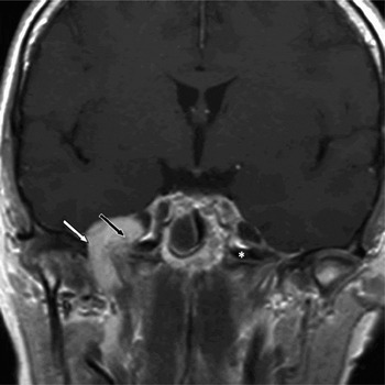
Magnetic resonance imaging (MRI) with gadolinium enhancement was performed (Figure 1). This showed a 2.7 × 2.1 × 1.7 cm lesion occupying the foramen ovale, extending medially into the cavernous sinus, slightly compressing Meckel's cave and extending posteriorly to impinge upon the petrous carotid artery. The inferior margin of the lesion extended to the pterygoid muscles. The lesion had a high-signal intensity on T2-weighted images and a low-signal intensity on T1-weighted images which were enhanced after gadolinium injection. A clouding of the right middle ear and mastoid air cells was also detected.
Coronal, T1-weighted magnetic resonance image with gadolinium enhancement, showing the tumour extending through the foramen ovale (white arrow). More than half of the tumour's bulk is located in Meckel's cave (black arrow). The asterisk indicates the internal carotid canal.

At this time, the differential diagnosis included schwannoma, meningioma, metastasis, lymphomatous infiltration and haemangioma.
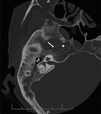
In order to evaluate the bony involvement of this tumour, computed tomography (CT) scanning of the temporal bone was completed. This revealed complete clouding of the right mastoid air cells and the middle ear secondary to an expanding lesion compressing the right eustachian tube, as well as lysis of the horizontal petrous segment of the carotid canal. Lysis of the anterior aspect of the sphenoid bone's right greater wing was also noticed at the level of the ovale and rotundum foramina (Figure 2).
Axial computed tomography scan showing clouding in the mastoid air cells (M). Partial lysis (arrow) of the petrous carotid canal (*) is also seen.

In order to complete the investigation and to exclude a neoplastic tumour, a complete body CT scan, positron emission tomography (PET) scanning and mammography were performed. No other tumours were found; moreover, the PET scan showed that the lesion in Meckel's cave did not demonstrate any hypermetabolism and was thus less likely to be malignant.
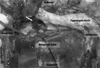
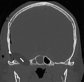
At surgery, an extradural, subtemporal approach was used to expose the tumour. The greater superficial petrosal nerve was sacrificed to prevent facial nerve stretching and paralysis. The middle meningeal artery was controlled by two vascular microclips and sectioned. The tumour was slightly adherent to the petrous carotid artery. The location of the latter was confirmed using intra-operative Doppler ultrasound. The tumour mainly involved the mandibular branch of the trigeminal nerve, bulging through the foramen ovale and extending to the maxillary branch of the trigeminal nerve. The foramen ovale was drilled laterally and anteriorly in order to increase exposure of the tumour. Both invaded mandibular and maxillary branches of the trigeminal nerve were severed (Figure 3). Complete macroscopic removal was achieved. The widened foramen ovale was completely filled with a fat graft (Figure 4).
Superior intra-operative view of the surgically enlarged foramen ovale (arrow) remaining after tumour removal.

Post-operative, coronal computed tomography scan showing the surgically enlarged foramen ovale (arrow) filled with a fat graft.

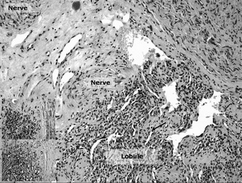
Histopathological examination of the surgical specimen revealed that the lesion possessed a lobular architecture consisting of capillary-like vessels surrounded by CD 31 and CD 34 positive endothelial cells. Those endothelial cells were surrounded by a monolayer of pericytes. This architecture is typical of a capillary lobular haemangioma, which is a benign tumour. The vessels' lumens were slit-like (Figure 5).
Main picture: lobular lesion with multiple, capillary-sized vessels with slit-like lumens (arrow) (H&E; ×200 total magnification). Upper inset: CD 34 immunostaining, showing CD 34 positive cells lining the capillary-sized vessels (×400 total magnification). Lower inset: smooth muscle actin (SMA) staining is positive in a monolayer of pericytes surrounding the endothelial cells (×400 total magnification). These findings are typical of a capillary lobular haemangioma.

The post-operative course was uneventful, and the patient was discharged five days later. Her pre-operative facial pain had resolved.
Discussion
Capillary haemangiomas rarely involve the central or peripheral nervous systems, being most frequently encountered in the skin and other soft tissues. Very few cases of capillary haemangiomas involving the cranial nerves have been described in the literature.Reference Brazis, Wharen, Czervionke, Witte and Jones12, Reference Quevedo, Delvalle, Higa, Iffenecker, Quillard and Sterkers13 However, cavernous haemangiomas of Meckel's cave have been reported more frequently. One such case report described a capillary haemangioma of the trigeminal nerve, involving only the mandibular branch, with palsy of the ipsilateral VIth cranial nerve.Reference Brazis, Wharen, Czervionke, Witte and Jones12 Our patient represents the first reported case of a trigeminal nerve haemangioma invading the maxillary branch of the nerve and eroding the petrous carotid canal. Capillary haemangiomas are best identified through MRI. Typically, they present as either iso- or hypointense on T1-weighted images, and are enhanced in a strong, homogeneous manner after gadolinium injection. On T2-weighted images, they present as hyperintense lesions.Reference Shin, Lee, Jeon and Park14
The differential diagnosis of trigeminal nerve haemangioma should include various hamartomatous and neoplastic lesions: schwannoma, meningioma, paraganglioma, ependymoma, cavernous angioma, angiolipoma, solitary fibrous tumour, lymphoma, haemangiopericytoma and haemangioendothelioma. Considering the non-specific nature of the capillary lobular haemangioma as visualised by current imaging techniques, all lesions included in the above differential diagnosis should be kept in mind.
The definitive diagnosis of trigeminal nerve haemangioma is made based upon histological findings. Pathologically, these lesions typically present as a proliferation of capillary-sized vessels lined by flattened, benign endothelial cells with positive CD 34 immunoperoxidase staining. Considering the location of the tumour, a paraganglioma should be included in the differential diagnosis, but the absence of differentiated neuroendocrine cells excludes this diagnosis.
Our patient's main complaints were isolated, severe, right-sided facial pain, numbness, and a mild hearing impediment on the right side. No other neurological symptoms or signs were found. Our patient's severe pain is easily explained by the tumour's nerve compression, while the episodic exacerbation of pain probably resulted from increased nerve irritation secondary to microscopic, intra-tumoural haemorrhages. The prominent pain reported by our patient, compared with the lesser pain reported in cases of trigeminal nerve schwannoma, might be due to compression rather than invasion of the nerve as such. Even though the tumour extended through the foramen ovale to the pterygoid muscle, there was no pterygoid weakness; these muscles were not invaded. The patient had serous otitis media secondary to eustachian tube involvement by the tumour. This feature should alert the physician, and warrant more advanced investigation (beyond standard flexible nasal endoscopy).
• Although lobular capillary haemangiomas of the trigeminal nerve are very rare tumours, they should always be considered in the differential diagnosis of lesions involving Meckel's cave and the foramen ovale
• Patients with severe, chronic facial pain should be investigated to exclude a tumour involving the trigeminal nerve
• The definitive diagnosis is made only after tumour removal and histopathological examination
Considering the lack of specificity of current, standard imaging techniques (i.e. MRI and CT scanning), a wide range of differential diagnoses should be considered, as aforementioned. Therefore, the investigation of such a tumour presentation should include a total body CT scan, a PET scan and a mammography to exclude any metastatic or primary tumours. However, only removal of the tumour and histopathological examination will confirm its true nature.
Piecemeal removal of the tumour was achieved in our patient without major bleeding, which is unusual considering previous reports.Reference Pearl, Takei, Tindall, O'Brien, Payne and Hoffman15 This is probably explained by the fact that, during tumour exposure, all vessels nourishing the tumour were cauterised; thus, a careful dissection when exposing the tumour might assist its bloodless removal. In our opinion, pre-operative embolisation is not warranted. Obliteration of the widened foramen ovale with a fat graft after tumour removal is important to prevent extradural pneumoencephalus via the eustachian tube, which is exposed to the intracranial space by tumour invasion and by drilling of the foramen ovale. The surgeon should bear in mind that the sphenoid sinus may be very large laterally and could be easily opened during operative widening of the foramen ovale.
Conclusion
Although lobular capillary haemangiomas of the trigeminal nerve are very rare tumours, they should always be considered in the differential diagnosis of lesions involving Meckel's cave and the foramen ovale. Patients with severe, chronic facial pain should be investigated to exclude a tumour involving the trigeminal nerve. The definitive diagnosis is made only after tumour removal and histopathological examination.