Implications
The mycotoxin deoxynivalenol (DON) is a frequently detected compound produced by moulds, which grow on wheat and other cereals during pre-harvest. The main effects of DON in broilers are reduced feed consumption and weight gain. The measurement of the gene expression in the liver gave indications on the functional mechanism of DON. Genes related to altered nutrient uptake to the cell, detoxification, altered protein synthesis rate and DNA repair mechanisms could be detected. With the description of new pathways, a better understanding of the effects of DON on the metabolism of broiler chicks can be expected.
Introduction
About 25% of the world's food crops are contaminated with mycotoxins. The most frequently occurring and widespread mycotoxin in feedstuffs for poultry in European countries is deoxynivalenol (DON) (Awad et al., Reference Awad, Bohm, Razzazi-Fazeli, Hulan and Zentek2004). The mycotoxin-contaminated wheat used in this study contained mainly DON, in addition to a very low contamination with T-2 toxin and zearalenone (ZON) in relation to maximal guidance values in the European Union (EU). DON is a type B trichothecene, a secondary metabolite produced by Fusarium species mainly from Fusarium graminearum and Fusarium culmorum. Owing to the growth of the fungi on maize, wheat and other cereals, poultry feed can contain DON (Rotter et al., Reference Rotter, Prelusky and Pestka1996; Awad et al., Reference Awad, Bohm, Razzazi-Fazeli, Hulan and Zentek2004). In the EU, the maximal permitted value for feed contamination is 5 mg DON/kg feed in poultry nutrition. At that level, no damage to the birds is expected (European Union, 2006). However, decreased feed intake, weight gain and reduced peripheral blood monocytes have been observed in broilers fed 9.3 mg/kg DON in the grower phase (Swamy et al., Reference Swamy, Smith, Karrow and Boermans2004). DON fed to pigs either in a purified form or in the form of naturally contaminated corn led to reduced weight gain and feed intake. Until the end of the experiment, the animals receiving the purified DON recovered from the growth depression in contrast to the animals receiving naturally contaminated corn. The differences between naturally DON-contaminated feed sources and the feed spiked with purified DON might be caused by fungal components present in the naturally mycotoxin-contaminated feed (Prelusky et al., Reference Prelusky, Gerdes, Underhill, Rotter, Jui and Trenholm1994). We decided to use naturally mycotoxin-contaminated wheat for the experiment to reproduce the effects that occur in the commercial broiler production. It was suggested that the DON tolerance of broilers results from poorer toxin bioavailability and rapid elimination from the body. DON is rapidly absorbed in the upper part of the gastrointestinal tract and is also quickly cleared from the body of the chickens. It has been suggested that an efficient clearance takes place due to a hepatic or renal first-pass effect in chickens. The oral administration of 14C-DON to chickens showed high radioactivity in the liver and bile, with more than 90% of the original label occurring in the excreta before 48 h (Awad et al., Reference Awad, Ghareeb, Bohm, Razzazi, Hellweg and Zentek2008). Another experiment showed that in hens administered 3H-DON, an overwhelming amount of radioactivity was detected in the urine, demonstrating the absorption of DON by the digestive system (Lun et al., Reference Lun, Moran, Young and McMillan1989). The main DON metabolite detected in urine and faeces was de-epoxy DON (DOM-1) (Yoshizawa et al., Reference Yoshizawa, Cote, Swanson and and Buck1986). Therefore, the liver, as a direct target of DON, was chosen to analyse the gene expression with microarrays. It has been suggested that cells and tissues with high protein turnover rate, such as the liver and small intestine, are most affected by DON (Awad et al., Reference Awad, Ghareeb, Bohm, Razzazi, Hellweg and Zentek2008). It was also observed that fast-growing broilers are more susceptible to DON than laying hens (Rotter et al., Reference Rotter, Prelusky and Pestka1996). Pestka (Reference Pestka2007) claimed that DON is metabolised in all species, and that it does not accumulate. The main effect of DON is the binding to the 60 S ribosomal subunit and the subsequent prevention of polypeptide chain initiation or elongation (Ueno, Reference Ueno1984). Further, DON affects the immune system either as an immunosuppressive or immunostimulative, depending on the dose and exposure regime. The immunosuppression was explained mainly by the binding capacity of DON to the ribosomes and by the inhibition of protein synthesis (Bondy and Pestka, Reference Bondy and Pestka2000). However, there is little or no information available about the involvement of DON in the pathways of immune suppression, nutritional uptake or detoxification within the living animal. Most of the studies at the cellular level focused on the acute effects of DON administration. Long-term effects of DON-contaminated cereals seem to be important for animal production because of a regular occurrence of DON-contaminated feed.
The microarrays used in our experiment contained 37 000 probe sets corresponding to more than 28 000 genes. Especially when little is known about the molecular effects of a substance, the use of microarrays as a screening method provides an invaluable starting point. The sequence of the probe sets was designed according to the sequence information from all transcribed genes, including the ones with an unknown function. The profiling of gene expression with microarrays should help gain insight into the effects and metabolism-related genes upon DON administration. In addition, the expression of genes, with a biological relevance to DON administration, was verified with reverse transcription (RT) qPCR in the liver and the jejunum, to cover the main target organs of DON. Possible marker genes and affected pathways should be identified to achieve a better understanding of how chronic and low doses of mycotoxin-contaminated feed uptake affects liver and the small intestine cells in broilers. In addition, the concentration at which DON influences the gene expression in the liver should be reviewed.
Material and methods
Experimental design, birds and diets
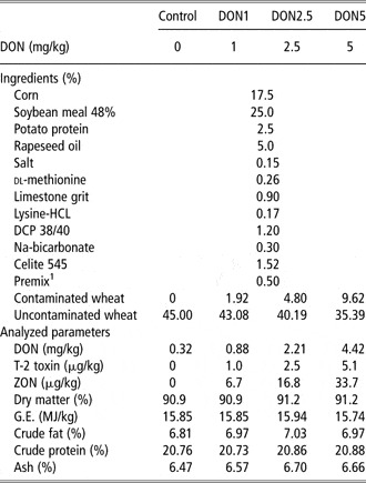
Twenty 1-day-old male ROSS 308 broiler chicks were obtained from the commercial hatchery Erb Brüterei AG (Oberdiessbach, Switzerland). The birds were weighed at the beginning of the experiment and separated into four treatment groups according to similar group weight. Five broilers per treatment group per cage were raised together for 23 days. A standard diet (Table 1) was formulated according to the National Research Council (1994) and prepared for all groups in the same process. DON (52 mg/kg) was added to the basic feed mixture of the treatment groups in the form of mycotoxin-contaminated wheat. DON was determined in the diets using LC–MS/MS; the other mycotoxins were determined in the contaminated wheat because of their low concentration, which was close to or below the detection level in the diet. T-2 toxin and ZON concentrations of the contaminated wheat were determined using LC–MS/MS; ochratoxin A, fumonisin B1 and B2 and aflatoxin B1, B2, G1 and G2 were determined with HPLC-fluorescence derivatisation in an external laboratory (UFAG; Sursee, Switzerland). The pelleted feed and water were provided ad libitum from day 1 onwards. The animal experiment was performed in accordance with the regulations of the cantonal veterinary office in Zurich.
Broiler diets

DON = deoxynivalenol; DCP = di-basic calcium phosphate; ZON = zearalenone; G.E. = gross energy.
Contaminated wheat (52 mg DON/kg), uncontaminated wheat (0.58 mg DON/kg).
Maximal allowed contamination per kg feed of poultry in European Union (2006; percentage in group DON5 of maximal allowed concentration): DON: 5 mg/kg (88.5%), T-2: 150 μg/kg (3.4%), ZON: no recommendations. Mycotoxins below the detection limits: aflatoxin B1, B2, G1, G2 (1.3 μg/kg feed), ochratoxin A (1 μg/kg feed) and fumonisin B1, B2 (10 μg/kg feed).
1Premix: diet from Aeschbacher et al. (Reference Aeschbacher, Messikommer, Meile and Wenk2005).
The total cage weight and feed intake were monitored on a weekly basis. Total water intake per cage was recorded for 4 days a week. Feed samples were taken from the principal components, the control and the treatment diet before and after pelleting and on days 1 and 23 of the experiment. All the feed samples were stored in a 4°C cooler. Excreta samples were taken from days 8 to 11 and from days 15 to 18 from all four cages and stored at −20°C. Dry matter, ash, gross energy, crude fat and crude protein content were determined in all feed and excreta samples (Table 1). Apparent metabolisable energy intakes (AME) and nitrogen (N) retention were calculated according to the method described by Scott and Hall (Reference Scott and Hall1998). After 23 days, broilers were weighed individually and slaughtered by cervical dislocation. Liver samples were removed immediately and washed, and from the middle portion of the left liver lobe, a slice of about 1 cm width was cut and frozen in liquid N. In a similar way, a slice of the jejunum was collected; first 7 cm below the angle of Treitz, a 4-cm-long piece from the jejunum was cut out, and then the lumen was opened and rinsed with 0.9% NaCl before it was also frozen in liquid N.
High-density oligonucleotide array hybridisation
Total RNA was isolated from 12 frozen liver samples with TRIZOL reagent (Invitrogen, Basel, Switzerland). From all four groups, three samples were selected according to the weight of the birds. The heaviest and lightest birds were excluded. The samples were homogenised in a mortar filled with liquid N. After isolation, RNA concentration and purity were determined with an ND-1000 (NanoDrop Technologies, Wilmington, DE, USA) and further cleaned up with the RNeasy Mini Kit (Qiagen, Hilden, Germany). Before RT, the RNA integrity was determined using a Bioanalyzer 2100 (Agilent Technologies, Basel, Switzerland). The integrity number was calculated using the Bioanalyzer 2100 software with a value between 1 (for totally degraded RNA) and 10 (for undegraded RNA). For the microarray experiments, only samples with a value higher than 9 were taken. Labelled cRNA probes were generated according to the protocol from Affymetrix using the GeneChip One-Cycle cDNA Synthesis Kit (Affymetrix, Santa Clara, CA, USA). The procedure consisted of RT of 7 μg total RNA with a T7 oligo (dT) promoter primer and a second-strand cDNA synthesis, followed by an in vitro transcription to biotin-labelled cRNA. Finally, the labelled cRNA was fragmented and hybridised at 45°C for 16 h on the Chicken Genome Arrays (Affymetrix, Santa Clara, CA, USA). For every sample a single microarray was used. The arrays were washed and stained using the GeneChip Fluidics Station and scanned with a GeneChip Scanner 3000 according to the manufacturer's protocol (Affymetrix, Santa Clara, CA, USA). The GeneChip operating software (GCOS, Affymetrix, Santa Clara, CA, USA) controlled the scanner and the fluidics station. The data were analysed with the Microarray Suite version 5.0 (MAS 5.0) using Affymetrix default analysis settings and global scaling as a normalisation method. The image data on each individual chip were scaled to target intensity 500.
RT and qPCR
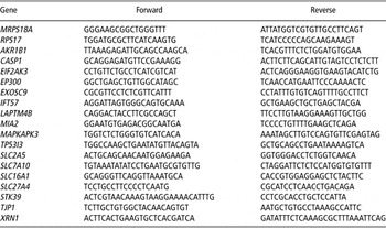
To verify the results from the microarray experiment, the total RNA was extracted from the remaining liver and the jejunum samples. RNA extraction, quantification and quality control were performed in the same manner as the microarray samples. The experiments for 20 samples per tissue were conducted in two steps: first, the RT was performed using the High-Capacity cDNA Reverse Transcription Kit (Applied Biosystems, Rotkreuz, Switzerland) and then the real-time PCR (qPCR) was conducted with adjusted amounts of cDNA. The genes for the qPCR were selected after the gene expression analysis and the literature review, depending on their possible biological relevance in relation to mycotoxin-contaminated feed. Primers were designed with Primer Express 3.0 software (Applied Biosystems, Rotkreuz, Switzerland) on two adjoining exons in order to exclude a possible amplification due to genomic DNA contamination. The two endogenous controls mitochondrial ribosomal protein S18A (MRPS18A) and ribosomal protein S17 (RPS17) turned out to perform the best for adjusting the cDNA concentration, compared with the traditional and other selected control genes such as GAPDH, β-actin, DHRS7, RPS28, APOB and LDHA. Genes and the corresponding primer pairs are indicated in Table 2. The 20-μl qPCR reaction contained 500 nM of forward and reverse primers, 100 ng of sample cDNA and 10 μl Fast SYBR Master Mix (2X; Applied Biosystems, Rotkreuz, Switzerland). The PCR amplification was performed on the 7500 Fast real-time PCR System with SDS Software 1.3.1 (Applied Biosystems, Rotkreuz, Switzerland) and repeated four times separately. The cycling programme was 20 s at 95°C and then 40 cycles of the following: denaturation for 3 s at 95°C, annealing and extension at 60°C for 30 s. The results were evaluated with the comparative Ct method (2−△△Ct) with the SDS software.
Primer sequence for qPCR

5′–3′ direction.
Statistical analysis
The alterations in the expression profiles between the control and the treatment groups were analysed using the GeneSpring 7.3 software (Agilent Technologies, Basel, Switzerland). Normalisation was carried out with GeneSpring 7.3 and the default settings as follows: values below 0.01 were set to 0.01; each measurement was divided by the 50th percentile of all measurements in that sample and each gene was divided by the 50th percentile of its measurements in all the samples. The pre-filtering was performed according to Pepper et al. (Reference Pepper, Saunders, Edwards, Wilson and Miller2007) as follows: the genes had to be present or marginal in all samples, and the raw value had to be above the 50th percentile, of all present and marginal genes, in all samples of a group to remove genes with a low signal strength. The gene lists from the four groups were merged together, and Student's t-test was performed between the control and each treatment group (P < 0.05). Further, genes were grouped into seven subgroups: first, genes with significant changed expression in only one group (DON1, DON2.5, DON5) compared with the control group; then, genes within two groups (DON1/2.5, DON2.5/5, DON1/5) compared with the control group and genes within all three groups (DON1/2.5/5) compared with the control. Finally, for every subgroup a t-test was performed and the P-values are indicated in Tables 4 and 5. The fold changes were listed for every comparison. The final gene list was separated into groups of up- and downregulated genes. The microarray raw data have been deposited into the Gene Expression Omnibus (National Center for Biotechnology Information, http://www.ncbi.nlm.nih.gov/geo) with accession number GSE25185. Genes with an average raw value above the 80th percentile, of all present and marginal genes, in the group with the significantly changed expression are indicated with an asterisk (Tables 4 and 5). The average raw value was indicated to estimate the importance of a gene in cellular processes. All the other measured parameters were analysed using Student's t-test to determine significance (P < 0.05).
Results
Diet characterisation and performance of broilers
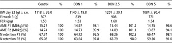
The DON concentration of the control diet was 0.32 mg/kg feed, which was close to the detection level of 0.2 mg DON/kg. The DON concentration in DON1 was 0.89 mg/kg, 2.21 mg/kg for DON2.5 and 4.43 mg/kg for DON5. The calculated T-2 toxin and ZON concentrations in group DON5 were low, with values of 5.1 and 34.6 μg/kg feed, respectively. The other measured mycotoxin concentrations were below the detection limit. The performance of the broilers within the groups was balanced, and they did not show an abnormal behaviour or other indications due to DON contamination. The progression of growth and the feed intake were within the range of expectation (Table 3). No behavioural or macroscopic pathological alterations were detected in the appearance of liver, stomach, intestine, spleen, heart, muscles and lungs of the broilers receiving the mycotoxin-contaminated feed.
Performance parameters

DON = deoxynivalenol; FI = feed intake; FCR = feed conversion rate (g gain/g feed); AME P1/2 = apparent metabolisable energy in MJ/kg dry matter and % relative to the control; P1/2 = period 1 or 2.
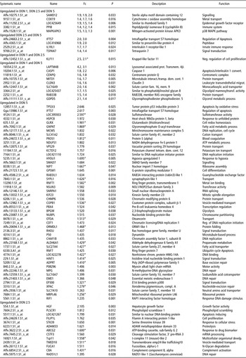
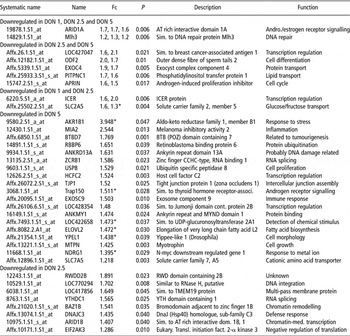
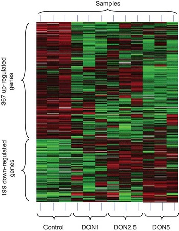
The parameters for the RNA quality and quantity, as well as the microarray quality parameters, were within the requirements. In Tables 4 and 5, a summary of genes with an altered expression are sorted according to the DON groups. The expression of a total of 566 genes was significantly altered in DON groups compared with the control group (supplementary files). The heatmap of the cluster analysis of all significantly altered genes between the control and the DON-containing groups is shown in Figure 1.
Upregulated genes in DON group

DON = deoxynivalenol; Fc = fold change of groups DON1, DON2.5 and DON5; P = P-value of Student's t-test.
The systematic name of the probe sets in chicken is the species code Gga., followed by the identifier (e.g. GgaAffx.5975.1.S1_at or Gga.2439.1.S1_at).
*Raw value above 80th percentile.
Downregulated genes in DON groups

DON = deoxynivalenol; Fc = fold change of groups DON1, DON2.5 and DON5; P = P-value of Student's t-test.
The systematic name of the probe sets in chicken is the species code Gga., followed by the identifier (e.g. GgaAffx.5975.1.S1_at or Gga.2439.1.S1_at).
*Raw value above the 80th percentile.
Heatmap of cluster analysis. Heatmap: green, upregulated genes; black, normal expression; red, down-regulated genes. The regulation is displayed according to significant alterations between control group and DON-containing groups. Each horizontal line corresponds to a normalised expression of a gene from the respective sample.

The entire list of up- and downregulated genes is available in the section of supplementary files. In the DON groups, 367 genes were upregulated. Of these, 230 genes have been specified in relation to their function or structure, and for 138 genes only the transcript sequence is known. The average expression of 152 upregulated genes, or 41% of the upregulated genes, was above the 80th percentile of the raw value of all genes, which is indicated by an asterisk (*) in the column ‘Fold change’ (Tables 4 and 5). The fold change of 28 genes was above two. Venn diagrams are presented in the section of supplementary files with the distribution of significantly altered genes between the treatment groups. A summary of discussed genes with an upregulated expression in the DON groups is presented in Table 4.
In Table 5, the most important and significantly downregulated genes are indicated according to the DON groups. Of 199 significantly downregulated genes, the function of 139 genes was known. Sixty genes were unknown or no function could be assigned to them; 14 genes had a fold change higher than two; and 45 genes, or 32% of the downregulated genes, had a raw value above the 80th percentile.
RT and qPCR
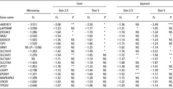
The results from the microarray experiment were verified with RT qPCR, and the results are shown in Table 6. The alterations could be confirmed for 9 of the 15 genes in the liver. A total of 10 of 17 genes showed a significantly altered expression in the liver and the jejunum. The correlation between the fold changes of the qPCR experiment and the microarray analysis in group DON5 was 0.75. XRN1 and SLC16A1 expression were measured because of their biological relevance. The genes AKR1B1, MIA2, SLC2A5, SLC7A10 and LAPTM4B were significantly regulated in the liver, measured with qPCR and microarrays, as well as in the jejunum tissues. SLC27A4, CASP1 and EP300 had a significant altered expression in the microarray experiment in the liver and in the qPCR experiment in the jejunum. The alteration of the expression for the genes EXOSC9, TJP1, EIF2AK3 and IFT57 could be confirmed only in the liver by qPCR. SLC16A1 was not significantly altered in any measurement in the liver, but showed a decreased expression in the jejunum groups. The reduced expression of XRN1 did not reach significance in the microarray analysis (P < 0.08), but was significantly reduced in the qPCR measurement. The expression alteration of the genes MAPKAPK3, STK39 and TP53I3 could not be confirmed with qPCR in any tissues. Taking the liver and jejunum groups together, 14 of 17 expression alterations could be confirmed with qPCR.
RT qPCR results

RT = reverse transcription; DON = deoxynivalenol; Fc = fold change; P = P-value of Student's t-test.
n = 5 animals per treatment group.
Fc and P were measured between control and the corresponding DON-containing group.
1Raw value above 80th percentile.
*P < 0.05; **P < 0.01; ***P < 0.001.
Discussion
The use of naturally mycotoxin-contaminated wheat resulted primarily in a high DON contamination (4.425 mg/kg) and a low calculated contamination with T-2 (5.1 μg/kg) and ZON (33.7 μg/kg) in the final feed mixture of DON5. Because of the low calculated concentrations of T-2 and ZON approximately or below the detection level, they were measured in naturally mycotoxin-contaminated wheat. The other measured mycotoxins in the mycotoxin-contaminated wheat were below the detection limit. The maximal allowed concentration for DON in poultry feed is 5 mg/kg feed and 150 μg/kg feed for T-2. No maximal guidance values exist for ZON (bovine: 500 μg/kg feed), because of the high tolerance of poultry. The maximal permitted mycotoxin concentrations are related to the level at which no damage to the birds is expected (EFSA, 2004). The mycotoxin analysis in the final feed mixture showed, in comparison with the maximal permitted concentration, a proportion of 88.5% of DON, 3.4% of T-2 and 6.7% of ZON (bovine guidance value) in the feed of group DON5. Because of the high proportion of DON and the low amount of T-2 and ZON, we considered the observed alterations at the transcriptional level due to the effects of DON. Most of the reported effects have been investigated mainly in cell cultures or model organisms such as mice. In our experiment, we showed for the first time similar alterations in the gene expression in broilers.
The growth, feed intake and feed conversion rates were within the normal range in our experiment. Although BW and feed intake were numerically the lowest in group DON5 and the highest in group DON2.5, this was statistically not significant. It has been mentioned by Awad et al. (Reference Awad, Ghareeb, Bohm, Razzazi, Hellweg and Zentek2008) that moderately high concentration levels of DON can even have growth-promoting effects. Further, the increased s.e. in BW of group DON5 indicates that different sensitivity towards DON-contaminated feed might exist, depending on the genetic background of the animals.
Nutrient transport
The gene expression analysis has shown that the nutrient uptake into the cell seems to be influenced by the mycotoxin-contaminated feed. The passive d-fructose transporter with a very low glucose affinity, SLC2A5 (fc: −1.259, DON2.5), had a decreased expression in the liver (fc: −1.54, DON2.5) and the jejunum (fc: −1.94, DON2.5/5). The numerical decrease in AME by 5.9% might be a further indication of a reduced monosaccharide uptake in the jejunum. Maresca et al. (Reference Maresca, Mahfoud, Garmy and Fantini2002) showed in their study that fructose uptake into the cell and corresponding transporter SLC2A5 were inhibited by 42% in the intestinal cell line HT-29-D4 cells because of DON administration. Our findings indicate that similar effects occur in the jejunum of broilers in the living animal, and to a lesser extent in the liver. The gene SLC27A4 (fc: +1.427, DON5) is a palmitate transporter and the increased expression could not be confirmed with qPCR in the liver. However, the expression was significantly reduced in the jejunum (fc: −1.87, DON5). In primary enterocytes treated with SLC27A4 antisense, palmitate and oleate uptake were reduced simultaneously (Stahl et al., Reference Stahl, Hirsch, Gimeno, Punreddy, Ge, Watson, Patel, Kotler, Raimondi, Tartaglia and Lodish1999). Maresca et al. (Reference Maresca, Mahfoud, Garmy and Fantini2002) reported an increased palmitate uptake by 35% in their study after challenging HT-29-D4 cells with DON. We assume that chronic exposure to DON can invert the regulation or that another functional mechanism on the transport activity is responsible for the discrepancy between the decreased expression of SLC27A4 in the jejunum and the increased palmitate uptake reported by Maresca et al. (Reference Maresca, Mahfoud, Garmy and Fantini2002). The SLC7A10 protein (fc: +1.26, DON5) transports d-serine, glycine and l-isomers of alanine, serine, threonine and cysteine (Nakauchi et al., Reference Nakauchi, Matsuo, Kim, Goto, Chairoungdua, Cha, Inatomi, Shiokawa, Yamaguchi, Saito, Endou and Kanai2000). The expression change was confirmed with qPCR in the liver (fc: +1.49, DON5) and the jejunum (fc: +2.52, DON5). Further, the N retention was numerically reduced by 9%, which gives an indication on the amino acid uptake. The decrease in l-serine uptake in the cell culture study by Maresca et al. (Reference Maresca, Mahfoud, Garmy and Fantini2002) is in disagreement with our observation that SLC7A10 showed an upregulated expression, but is in agreement with the reduced N retention in group DON5. The discrepancy could indicate a direct inhibition of this l-serine transporter by DON, resulting in the upregulation of SLC7A10. SLC7A5 (fc: −1.218, DON5) is a transporter of neutral amino acids with branched or aromatic side chains such as leucine (Kanai et al., Reference Kanai, Segawa, Miyamoto, Uchino, Takeda and Endou1998). In an incorporation study with mice, the 14C leucine incorporation into the liver was decreased at 20 mg DON/kg feed. It was assumed that DON inhibited the protein synthesis, and therefore the reduction of the 14C leucine amount occurred (Robbana-Barnat et al., Reference Robbana-Barnat, Loridon-Rosa, Cohen, Lafarge-Frayssinet, Neish and Frayssinet1987). The addition of 0.5 and 5 μg DON/ml to Caco-2 cells led to a decreased [3H] leucine incorporation and a parallel decrease of cellular protein content (De Walle et al., Reference De Walle, Sergent, Piront, Toussaint, Schneider and Larondelle2010). The extent to which leucine uptake into the cell is responsible for the decrease of the protein synthesis remains unclear. In a related, unpublished study, SLC16A1 expression was significantly altered, and therefore the test was also used to verify the expression in the jejunum in this study. SLC16A1 (fc: −1.87, DON5) had a decreased expression in the jejunum, and it transports monocarboxylates such as butyrate, lactate and pyruvate (Lambert et al., Reference Lambert, Wood, Ellis and Shirazi-Beechey2002). It has been suggested that effects of DON on membrane transport is a result of specific modulation of the transporters and is not due to cell damage (Maresca et al., Reference Maresca, Mahfoud, Garmy and Fantini2002), which is supported by our observations, especially because SLC7A10 was upregulated. The alteration of these five gene expression levels influences the nutrient uptake into the cell and might have implications for the growth rate of birds.
Detoxification
AKR1B1 (fc: −3.511, DON2.5 and 5) had a decreased expression in the liver, measured with microarray and qPCR experiments (fc: −2.5, DON2.5 and 5), as well as in the jejunum (fc: −2.49, DON5). AKR1B1 reduces reactive aldehydes and ketones to primary and secondary alcohols. Reactive oxygen species also target polyunsaturated fatty acids that form lipid hydroperoxides, which are finally decomposed to form reactive lipid aldehydes, which are highly mutagenic. AKR1B1 detoxifies those lipid aldehydes that can react with bases in DNA to form etheno- and heptano-etheno-DNA adducts (Jin and Penning, Reference Jin and Penning2007). The downregulation of AKR1B1 by chronic DON exposure might also be in relation to several upregulated DNA repair genes, which are discussed in the section below. In the human hepatoma-derived cell line HepG2, the expression of AKR1B1 was significantly reduced after the cells were challenged with a series of cytotoxic substances (Kawata et al., Reference Kawata, Yokoo, Shimazaki and Okabe2007). In conclusion, AKR1B1 seems to be an interesting in vivo biomarker for DON-induced alteration of gene expression.
DNA repair
Six genes were significantly upregulated in DON5 and one in DON2.5, which are responsible for DNA damage repair and recognition, showing a possible effect of DON on DNA integrity. The six genes correspond to 4.1% of significantly altered genes, with a known function, in group DON5. The protein is involved in DNA repair and has a critical role in signalling DNA single-strand breaks (Plummer and Calvert, Reference Plummer and Calvert2007). Shifrin and Anderson (Reference Shifrin and Anderson1999) showed that the addition of DON to the Jurkat human lymphoid cell line led to the activation of pro-caspase-3, leading to a higher cleavage of PARP1, a substrate of caspase-3. In murine macrophages, J774A.1, an increased amount of PARP1 and significantly increased amount of cleaved PARP1 protein resulted after the challenge with DON (Marzocco et al., Reference Marzocco, Russo, Bianco, Autore and Severino2009). The upregulation of PARP1 therefore shows that similar effects might occur in the liver of broilers. MPG (fc: +1.406, DON5) is responsible for the recognition and excision of alkylated purine bases (O'Brien and Ellenberger, Reference O'Brien and Ellenberger2004). EME1 (fc: +1.366, DON5) cleaves branched DNA structures. In the absence of EME1, chromosomal aberrations occurred and led to genomic instability (Abraham et al., Reference Abraham, Lemmers, Hande, Moynahan, Chahwan, Ciccia, Essers, Hanada, Chahwan, Khaw, McPherson, Shehabeldin, Laister, Arrowsmith, Kanaar, West, Jasin and Hakem2003). The gene RAD51L1 (fc: +1.395, DON2.5) functions in homologous recombination and DNA double-strand break repair (Miller et al., Reference Miller, Yoshikawa, McConnell, Clark, Schild and Albala2002). CHAF1B (fc: + 1.435, DON5) is part of the complex CAF-1 and the only subunit available on the Chicken Genome Array. The complex is related to nucleotide excision repair and to the repair of single-strand breaks (Mello et al., Reference Mello, Sillje, Roche, Kirschner, Nigg and Almouzni2002). RIF1 (fc: +1.235, DON5) functions as a DNA damage response factor (Silverman et al., Reference Silverman, Takai, Buonomo, Eisenhaber and de Lange2004). XPAC (fc: +1.279, DON5) is involved in nucleotide excision repair (Shimamoto et al., Reference Shimamoto, Tanimura, Yoneda, Kobayakawa, Sugasawa, Hanaoka, Oka, Okada, Tanaka and Kohno1995). In male broiler chickens fed with 10 mg DON/kg feed, a comet assay was performed in spleen leukocytes, which detected aberrations, and a significantly higher amount of DNA breaks were found (Frankic et al., Reference Frankic, Pajk, Rezar, Levart and Salobir2006). Interestingly, all the DNA repair-related genes mentioned above were only upregulated in group DON5 with the exception of RAD51L1, which was upregulated in DON2.5. Genes such as PARP1, MPG, EME1, XPAC and CHAF1B belong either to the base excision repair or to the nucleotide excision repair pathway. Under reduced activity of AKR1B1, the anchorage of lipid aldehydes to the DNA might have occurred with a higher frequency, and therefore the discussed single-strand repair genes might have shown a higher expression in the group DON5. A single-strand break or modified nucleotides do not lead necessarily to double-strand breaks or, as a result, to detection in the comet assay. Continuing, it seems that the activation of DNA damage-related genes is induced only by a concentration of 4.42 mg DON/kg feed.
Translation initiation
The expression decrease of EIF2AK3 (fc: −1.286, DON2.5/5) could be confirmed in the liver with qPCR (fc: −1.70). EIF2AK3 is a target for transcriptional upregulation of the unfolded protein response and phosphorylates eIF2α, a translation initiator (Yang et al., Reference Yang, Li and Pestka2000). The phosphorylation of eIF2α inhibits translation initiation and protein synthesis. EIF2AK3 activation and the subsequent eIF2α phosphorylation prevent the cell from the accumulation of incorrectly folded or unfolded proteins (Ron, Reference Ron2002). DNAJC3 (fc: −1.435, DON2.5) is induced during endoplasmic reticulum (ER) stress (van Huizen et al., Reference van Huizen, Martindale, Gorospe and Holbrook2003). DNAJC3 binds to EIF2AK3 and disrupts the activity of EIF2AK3. Subsequently, eIF2α phosphorylation by EIF2AK3 is reduced, stimulating the eIF2α-induced protein synthesis (Gale et al., Reference Gale, Blakely, Hopkins, Melville, Wambach, Romano and Katze1998). Our results are consistent with the publication of Yang et al. (Reference Yang, Li and Pestka2000), which showed reduced expression level of DNAJC3 after 24 h in murine thymoma cell line EL-4 treated with DON. The binding of DON to the 60 S ribosomal subunit and the subsequent translation inhibition might be counterbalanced by the downregulation of EIF2AK3 and DNAJC3. Pestka (Reference Pestka2010) assumed that EIF2AK3 might have an important role in the signalling after the challenge with DON, because of influenced up- and downstream signal transducers. The downregulation of EIF2AK3 in our experiment confirms this assumption. The ribotoxic stress response seems to have some pathway elements similar to the ER stress response, but with inverse gene expression regulation.
Stabilisation of mRNA
The expression change of EXOSC9 (fc: −1.503, DON5) was confirmed with qPCR (fc: −1.61) in the liver. EXOSC9 is a subunit of the exosome, a 3′–5′ exoribonuclease complex. This complex processes ribosomal and small nuclear RNAs and degrades mRNAs. In HeLa extracts, the addition of RNA containing an adenylate/uridylate-rich element (ARE) increased their 3′–5′ exonucleolytic decay rate. The inactivation of EXOSC9 highly decreased the efficiency of the exonucleolytic decay, showing the central role of EXOSC9 in mRNA decay (Mukherjee et al., Reference Mukherjee, Gao, O'Connor, Raijmakers, Pruijn, Lutz and Wilusz2002). In human HEp-2 cells, it has been shown that the knockdown of EXOSC9 led to a twofold increase in β-globulin containing an ARE (van Dijk et al., Reference van Dijk, Schilders and Pruijn2007). The 5′–3′ exoribonuclease XRN1 (fc: −1.796, DON5) was numerically downregulated in the microarray experiment and significantly downregulated in qPCR experiment (fc: −1.25) in the liver. XRN1 is a component of the alternative 5′–3′ mRNA decay pathway (Schilders et al., Reference Schilders, van Dijk, Raijmakers and Pruijn2006). The knockdown of XRN1 led to a 3.5-fold increase of the β-globulin-ARE mRNA. Therefore, EXOSC9 and XRN1 seem to be involved in the turnover of ARE-containing transcripts (van Dijk et al., Reference van Dijk, Schilders and Pruijn2007). In his review, Pestka (Reference Pestka2008) summarised the genes with a lower mRNA decay rate upon DON administration, which are COX-2, tumour necrosis factor (TNF)-α and interleukin (IL)-6 in macrophages and IL-2 in EL-4T-cells. It has been reported that all four mRNAs are stabilised by ARE in the 3′ untranslated region (Chen et al., Reference Chen, Gherzi, Ong, Chan, Raijmakers, Pruijn, Stoecklin, Moroni, Mann and Karin2001, Paschoud et al., Reference Paschoud, Dogar, Kuntz, Grisoni-Neupert, Richman and Kuhn2006, Rajasingh et al., Reference Rajasingh, Bord, Luedemann, Asai, Hamada, Thorne, Qin, Goukassian, Zhu, Losordo and Kishore2006). The downregulation of EXOSC9 and XRN1 might be the reason for increased mRNA stability of several ARE-containing genes in DON-challenged cell cultures. Further, the reduced mRNA depletion, due to XRN1 and EXOSC9 downregulation, might have also led to higher mRNA levels in general. In the final gene list (supplementary files), a disequilibrium exists with 367 upregulated genes and 199 downregulated genes. The possible higher mRNA amount might compensate for the possibly reduced translation, due to binding to the 60 S ribosomal subunit by DON. EXOSC9 and XRN1 seem to be interesting biomarkers for mRNA stabilisation and the decay rate of ARE-containing mRNA.
Tight junctions
Cells are connected by intercellular complexes such as tight junctions and adherens junctions. In Caco-2 cells, it has been shown that the transepithelial electrical resistance (TEER) decreased to 30% after 24 h of DON incubation at a concentration of 2 μg/ml. It was mentioned that TEER reflects the functional tight junctions made of transmembrane proteins (Sergent et al., Reference Sergent, Parys, Garsou, Pussemier, Schneider and Larondelle2006). Among the proteins mentioned was TJP1 (fc: −1.52, DON5), which was downregulated in our experiment in the liver, which was also measured with qPCR (fc: −1.66). In porcine intestinal epithelial cells (IPEC-1) and -J2 cells, the treatment with 2 μg DON/ml led to disintegrated TJP1 structure and the total amount of TJP1 moderately decreased, which was measured with the Western blot analysis (Diesing et al., Reference Diesing, Nossol, Panther, Walk, Post, Kluess, Kreutzmann, Danicke, Rothkotter and Kahlert2011). It was reported that TJP1 interacts with CLDN3 (fc: +1.637, DON1/5; Harhaj and Antonetti, Reference Harhaj and Antonetti2004). Pinton et al. (Reference Pinton, Nougayrede, Del Rio, Moreno, Marin, Ferrier, Bracarense, Kolf-Clauw and Oswald2009) have shown a decreased CLDN3 protein amount in IPEC-1 after the treatment with DON for 48 h with a parallel decrease of TEER. Therefore, the upregulation of CLDN3 might counterbalance the reduction caused by DON over a feedback mechanism. The decrease of TEER shows the acute effects in cell cultures. The extent to which the chronic administration of DON leads to the same effects has not been reported. The significant expression change of TJP1 and CLDN3 indicates that similar effects, such as the decreased TEER in cell cultures, might also occur in the liver of living animals.
Immune-related genes
The expression of MIA2 (fc: −2.544, DON5) was significantly decreased, measured with qPCR in the liver (fc: −1.70) and the jejunum (fc: −1.35). MIA2 expression is regulated by IL-6 and transforming growth factor-beta, which increased the expression level of MIA2 in human hepatocytes and in HepG2 cells (Bosserhoff et al., Reference Bosserhoff, Moser, Scholmerich, Buettner and Hellerbrand2003). The downregulation of MIA2 in the liver and the jejunum may indicate a reduced amount of IL-6 in the liver cells of broilers. In murine macrophages, it has been shown that IL1RL1 (fc: +1.661, DON2.5/5) exerted anti-inflammatory effects by the negative regulation of the IL-6 production (Takezako et al., Reference Takezako, Hayakawa, Hayakawa, Aoki, Yanagisawa, Endo and Tominaga2006), which affirmed the previous results with the decreased MIA2 expression. In vitro experiments with the addition of DON exhibited that the half-life of the ARE-containing IL-6 and TNF-α mRNA and the amount of the respective proteins were increased within 24 h (Wong et al., Reference Wong, Schwartz and Pestka2001). IL-6 seems to be critical for the regulation of immunoglobulin (Ig)A. In broilers fed with 7 to 9.2 mg DON/kg feed, the biliary IgA concentration was decreased (Swamy et al., Reference Swamy, Smith, Cotter, Boermans and Sefton2002). Azcona-Olivera et al. (Reference Azcona-Olivera, Ouyang, Murtha, Chu and Pestka1995) showed an increased expression of IL-1β, IL-6 and TNF-α mRNA in mice exposed to a 25 mg DON/kg diet. Four hours after the administration of DON, the cytokines were again only slightly upregulated in the liver (Azcona-Olivera et al., Reference Azcona-Olivera, Ouyang, Murtha, Chu and Pestka1995). This may also explain the findings that IL-1β, IL-6 and TNF-α mRNA were unchanged in our experiment. IL1RL1 might be an antagonist to DON by stabilising IL-6 and TNF-α levels. Especially in long-term administration of DON, the normalisation of the mRNA and cytokine levels might be economical. The upregulation of pro- and anti-inflammatory factors may influence the sensitivity of the immune system and act as immune stimulators. LAPTM4B (+3.058, DON1/5) expression increase could be confirmed in the liver (fc: +1.77, DON5) and the jejunum (fc: +1.31, DON2.5). LAPTM4B is a tetra-transmembrane glycoprotein localised on endosomes and lysosomes (Liu et al., Reference Liu, Xiong, Wei, Yang and Zhou2009). LAPTM4B was also upregulated in the early phase of liver regeneration in rats (Xu et al., Reference Xu, Chang, Yuan, Li, Han, Yang, Zhao, Li, Zhang, Rahman and Zhang2005). Therefore, LAPTM4B might be used as a biomarker for liver regeneration.
Conclusion
We could confirm several results in the living animal from previous studies, which were performed mainly in cell cultures. The main effects with economic impact might be the altered expression of the solute carrier transcripts, which transport d-glucose/d-fructose, palmitic acid, monocarboxylates, l-serine, leucine and other amino acids. Further, the translation initiation-related genes EIF2AK3 and DNAJC3, as well as the mRNA stabilisation genes, could have an important influence on the protein synthesis rate and on the growth of broilers. The upregulation of DNA repair proteins indicates possible mutagenic effects of DON or its decomposition products. The detoxification enzyme AKR1B1 is an indicator that, at 2.21 mg DON/kg feed, the concentration might be high enough to show cytotoxic effects. The verification of the results in the jejunum from the microarray and the qPCR analysis in the liver increases the reliability of the DON-regulated genes. In general, effects on the level of RNA expression cannot be directly transposed from one organ to the other. Therefore, the parallel alterations in the liver and the jejunum might indicate a general functional mechanism of DON on the different relative amounts of transcripts. The maximum-allowed DON contamination in poultry feed in the European Union (2006) is 5 mg DON/kg feed. Significantly altered gene expression was observed at a concentration of 2.21 mg DON/kg feed.
Acknowledgements
The authors thank Dr Ruth Messikommer, Dr Christine Zuberbühler, Dr Peter Spring, Dr Hannes Jörg, Prof. Peter Vögeli, Rolf Bickel, Birgit Prause, Monika Haubitz and Urs Schuler for their expert assistance in the animal experiment, and to Claude Chaubert for the help with the mycotoxin analysis. The authors also thank Dr Ulrich Wagner and Andrea Patrignani from the Functional Genomics Center, Zurich for their invaluable help with the microarray experiments and bioinformatics. This study was funded by Alltech Inc., Nicholasville, KY, USA.