Introduction
As part of the multibillion aquarium trade industry, annually more than 20 million marine ornamental fish of over 1800 species are sold to hobbyists around the world (Wabnitz et al., Reference Wabnitz, Taylor, Green and Razak2003; Rhyne et al., Reference Rhyne, Tlusty, Schofield, Kaufman, Morris and Bruckner2012; Palmtag, Reference Palmtag, Calado, Olivotto, Oliver and Holt2017). The continuous expansion of this industry has caused concerns, because it has been estimated that more than 90% of marine ornamental fish are sourced from wild collected specimens (Wabnitz et al., Reference Wabnitz, Taylor, Green and Razak2003; Olivotto et al., Reference Olivotto, Planas, Simões, Holt, Avella and Calado2011; Palmtag, Reference Palmtag, Calado, Olivotto, Oliver and Holt2017). In addition, while the sensitive coral reef ecosystem is facing severe challenges such as coral bleaching (Hughes et al., Reference Hughes, Kerry, Álvarez-Noriega, Álvarez-Romero, Anderson, Baird, Babcock, Beger, Bellwood, Berkelmans, Bridge, Butler, Byrne, Cantin, Comeau, Connolly, Cumming, Dalton, Diaz-Pulido, Eakin, Figueira, Gilmour, Harrison, Heron, Hoey, Hobbs, Hoogenboom, Kennedy, Kuo, Lough, Lowe, Liu, McCulloch, Malcolm, McWilliam, Pandolfi, Pears, Pratchett, Schoepf, Simpson, Skirving, Sommer, Torda, Wachenfeld, Willis and Wilson2017), the practice of destructive collection methods (e.g. cyanide fishing) in tropical developing countries are particularly controversial, as such practice threatens non-target organisms including corals (Barber and Pratt, Reference Barber and Pratt1998; Olivotto et al., Reference Olivotto, Cardinali, Barbaresi, Maradonna and Carnevali2003). Aquaculture of marine ornamental fish is therefore perceived as a sustainable alternative to reduce the fishing pressure on the wild stocks and help protect fragile coral reefs ecosystem (Madhu et al., Reference Madhu, Madhu and Retheesh2016; Palmtag, Reference Palmtag, Calado, Olivotto, Oliver and Holt2017). Unfortunately, to date, there are only less than 30 species of marine ornamental fish that can be produced in commercial scale due to a major bottleneck: larviculture (Holt, Reference Holt, Cato and Brown2003; Callan et al., Reference Callan, Burgess, Rothe and Touse2018).
The family of marine angelfish, Pomacanthidae, is listed among the top 10 most-traded fish families with a contribution of 8% of the total value of marine ornamental fish (Green, Reference Green, Cato and Brown2003). Marine angelfish are distributed in tropics and subtropics waters around the world, and the family consists of 84 reef-associated species and includes seven genera (Allen et al., Reference Allen, Steene and Allen1998; Pyle, Reference Pyle2003). Among these, the genus Centropyge, with 32 species, accounts for more than one-third of all species in Pomacanthidae (Pyle, Reference Pyle2003). Centropyge spp. are distinguished from other species of the same family by the shape and spine of the pre-orbital surface and their relatively small body size, which gives rise to its common name, the dwarf angel fish (Gaither et al., Reference Gaither, Schultz, Bellwood, Rocha, Pyle, Di Battista and Bowen2014). Indeed, they are highly sought-after by aquarium hobbyists because of their vibrant colours, relatively small adult sizes and quick adaptation to captive environment. However, the larviculture of Centropyge has been proven to be very difficult (Olivotto et al., Reference Olivotto, Holt, Carnevali and Holt2006; Baensch and Tamaru, Reference Baensch and Tamaru2009), and it remains a major hurdle for commercial production of these fish.
The bicolor angelfish, Centropyge bicolor, and the coral beauty angelfish, Centropyge bispinosa, are especially popular in the aquarium trade, and both are sold at an average retail price of around US$30 per fish. Both species are widely distributed throughout the Indo-Pacific region in shallow water in depths of 1–25 m (Pyle, Reference Pyle, Carpenter and Niem2001). Despite their popularity in the aquarium trade, successful captive breeding techniques have not been reported for both species. In fact, detailed description of their embryonic and early larval development is also lacking. The embryonic and early larval development has been described for several species from the same genus: Centropyge acanthops, Centropyge argi, Centropyge fisheri (Bauer and Bauer, Reference Bauer and Bauer1981), Centropyge ferrugatus (Hioki et al., Reference Hioki, Suzuki and Tanaka1990), Centropyge flavissima (Olivotto et al., Reference Olivotto, Holt, Carnevali and Holt2006), and Centropyge interruptus (Hioki and Suzuki, Reference Hioki and Suzuki1987), but the early ontogeny of C. bicolor has never been documented. Although the embryonic development of C. bispinosa and five other Centropyge species was briefly described by Bauer and Bauer (Reference Bauer and Bauer1981), the description was not detailed as the study was focused indeed on the spawning behaviour and strategies of the Centropyge.
Investigating the development of embryos and early larvae is important for not only having a better understanding of fish early life history and physiological features, but also providing some useful clues for developing captive rearing techniques. Therefore, the present study aimed to describe the main morphological characteristics of the embryonic development and early larvae of C. bicolor and C. bispinosa in captivity.
Materials and methods
The eggs used for the description of the embryonic development of each angelfish species were obtained by natural spawning of a broodstock pair (C. bicolor: male: total length (TL) = 11.5 cm, wet weight (W) = 40.6 g; female: TL = 9.2 cm, W = 21.0 g; C. bispinosa: male: TL = 10.1 cm, W = 23.9 g; female: TL = 8.5 cm, W = 17, 2 g) kept at the Marine and Aquaculture Research Facility, James Cook University, Townsville, Australia. Each pair was kept separately in 1000 L tall cylindrical tanks connected to a recirculating system equipped with 25 μm sediment filters, biofilters, protein skimmer and a ultraviolet (UV) light sanitizer. The seawater was maintained at temperature 26–28°C, salinity 32–35 ppt and pH 7.8–8.0. The tanks were located outdoor, the fish were therefore exposed to a natural photoperiod condition. Each pair of angelfish broodstock was fed at least twice a day with pellets, frozen mysis shrimp, and a home-made wet seafood diet as described by Moorhead and Zeng (Reference Moorhead and Zeng2011).
Approximately 3500 newly fertilized floating eggs from C. bicolor and 1200 eggs from C. bispinosa were collected at dusk (±30 min before sunset) using an egg collector (15 L bucket with a mesh of 250 μm on the sides) placed at the water outlet of the tank. Eggs of each species collected were transferred to three 500 ml glass beakers as triplicates, and the beakers were kept in a water bath with gentle aeration and submerged thermostatic heaters. Throughout the incubation period, the seawater was kept 26.0 ± 0.2°C, salinity 33 ppt, pH 7.8 and dissolved oxygen 7.2 ± 0.1 mg/l. Data are presented as mean ± standard deviation.
Samples of eggs (n = 15) and larvae of each species (n = 10) were placed on a glass slide under a light microscope (Olympus BX53) equipped with a camera (Olympus DP73) connected to a computer. Once the photographs were taken via the software cellSens Standard 1.7, the diameters of eggs and TLs of larvae were measured based on the photographs taken using the same software. Main stages of embryonic development (cleavage, blastula, gastrula, segmentation and hatching) were observed and also photographed under a ×10 objective lens at regular intervals. The classification of the events was based on the morphological characteristics described by Fujimoto et al. (Reference Fujimoto, Kataoca, Otani, Saito, Aita, Yamaha and Arai2004).
Results
Newly released and fertilized eggs
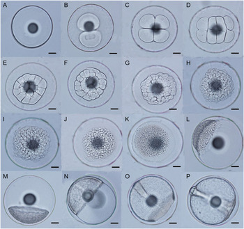
The fertilization rate and hatching rate of C. bicolor eggs were 82% and 85%, respectively; while the eggs of C. bispinosa had a slightly lower fertilization rate of 73% and hatching rate of 81%. Newly released and fertilized eggs for both species were transparent, pelagic and spherical. The mean diameter of the fertilized eggs was 703.6 ± 7.8 μm and 627.6 ± 7.8 μm (n = 15) for C. bicolor and C. bispinosa, respectively. The eggs of both species also showed several other common features, including having narrow perivitelline space, smooth and thin chorion, a homogenous and non-segmented yolk, and a similar-sized single oil globule with diameter of 177.5 ± 6.5 μm for C. bicolor and 172.7 ± 8.1 μm for C. bispinosa (Fig. 1A).
Embryonic development of the bicolor angelfish Centropyge bicolor incubated at 26°C. (A) Newly fertilized eggs. (B) Beginning of cleavage, two cells (20 min after fertilization (AF)). (C) 4 cells (35 min AF). (D) 8 cells (50 min AF). (E) 16 cells (1 h AF). (F) 32 cells (1 h 15 min AF). (G) 64 cells (1 h 20 min AF). (H) Beginning of blastula, 128 cells (1 h 30 min AF). (I) 256 cells (1 h 40 min). (J) 512 cells (2 h AF). (K) 1024 cells (2 h 30 min AF). (L) Beginning of gastrulation (2 h 40 min AF). (M) 20% epiboly (4 h 30 min AF). (N) 50% epiboly (6 h AF). (O) 90% epiboly (7 h AF). (P) 100% epiboly, blastopore closure (7 h 30 min AF). Scale bars in all images represent 100 μm.

Embryonic development
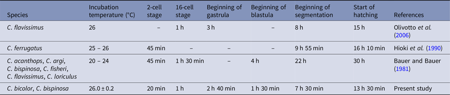
Overall, the embryonic development of C. bicolor and C. bispinosa showed high similarity and synchronization in major developmental events including cleavage, blastula, gastrula, segmentation and hatching. Consequently, the general description of embryonic development below referred to both species with only distinct differences between the two species specified. Additionally, the major embryonic development events of C. bicolor and C. bispinosa were summarized and are compared in Table 1.
Embryonic development events of Centropyge bicolor and Centropyge bispinosa

Cleavage
Cleavage is defined as the period when the blastomeres divide sequentially and double the cell numbers in each division until the blastodisc has 64 blastomeres (Fujimoto et al., Reference Fujimoto, Kataoca, Otani, Saito, Aita, Yamaha and Arai2004). Similar to that of other teleost embryos, the pattern of cleavage observed in the embryos of C. bicolor and C. bispinosa was meroblastic. In fact, the observed cleavage patterns of C. bicolor and C. bispinosa were the same from the first to the sixth cleavage. The first cleavage occurred 20 min after fertilization (AF), giving rise to two cells (Fig. 1B). The subsequent blastomeres divisions appeared to occur within shorter time intervals: the 4-cell stage occurred at 35 min AF (Fig. 1C), 8-cell stage at 50 min AF (Fig. 1D), 16-cell stage at 1 h AF (Fig. 1E), 32-cell stage at 1 h 15 min AF (Fig. 1F) and 64-cell stage at 1 h 20 min AF (Fig. 1G).
Blastula
Development during blastula stage took approximately 1 h 10 min for both C. bicolor and C. bispinosa. It commenced from the seventh cleavage when 128 blastomeres were formed and developed up to 1024 blastomeres, forming the basis of the spherical-shape blastodisc seen at the animal pole (Fujimoto et al., Reference Fujimoto, Kataoca, Otani, Saito, Aita, Yamaha and Arai2004). In the present study, the yolk syncytial layer between the periblast and the blastodisc border was already visible at 512-cell stage (2 h AF; Fig. 1J) for both species. The embryos of both species reached the end of blastula period by 2 h 30 min AF (1024-cell stage; Fig. 1K).
Gastrula
The gastrula period for both C. bicolor and C. bispinosa started at around 2 h 40 min AF (Fig. 1L). At this stage, the blastoderm grow by gradually spreading across and eventually completely covering the yolk, a process known as epiboly. Approximately 20% of the yolk was covered at 4 h 30 min AF (Fig. 1M) and 50% of the yolk was covered at 6 h AF (Fig. 1N). The development of the gastrula reached around 90% at 7 h AF. At this stage, the embryonic axis of both species, which defined the embryo position, differentiated and appeared as a dark line sitting along the yolk in the perivitelline space (Fig. 1O). At 7 h 30 min AF, the edges of the blastoderm merged and the blastoderm completely covered the yolk, marking the closure of the blastopore and the end of epiboly (Fig. 1P).
Segmentation
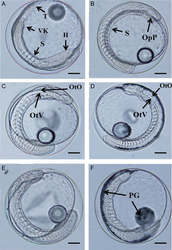
The appearance of the somites marked the transition to the segmentation stage, and the somatogenesis in this period continued until the onset of hatching. With the closure of the blastopore, the difference between the cephalic (head, H) and caudal (tail, T) regions of the embryonic axis became evident and four pairs of somites (S) were observed for the first time at 7 h 30 min AF (Fig. 2A). Furthermore, pairs of somites increased in number and started changing into a formation that resembles a ‘V’ (Fig. 2B). Such V-shaped somite pairs were initially seen in the embryos of C. bicolor and C. bispinosa at 10 h AF and 10 h 30 min AF, respectively. At 12 h 40 min AF, the number of V-shaped somites in the embryos of both species reached and stabilized at 19 until hatching.
Embryonic development of the bicolor angelfish Centropyge bicolor and the coral beauty angelfish Centropyge bispinosa during segmentation. (A) 7 h 30 min AF, showing the presence of rudimentary somites (S), Kupffer’s vesicle (VK), and clear differentiation of head (H) and tail (T) on embryonic axis of C. bicolor. (B) 10 h 30 min AF, showing the formation of V-shaped somites (S) and appearance of optic primordium (OpP) in an embryo of C. bicolor. (C) 11 h 40 min AF, showing differentiation of otoliths (OtO) inside otic vesicle (OtV) in an embryo of C. bicolor. (D) 12 h 40 min AF, appearance of otoliths (OtO) and otic vesicle (OtV) in an embryo of C. bispinosa. (E) Low number of pigmentation in an embryo of C. bicolor prior to hatching. (F) Intense pigmentation (PG) in the dorsal region of embryonic axis and the oil globule in an embryo of C. bispinosa prior to hatching. Scale bars in all images represent 100 μm.

At 10 h 30 min AF, the optic primordium (OpP) was visible for the first time for both C. bicolor and C. bispinosa, and it appeared as two rounded vesicles in the cephalic region of the embryo axis (Fig. 2B). The first signs of embryonic pigmentation occurred at 9 h 30 min AF for both species; however, the pigmentation appeared to be much more intense in C. bispinosa than that in C. bicolor, and such visual difference in pigmentation became very obvious prior to hatching. That is, the embryos of C. bispinosa at the verge of hatching were strongly pigmented and the melanophores concentrated in the oil globule and the dorsal portion of the embryonic axis (Fig. 2F) while the embryos of C. bicolor displayed much lower number of melanophores (Fig. 2E). The muscle contraction of embryos was observed for the first time at around 11 h 30 min AF in both C. bicolor and C. bispinosa. For C. bicolor, the otic vesicle (OtV) emerged as a rudimentary circular structure at 11 h 40 min AF at a position posterior to the OpP; indeed, otoliths (OtO) were also seen inside the otic vesicle at this stage (Fig. 2C). The same development was observed 1 h later (12 h 40 min AF) in the embryos of C. bispinosa (Fig. 2D). By 13 h AF, the rudimentary heart of both species displayed peristaltic movement, which promoted the axial artery dilation, suggesting that the embryos were on the verge of hatching.
Early larvae
The onset of hatching started at 13 h 30 min AF, and within 40 min, all larvae had hatched. The description of early larval development of C. bicolor and C. bispinosa was summarized in Table 1. The mean TLs of newly hatched larvae (n = 10) was 1.52 ± 0.2 mm for C. bicolor and 1.25 ± 0.2 mm for C. bispinosa. At this stage, the larvae of both species were primitive, lacking pigmented eyes, mouth and functional digestive system (Figs 3A and 4A). There was a 24-h difference in the mouth opening time between C. bicolor and C. bispinosa. By 72 h after hatching (AH), the C. bicolor larvae had already possessed an open mouth, pigmented eyes, a rudimentary digestive tract, pectoral fin and some swimming capability (Fig. 4B, C), and the larvae grew to a mean TL of 2.63 ± 0.84 mm (n = 10). However, such larval characteristics were not observed in C. bispinosa until 96 h AH (Fig. 3B, C) when the C. bispinosa larvae were measured in a mean TL of 2.56 ± 0.42 mm (n = 10). At this developmental stage, the yolk of both species had been almost completely absorbed, with half of the oil globule remained (Figs 3C and 4C).
Early larval development of Centropyge bispinosa. (A) A newly hatched larva with a big yolk sac and a single pigmented oil globule. (B) Presence of pectoral fin by 96 h after hatching (AH). (C) Larva with pigmented eyes, open mouth, half oil globule, rudimentary digestive tract and some pigmentation in the mid-body region at 96 h AH.

Early larval development of Centropyge bicolor. (A) A newly hatched larva with a big yolk sac and a single pigmented oil globule. (B) Presence of pectoral fin by 72 h after hatching (AH). (C) Larva with pigmented eyes, open mouth, half oil globule, rudimentary digestive tract and some pigmentation in the mid-body region at 72 h AH.

Discussion
The embryonic and early larvae development have been described in detail for species from different genera of family Pomacanthidae, including Apolemichthys (Hioki and Suzuki, Reference Hioki and Suzuki1995a), Chaetodontoplus (Fujita and Mito, Reference Fujita and Mito1960; Arai, Reference Arai1994; Hioki and Suzuki, Reference Hioki and Suzuki1995b; Leu et al., Reference Leu, Meng, Huang, Tew, Kuo and Liou2010; Leu et al., Reference Leu, Sune and Meng2015), and Genicanthus (Suzuki et al., Reference Suzuki, Hioki, Tanaka and Iwasa1979; Hioki et al., Reference Hioki, Suzuki and Tanaka1982, Reference Hioki, Tanaka and Suzuki1995). However, the description of early ontogeny of species from the genera Pomacanthus (Moe, Reference Moe1976; Kelley, Reference Kelley1995; Leu et al., Reference Leu, Liou, Wang, Yang and Meng2009) and Centropyge (Bauer and Bauer, Reference Bauer and Bauer1981; Hioki and Suzuki, Reference Hioki and Suzuki1987; Hioki et al., Reference Hioki, Suzuki and Tanaka1990; Olivotto et al., Reference Olivotto, Holt, Carnevali and Holt2006) was mostly superficial. Therefore, this study focused on documenting the embryonic development and early larval characteristics of two Centropyge species, the bicolor angelfish C. bicolor and the coral beauty angelfish C. bispinosa in detail to fill the knowledge gap.
In the present study, the mean diameters (D) of fertilized eggs and mean TL of newly hatched larvae of C. bicolor (D = 0.70 mm, TL = 1.52 mm) and C. bispinosa (D = 0.63 mm, TL = 1.25 mm) were similar. Leu et al. (Reference Leu, Sune and Meng2015) compiled the characteristics of the eggs and larvae of 17 Centropyge species, and the mean egg diameters and TL of C. bicolor and C. bispinosa reported from the present study were well within the ranges of the egg diameters (0.58–0.75 mm) and TL (1.13–1.70 mm) of other Centropyge species.
Overall, the embryonic development patterns of C. bicolor and C. bispinosa were similar, and the developmental events largely occurred synchronously from fertilization to the beginning of segmentation. However, after segmentation started, certain development events occurred at different times between the two species with C. bicolor appeared to have slightly faster development than C. bispinosa. For example, the appearance of V-shaped somites and otic vesicle in the embryos were observed 30 min AF and 1 hour AF earlier, respectively, in C. bicolor than in C. bispinosa (Table 1). The pigmentation patter is an easy to observe feature that demonstrated differences between the embryos of C. bicolor and C. bispinosa. Based on the colour of the embryos, the number of melanophores was substantially higher in the embryos of C. bispinosa (Fig. 2F) than that of C. bicolor (Fig. 2E) prior to hatching, making it possible to distinguish the embryos of the two species. Bauer and Bauer (Reference Bauer and Bauer1981) also reported limited pigmentation in the embryos of C. flavissimus, and C. acanthops, which appeared to be similar to what was observed in C. bicolor in this study. Conversely, dense pigmentation on embryos as observed in C. bispinosa were also reported in other angelfish species from the same genus, such as C. argi (Bauer and Bauer, Reference Bauer and Bauer1981), C. fisheri (Bauer and Bauer, Reference Bauer and Bauer1981), and C. ferrugatus (Hioki et al., Reference Hioki, Suzuki and Tanaka1990). Therefore, the embryonic pigmentation pattern appears to be a species-specific feature for the genus Centropyge.
C. bicolor and C. bispinosa also had a similar embryonic developmental pattern when compared with that of C. flavissimus and C. ferrugatus (Table 2). This finding is not very surprising, as past studies had reported similar reproductive biology among species from the genus Centropyge (Moyer and Nakazono, Reference Moyer and Nakazono1978; Bauer and Bauer, Reference Bauer and Bauer1981; Olivotto et al. Reference Olivotto, Holt, Carnevali and Holt2006; Baensch and Tamaru, Reference Baensch and Tamaru2009; Callan et al., Reference Callan, Laidley, Forster, Liu, Kling and Place2011). However, the embryonic development of C. bispinosa and five other Centropyge species reported by Bauer and Bauer (Reference Bauer and Bauer1981) appeared to be much slower when compared with the results of the present study, as well as those by Hioki et al. (Reference Hioki, Suzuki and Tanaka1990) and Olivotto et al. (Reference Olivotto, Holt, Carnevali and Holt2006), particularly from the beginning of segmentation onward (Table 2). A likely explanation for such difference was the different incubation temperatures used by these studies. While Hioki et al. (Reference Hioki, Suzuki and Tanaka1990), Olivotto et al. (Reference Olivotto, Holt, Carnevali and Holt2006) and the present study incubated the embryos at around 26°C, Bauer and Bauer (Reference Bauer and Bauer1981) used a wider and lower range of incubation temperature of 20–24°C. It is well known that temperature is a highly influential environmental factor on the embryonic developmental rate of marine fish (Kamler, Reference Kamler2002; Yang and Chen, Reference Yang and Chen2005; Shi et al., Reference Shi, Zhang, Zhu, Liu and Zang2010). Therefore, the low incubation temperature used by Bauer and Bauer (Reference Bauer and Bauer1981) probably delayed embryonic development. Future studies could further verify the effects of incubation temperature on the embryonic development as well as larval rearing success of Centropyge species.
Comparison of incubation temperatures and the timing (after fertilization) of various embryonic development events of Centropyge species

‘–’ Indicates the timing was not reported by the authors.
Newly hatched C. bicolor and C. bispinosa larva possessed a large yolk and an oil globule (Figs 3A and 4A), however the subsequent development time to first feeding larvae appeared to be different between C. bicolor and C. bispinosa. While it took 72 h AH for C. bicolor larvae to gain an opened mouth, pigmented eyes, pectoral fins, improved swimming ability and a rudimentary digestive tract, it took 96 h for C. bispinosa. Nevertheless, they are both within the reported range of time (3 to 4 days) for other Centropyge species to reach this stage (Bauer and Bauer, Reference Bauer and Bauer1981; Hioki et al. Reference Hioki, Suzuki and Tanaka1990; Olivotto et al. Reference Olivotto, Holt, Carnevali and Holt2006; Baensch and Tamaru, Reference Baensch and Tamaru2009; Leu et al. Reference Leu, Sune and Meng2015). Furthermore, it was observed for both C. bicolor and C. bispinosa that the yolk was almost depleted at this critical stage of mouth opening, when only approximately half of the oil globule remained (Figs 3C and 4C). The fact that yolk sac was consumed quicker than oil globule probably can be explained by the role of yolk sac as a primary energy source for early fish larvae (Fyhn and Govoni, Reference Fyhn and Govoni1995; Williams et al., Reference Williams, Papanikos, Phelps and Shardo2004). For the oil globule, it may play a role in facilitating vertical orientation in water column for early larvae via buoyance regulation (Leu et al., Reference Leu, Sune and Meng2015). At this stage, the depleting endogenous reserves combined with the development of more advanced features in the larvae, such as the presence of an opened mouth, pigmented eyes and functional digestive tract, marked the transition point from endogenous to exogenous feeding. Finding the suitable live feed and rearing environment hence become the key to supporting larval survival to the next phase.
In conclusion, the results of this study showed that the embryonic and early larval development of C. bicolor and C. bispinosa was similar although differences in timing of a few developmental events and pigmentation pattern were observed. Overall, the observed developmental pattern of both species also resembled that of other Centropyge species documented in the literature.
Acknowledgements
We thank Da Li for helping us edit the figures on this manuscript.
Financial support
The first author (RCM) was funded by the Federal Agency for Support and Evaluation of Graduate Studies in Education (CAPES) and received assistance in the funding of research and the sandwich doctorate scholarship within the project ‘Sustainability and Innovation of native marine species’, SCIENCES OF THE SEA 2 no. 43/2013, CAPES.
Conflicts of interest
None.
Ethical standards
Not applicable.