Introduction
Pelvic organs including rectum, bowel, bladder and uterus are subject to physiological changes in position, shape and volume.Reference Foroudi, Wong and Kron1, Reference Jadon, Pembroke and Hanna2 During radiotherapy, these variations result in discrepancies between the planned and actual treatment delivered, which can lead to geographical miss of the tumour and/or variable dose delivery to adjacent organs at risk (OAR). Day-to-day and during treatment, delivery variability is referred to as inter-fraction and intra-fraction motions, respectively. On-treatment image guidance using cone beam computed tomography (CBCT) and/or fiducial markers can guide couch shifts to correct for simple translations in organ position, but correcting for organ rotation and deformation remains challenging using current technology.Reference Nichol, Brock and Lockwood3–Reference van der Wielen, Mutanga and Incrocci5 This means that appropriate and consistent patient preparation and positioning strategies remain important.Reference McNair, Wedlake and Lips6 Organ motion may be of greater significance during intensity-modulated radiotherapy (IMRT), since more complex dose distributions and steeper dose gradients are used than during three-dimensional conformal radiotherapy (3D-CRT).Reference Jadon, Pembroke and Hanna2 This is especially relevant for the safe and effective delivery of highly conformal and hypofractionated treatments such as stereotactic ablative radiotherapy (SABR).Reference Martin, Thomas and Harden7 This review aims to provide an overview of the extent of pelvic organ motion and patient preparation and positioning methods for managing organ motion in the pelvis.
Methods
Literature searches were performed using PubMed (NCBI) for terms relating to pelvic organ motion and strategies to manage this motion. Further relevant articles were found by manually searching reference lists of relevant publications. Given the breadth of this topic, a systematic review was purposely not undertaken. Instead, to bring the best existing evidence into one article, systematic reviews which focus on one or more areas within the subject of managing internal pelvic organ motion are discussed, where these are available. In addition, individual higher quality studies, such as randomised controlled trials (RCTs) or well-conducted cohort studies, are specifically mentioned.
Additional individual studies addressing strategies for managing pelvic organ motion, judged to be of lower quality (see below), are included as an appendix (see Supplementary Material).
A hierarchy of evidence and recommendations grading scheme was applied using the Oxford Centre for Evidence-based Medicine—Levels of Evidence.8 Studies allocated level 1b included well-conducted RCTs (e.g., Mariados et al.Reference Mariados, Sylvester and Shah9). Individual cohort studies (e.g., Krol et al.Reference Krol, McColl and Hopman10) were allocated level 2b, unless judged to be of lower quality. We allocated a level of 2c for studies with small patient numbers (taken as <20 patients), studies that were retrospective or treatment planning studies without reference to clinical outcomes such as toxicity. Grade recommendation A was applied where level 1 studies were available and grade B where evidence was provided by level 2 studies.
Results
Extent of pelvic organ motion is described below for rectum, bladder and bowel. Strategies to manage this motion are then described. Motion management strategies were separated into similar themes, and the available evidence for each strategy considered. In total, four systematic reviews and seven RCTs were identified that addressed different methods of managing pelvic organ motion. Best level of evidence, alongside grade of evidence, is presented for each pelvic organ motion management strategy (see Table 1). Level and grade of evidence for individual studies, including those contained within the cited systematic reviews, are included in Supplementary Material.
Summary of strategies to manage pelvic organ motion and accompanying level of evidence and grade recommendation

Extent of Pelvic Internal Organ Motion
Rectum
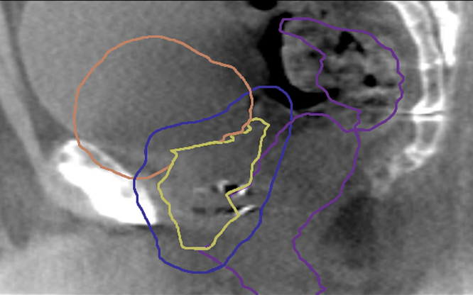
Rectal filling with faeces and gas is the predominant factor influencing rectal distension (see Figure 1). In prostate radiotherapy, rectal distension can result in significant and predominantly anterior–posterior displacements of the prostate gland.Reference Chen, Yang and Wang11, Reference Padhani, Khoo and Suckling12 Presence of rectal gas may also affect the delivered dose distribution during prostate IMRT.Reference Soukup, Söhn and Yan13 Retrospective studies have observed inferior biochemical and local control for patients with a distended rectum at the time of prostate radiotherapy planning.Reference de Crevoisier, Tucker and Dong14–Reference Heemsbergen, Hoogeman and Witte16 In rectal cancer radiotherapy, a systematic review of studies of mesorectal (containing the rectum and perirectal fat) motion found that the greatest displacements were anteriorly in the upper mesorectum.Reference Gwynne, Webster and Adams17 For hypofractionated courses of radiotherapy, such as short-course pre-operative radiotherapy in rectal cancer, an error on even a single fraction could potentially be significant.Reference Nijkamp, de Jong and Sonke18 A systematic review of pelvic organ motion in cervical radiotherapy observed that movement of the cervix and upper vagina is mainly related to rectal filling.Reference Jadon, Pembroke and Hanna2
Sagittal CBCT on-treatment image with contours from planning CT overlaid [clinical target volume prostate and seminal vesicles (yellow), planning target volume (blue), bladder (orange) and rectum (purple)]. Increase in bladder volume seen compared to planning with expansion superiorly and anteriorly. Increase in mid/upper rectal volume seen compared to planning due to faeces and gas with expansion anteriorly. Motion results in shift in prostate position compared to planning identified by displacement of fiducial markers.

Bladder
The main factor influencing bladder motion is bladder filling (see Figure 1). This causes more movement in the anterior and superior directions because expansion laterally and posteriorly is limited by the pelvic bones and rectum.Reference Pinkawa, Asadpour and Siluschek19 Filling may differ between diseased and healthy bladders, with cancer infiltration causing greater wall rigidity, resulting in asymmetry of bladder distension and smaller bladder capacity. Greater variation and magnitudes of motion are also noted in patients with bladder cancer.Reference Fokdal, Honoré and Høyer20, Reference McBain, Khoo and Buckley21 In prostate radiotherapy, deformation of the prostate by bladder (and rectal) filling is limited. However, significant deformations of seminal vesicles by the bladder may occur.Reference van der Wielen, Mutanga and Incrocci5, Reference Roeske, Forman and Mesina22 In cervical radiotherapy, bladder filling may alter the position of the tip of the uterus in both superior–inferior and anterior–posterior directions. In addition, bladder volume may be altered towards the end of a course of radiotherapy as a result of early radiation toxicities.Reference Jadon, Pembroke and Hanna2
Bowel
Bowel motion is under neurological and hormonal control and results in complex peristaltic waves of dilatation and relaxation.Reference Husebye23 Small bowel peristaltic waves have been shown to occur 11 times per minute with average amplitude of 7 mm. In addition to this oscillating motion, large changes in small bowel position and volume occur as a consequence of faeces and gas within the bowel and also vary with bladder filling.Reference Froehlich, Patak and von Weymarn24 Large bowel exhibits considerable variation in luminal diameter and is predominantly gas filled in the absence of faeces. Peristaltic movements may be less frequent for large than small bowel, but differences have also been observed between proximal and distal large bowel. In a cine magnetic resonance imaging (MRI) study, Buhmann et al. found peristaltic waves occurring six times per minute in the ascending colon compared with three times per minute in the descending and sigmoid colon.Reference Buhmann, Kirchhoff and Wielage25 There is considerable variation in the appearance of bowel both within and between patients, and a single CT image represents only an arbitrary shape and position of a mobile and distensible organ. It may be that only 20% of bowel occupies the same position throughout the treatment compared with at planning.Reference Hysing, Kvinnsland and Lord26, Reference Sanguineti, Little and Endres27
Strategies to Manage Pelvic Organ Motion
Levels of evidence
For each of the interventions discussed below, the best level of evidence is presented in Table 1. Individual studies have also been allocated a suggested level of evidence and are present in Supplementary Material. While some high-quality evidence does exist, for example RCTs, cohort studies form the majority of published evidence.
Patient preparation
To try to achieve reproducibility in the volume and position of pelvic organs, use of consistent patient preparation strategies to reduce organ motion should be applied both at planning and during treatment. Patient compliance with protocols may be greater at the time of planning with more directed patient education.Reference McNair, Wedlake and Lips6 In addition, radiotherapy toxicity may alter organ volume and position towards the end of treatment.Reference Jadon, Pembroke and Hanna2 Much of the published literature relating to rectal and bladder filling concerns prostate radiotherapy.
Diet and laxatives
McNair et al. performed a systematic review of interventions to empty the rectum or stabilise its volume.Reference McNair, Wedlake and Lips6 Low-fibre diets and reduced dietary consumption of fermentable carbohydrates (such as beans and pulses) to reduce rectal gas and diarrhoea in prostate radiotherapy did not appear successful. Several studies in the review examined the laxative milk of magnesia (MoM; magnesium hydroxide) in combination with dietary advice. There was some evidence to support reduction in rectal gas with use of MoM, but this did not always correlate with reduced prostatic motion. In addition, MoM appeared to be poorly tolerated by patients. An RCT of the laxative magnesium oxide compared with placebo concluded that magnesium oxide did not reduce prostatic motion, and there was a trend to worse quality of life with the laxative.Reference Lips, van Gils and Kotte28 Oates et al. investigated the effect of dietary intervention with a bulk-forming laxative in an RCT and found a non-significant trend to more consistent rectal volumes.Reference Oates, Schneider and Lim Joon29 At the level of the prostate, the combination therapy was associated with reduced rectal faeces and gas. However, this relationship was not observed in the superior rectum, where the greatest changes in volume occur.Reference McNair, Wedlake and Lips6, Reference Oates, Schneider and Lim Joon29
Other methods of altering bowel gas
The anti-foaming drug simeticone has been used to try to reduce rectal gas in prostate radiotherapy patients, although there is limited evidence for its benefit. While Madsen et al. described little intra-fraction prostatic motion when using simeticone, a rectal catheter was also inserted when rectal gas was seen which limited interpretation of the benefit from simeticone.Reference Madsen, Hsi and Pham30
Ki et al. performed a randomised study of probiotics containing Lactobacillus acidophilus compared to placebo in prostate radiotherapy. They found that the probiotic reduced rectal gas and variation in rectal volume from planning to treatment imaging. However, some patients had excessive rectal distension suggesting variability in outcome using this particular probiotic.Reference Ki, Kim and Nam31
Rectal emptying strategies
Rectal emptying tubes
McNair et al. also reviewed studies of rectal emptying, which has been advocated as a method of reducing variation in rectal filling.Reference McNair, Wedlake and Lips6 There was some evidence that rectal emptying tubes reduced rectal volume variation and prostatic motion during prostate radiotherapy. No RCTs have been performed. Disadvantages of rectal emptying tubes include the additional time taken for the procedure, staff training and patient compliance. Manual evacuation of the rectum, although found in one study to reduce rectal volume and prostatic motion, is unlikely to be tolerated during routine clinical practice.
Rectal enemas and suppositories
McNair et al. concluded that some studies using glycerine suppositories and micro-enemas demonstrated reduced anterior displacement of the rectum (and therefore anterior–posterior prostatic motion).Reference McNair, Wedlake and Lips6 However, most studies included only small numbers of patients and did not prospectively compare enemas to alternative interventions. Sabater et al. performed a prospective trial of 59 patients using enemas in vaginal brachytherapy for post-operative endometrial cancer, with the patient acting as their own control.Reference Sabater, Andres and Gascon32 Despite an overall 15% reduction in mean rectal volume following an enema, over one-third of patients had an increase in rectal volume, and no improvement in rectal dosimetry was observed. In external beam radiotherapy, the extent of rectal emptying, especially from patient self-administration of enemas or suppositories, may vary, with some patients requiring further rectal emptying.Reference McNair, Wedlake and Lips6 Superior rectal volume may have the greatest impact on prostatic displacement, but in some studies reviewed by McNair et al., rectal volume was measured at the level of the prostate gland (corresponding to the level of the mid rectum). Therefore, it is possible that superior rectal volume may not be reduced through the use of an enema or suppository, which acts more distally. Self-administration of enemas or suppositories was well tolerated by patients.Reference McNair, Wedlake and Lips6
Rectal displacement strategies
Endorectal balloons/devices
Previous studies of endorectal balloons (ERBs) in prostate radiotherapy, including one RCT, have demonstrated reduced anorectal toxicity through reduction in the volume irradiated and dose delivered to the anal and rectal walls.Reference Krol, McColl and Hopman10, Reference Wortel, Heemsbergen and Smeenk33 Wortel et al. suggested that patients tolerate ERBs.Reference Wortel, Heemsbergen and Smeenk33 However, ERB insertion may deform the prostate gland and increase treatment time. Therefore, outside of a clinical trial, it is possible that patient acceptance for daily insertion of an ERB might be lower. An RCT is currently investigating the use of a daily inserted rectal obturator (ProSpare) in prostate bed radiotherapy (ClinicalTrials.gov Identifier: NCT02978014). The trial is using smaller planning target volume (PTV) margins for patients allocated ProSpare to determine if this reduces rectal toxicity. In addition, steel markers within the device mean it can be used for treatment verification as an alternative to implanted fiducial markers.
Rectal spacers
The vast majority of the evidence for rectal spacers concerns prostate radiotherapy. Mok et al. performed a systematic review of rectal spacers inserted between the prostate and rectum.Reference Mok, Benz and Vallee34 Spacers are used to increase the distance between these structures and reduce both dose to the rectum and the volume of rectum irradiated to a significant dose. These are made from biodegradable materials such as polyethylene glycol, hyaluronic acid or collagen and can be injected using ultrasound guidance under local, epidural or general anaesthesia. Biodegradable balloons made of polyatic acid have also been used. Biodegradation occurs after around 6 months for polyethylene glycol spacers and polyatic acid balloons and 12 months for hyaluronic acid and collagen spacers. In the review by Mok et al., studies of spacers and balloons demonstrated good safety profiles and improvements in rectal dosimetry.Reference Mok, Benz and Vallee34 One RCT, comparing a hydrogel spacer with no spacer in prostate radiotherapy, found that spacer insertion was well tolerated, and late rectal toxicity was reduced from 7 to 2% for patients in the spacer group.Reference Mariados, Sylvester and Shah9 Further analysis of the trial at 3 years, including patient reported outcomes, was also reported.Reference Karsh, Gross and Pieczonka35 In addition to the improvements in late rectal toxicity, statistically significant differences in favour of the spacer group for urinary toxicity and minimally important differences in bowel, urinary and sexual quality of life domains were found. Potential disadvantages of spacers may include complications from insertion, patient discomfort and infection (although in the RCT by Mariados et al., the only procedure-related complication was mild transient perianal discomfort reported in 10% of patients). In addition, spacers have mainly been used in localised (T1 and T2) prostate cancers, and their role in locally advanced tumours remains uncertain.Reference Mariados, Sylvester and Shah9, Reference Mok, Benz and Vallee34 Nevertheless, it was recently reported that hydrogel spacer will be funded for patients in the United Kingdom as part of an NHS innovation and technology programme.36
Electromagnetic transponders
In prostate radiotherapy, implanted electromagnetic transponders such as the Calypso 4D localisation system (Calypso Medical Technologies, Seattle, WA, USA) can monitor for inter-fractional changes in prostate position.Reference Das, Liu and Jani37 In addition, these also permit real-time tracking, providing the potential to correct for intra-fractional prostate motion and gating of the radiation beam if intra-fraction motion exceeds a certain threshold. This could be especially useful for treatments requiring a high degree of conformality such as SABR or boosting of dominant intra-prostatic lesions. A retrospective study of electromagnetic transponders in 236 patients undergoing prostate radiotherapy observed that changes in intra-fractional prostate position were more likely the longer the treatment delivery time.Reference Tong, Chen and Li38 Variations of >3 mm were seen for 12% of the time taken to deliver fixed-field IMRT delivered within 10 minutes, compared to only 4% for more rapidly delivered volumetric modulated arc therapy (VMAT) treatments completed within 5 minutes. Using the real-time tracking system, the authors also observed changes in prostate position within 1 minute of patient setup. They speculated that this may occur due to patient relaxation on the treatment couch or passage of rectal gas. Since VMAT could be delivered within a few minutes, the group therefore suggested that there could be a benefit in watching for any initial prostate displacement before commencing treatment delivery. Potential drawbacks of electromagnetic transponders include need for implantation and specialist equipment and staff training. In addition, significant image artefacts are produced on MRI, which could limit their use within an MRI-based planning pathway. Patients with pacemakers, hip prostheses and larger patients are also unsuitable. Reference Das, Liu and Jani37
Bladder-filling protocols
Wiesendanger-Wittmer et al. performed a systematic review of strategies to reduce irradiated small bowel volume during pelvic radiotherapy, including patient positioning and bladder filling.Reference Wiesendanger-Wittmer, Sijtsema and Muijs39 They concluded that use of a drinking protocol to achieve a full bladder reduced the volume of small bowel irradiated during external beam radiotherapy for various pelvic cancers, especially for whole pelvis treatments. Many of the studies included in this review, however, did not specify the exact drinking protocol, which limited definition of the optimal bladder volume/drinking protocol. In a retrospective cohort study of 1,080 patients treated with 3D-CRT to the prostate, use of both an empty rectum and comfortably full bladder was associated with reduced biochemical and clinical relapse and risk of dying from prostate cancer.Reference Maggio, Gabriele and Garibaldi40 However, some full bladder protocols used for prostate radiotherapy have been shown to result in greater inter-fraction variation in prostate position compared to empty bladder protocols, especially in the superior and anterior directions, and therefore may be less reproducible.Reference Zellars, Roberson and Strawderman41 Jadon et al. reviewed studies in cervical cancer and observed that daily variation in bladder volume was common and maintaining a consistently large bladder volume may become more difficult later in a course of radiotherapy because of early radiation cystitis and intravenous fluid administered with chemotherapy.Reference Jadon, Pembroke and Hanna2 This may alter the position of the target and OAR. Because of this, the advice frequently given to patients is to maintain a comfortably full bladder. Since this statement is ambiguous, more specific instructions regarding bladder emptying and filling could help minimise differences in daily bladder volume.Reference Wiesendanger-Wittmer, Sijtsema and Muijs39 This approach is supported by an RCT by Mullaney et al. of two different drinking protocols in prostate radiotherapy. The group found that 540 mL (3 cups of water over 10 minutes) was associated with better reproducibility of bladder volume as assessed by bladder ultrasound than 1,080 mL (6 cups of water over 10 minutes).Reference Mullaney, O’shea and Dunne42 Studies of ultrasound bladder scanning have reported improved consistency of bladder volume during prostate radiotherapy.Reference Cramp, Connors and Wood43–Reference Ung, White and Mathlum45 This might be because measuring bladder volume encourages better patient compliance with drinking protocols.Reference Cramp, Connors and Wood43 A cohort study of 190 patients by Mullaney et al. found that bladder volume measured by ultrasound was strongly positively correlated with the bladder volume delineated on the radiotherapy planning CT scan.Reference Mullaney, O’shea and Dunne44 Different bladder-filling strategies may be necessary for whole pelvis treatments compared to the more limited volumes treated during prostate radiotherapy. Eminowicz et al. performed a cohort study comparing bladder volume measured at planning and on CBCTs performed during treatment for cervical cancer.Reference Eminowicz, Motlib and Khan46 They recommended that the ideal bladder volume at planning was 150–300 mL, since larger volumes were not reproducible throughout treatment. Shorter waiting times prior to delivery of radiotherapy on chemotherapy and post-chemotherapy days were also proposed to minimise bladder volume variation. Bladder ultrasound could be beneficial in maintaining consistency of bladder volumes throughout the course of whole pelvis treatments. Umesh et al. performed a cohort study of patients treated with cervical radiotherapy.Reference Umesh, Kumar and Chadha47 They found that a 300 mL bladder volume was tolerable throughout treatment and was achieved after a mean time of 65 minutes following bladder emptying and administration of 1,000 mL of water. A further benefit from ultrasound is the potential to reduce radiation dose from additional CBCT scans.Reference Mullaney, O’shea and Dunne44 Limitations to the use of ultrasound, however, may include imprecision of volume measurements, inter-operator variability in use and additional time needed within the patient pathway to perform the scan (especially if ultrasound was to be used to determine when a fixed bladder volume had been achieved).
Patient position and immobilisation
Belly board and prone position
Prone position has been used to displace small bowel superiorly out of the irradiated volume; however, evidence is less clear as to the clinical benefit for different tumour sites within the pelvis. The systematic review by Wiesendanger-Wittmer et al. examined the impact of patient positioning (supine, prone or prone with belly board) on irradiated small bowel volume.Reference Wiesendanger-Wittmer, Sijtsema and Muijs39 The authors concluded that prone position without a belly board could reduce the volume of irradiated small bowel compared to the supine position. They reported that the addition of a belly board led to further reductions in irradiated small bowel volume for both 3D-CRT and IMRT techniques. IMRT has been shown to result in better normal tissue sparing of small bowel, rectum and bladder in whole pelvis radiotherapy compared to 3D-CRT.Reference Portelance, Chao and Grigsby48 Addition of a belly board to IMRT allowed a further reduction in irradiated small bowel volume.Reference Wiesendanger-Wittmer, Sijtsema and Muijs39 This bowel-sparing benefit may also be observed in post-surgical patients where it might be expected that small bowel could be displaced inferiorly into a pelvic radiation field. The clinical benefit derived from small bowel sparing likely depends on the treatment indication. Extended whole pelvis treatments, such as those used in cervical cancer radiotherapy, would be expected to include larger volumes of small bowel than radiotherapy to the prostate or pre-operative rectum. It is known that for conventionally fractionated radiotherapy, acute and late bowel toxicity is related to the volume of bowel irradiated. However, since many of the studies examined by Wiesendanger-Wittmer et al. were retrospective, included small numbers of patients, used less conformal radiotherapy techniques and reported dosimetric rather than clinical endpoints such as rates of bowel toxicity; it is therefore difficult to be certain about the absolute clinical benefit from prone position and belly board.Reference Wiesendanger-Wittmer, Sijtsema and Muijs39 The major concerns about prone position relate to patient comfort, stability of patient position and reproducibility of setup.Reference Jadon, Pembroke and Hanna2 An RCT by Bayley et al. of prone versus supine position in 28 patients treated with prostate radiotherapy found that supine position was significantly more comfortable for patients and easier to set up.Reference Bayley, Catton and Haycocks49 Based on the studies reviewed, Wiesendanger-Wittmer found that prone position was associated with greater setup errors. The group concluded that modern image-guided radiotherapy (IGRT) techniques, such as online correction protocols, may help identify and permit correction of changes in internal anatomy and patient position.Reference Wiesendanger-Wittmer, Sijtsema and Muijs39 As Jadon et al. acknowledge in their review, however, application of simple translational shifts may be insufficient to account for internal motion organs within complex treatment volumes such as in cervical radiotherapy, and rotational errors are also not well compensated for by on-line correction protocols.Reference Jadon, Pembroke and Hanna2 Simply increasing PTV margins to account for this may negate the bowel-sparing benefits of IMRT. In the RCT performed by Bayley et al., prone position was associated with significantly greater anterior prostate inter-fraction motion and a larger PTV margin was, therefore, required to account for this.Reference Bayley, Catton and Haycocks49 Greater volumes of rectum, bladder and bowel were seen within the 50–95% isodoses as a result, although this study was performed using 3D-CRT rather than IMRT.
Discussion
Pelvic organ motion presents a challenge to safe and effective delivery of radiotherapy to a variety of primary sites both in terms of tumour control and toxicity. IGRT using online verification and volumetric imaging such as CBCT and/or fiducial markers may compensate for certain inter-fractional changes in volume or position, although this process remains a balance between PTV coverage and avoiding excess dose to OAR. In addition, certain movements including rotations and organ deformation as well as intra-fractional changes are not well corrected for using standard IGRT strategies.Reference Nichol, Brock and Lockwood3–Reference van der Wielen, Mutanga and Incrocci5
Organ motion may be more detrimental during IMRT than 3D-CRT because of the greater conformality and complex dose distributions used with IMRT. This is especially relevant to whole pelvis treatments such as those used in radical and post-operative gynaecological cancers, rectal cancers and node positive prostate cancers.Reference Gwynne, Webster and Adams17, Reference Eminowicz, Rompokos and Stacey50–Reference Hysing, Skorpen and Alber52 In whole pelvis IMRT, the large and complicated target volumes used may be impacted by motion of multiple pelvic organs which could result in undercoverage of the PTVs or overdose of OAR. Simply increasing internal target volume margins to account for organ motion may negate the conformality benefits of an IMRT-delivered treatment. Moreover, for cervical cancer, such large variations in uterine position may occur that even with relatively large margins there remains the potential for target volume undercoverage.Reference Eminowicz, Rompokos and Stacey50 Even for smaller target volumes, such as those used in localised prostate IMRT, organ motion may be detrimental given the small margins used. This would be particularly important for simultaneous integrated boost treatments, for example, boosting a dominant intra-prostatic lesion.Reference Guckenberger and Flentje53
Concerns about pelvic organ motion are especially relevant to SABR treatments where a high dose of radiation is given to a highly conformed volume in only a few fractions. A small margin from the gross tumour volume (GTV) to PTV is used with steep dose gradients and any deviation from this risks undercoverage of the tumour and/or overdose of adjacent critical OAR.Reference Martin, Thomas and Harden7 The unpredictability of pelvic organ motion, especially bowel with its potential for intra-fractional changes in position, could compromise the safe delivery of SABR. Further research is needed to establish the extent of inter- and intra-fractional bowel motion, its impact on delivery of SABR and strategies to best manage this motion.
Given the need to balance tumour control with normal tissue toxicity, there is considerable interest in adaptive radiotherapy. Various techniques have been described including reactive re-planning based on tumour shrinkage or other internal/external changes, selection of the most suitable plan from a library of plans and daily plan re-optimisation. Appropriate and consistent patient preparation and positioning, however, will still remain important in the era of adaptive radiotherapy, since widely different variations in internal anatomy would present a challenge to accurate and timely delivery of consistent treatments. In addition, organ motion artefacts, especially streak artefacts on CBCT resulting from moving bowel gas while the scan is acquired, may limit the identification of the target and adjacent OAR and thus make adapting the plan based on the position of these structures difficult.Reference Nijkamp, Pos and Nuver54, Reference Smitsmans, de Bois and Sonke55
Addressing intra-fractional changes in organ position will require real-time monitoring. Treatment could be interrupted or adapted if intra-fraction motion exceeded a certain threshold. This could be addressed by electromagnetic transponders, for example, using the Calypso system for prostate radiotherapy, or by MRI-delivered treatments such as the MR-Linac.Reference Das, Liu and Jani37, Reference Kerkmeijer, Fuller and Verkooijen56 However, the additional equipment and need for implantation may limit more general use of electromagnetic transponders and the complexities of rapid daily adaptive replanning at present represents a challenge to the routine use of the MR-Linac. An alternative could be Kilovoltage Intra-fraction Monitoring (KIM), which permits intra-fraction tracking of position of implanted prostate fiducial markers using the CBCT mounted on a standard linear accelerator without the need for additional equipment.Reference Keall, Nguyen and O’Brien57 KIM is being evaluated in a phase 2 trial of prostate SABR (ClinicalTrials.gov Identifier: NCT02397317).
Ensuring more consistent bladder and rectal volumes might appear a more straightforward approach to reducing organ motion. Despite significant interest and effort in investigating different methods of addressing variation in rectal and bladder filling, however, there is often conflicting evidence regarding the benefits of commonly undertaken interventions including bladder-filling protocols and rectal enemas.Reference McNair, Wedlake and Lips6, Reference Wiesendanger-Wittmer, Sijtsema and Muijs39 Levels of evidence and grades of recommendation for interventions to improve bladder, rectal and bowel motion have been allocated in this review (see Supplementary Material). While some RCTs were available, the majority of studies included in this review would be classed as cohort studies. Many of these are limited to a single centre and have included small patient numbers without randomisation, meaning that findings may not be more generally applicable.
While there may be some evidence to support more complex interventions, including rectal emptying tubes or use of ERBs and rectal spacers, the potential benefits have to be balanced against patient discomfort and acceptability, the need for additional procedures and increased treatment times. This may be especially relevant in the setting of prostate radiotherapy, where use of IMRT has already resulted in low rates of rectal and urinary toxicities.Reference Zelefsky, Levin and Hunt58
Bowel motion remains a concern and may not be reduced by interventions directed towards the bladder and the rectum. Some studies of bladder filling and use of prone patient positioning (with or without a belly board) have observed reduced dose to small bowel but have not necessarily demonstrated definitive clinical improvements in bowel toxicity.Reference Wiesendanger-Wittmer, Sijtsema and Muijs39 For SABR treatments of oligometastatic pelvic nodal disease, the node (and adjacent bowel) might be sufficiently distant to the bladder that bladder filling does not displace bowel away from the treatment volume. In addition, given the ablative doses used with SABR, the maximum dose to any loop of bowel close to the PTV is likely to be a more relevant constraint than the volume of bowel receiving a certain dose. Issues of stability and reproducibility of patient position when prone would also be of concern, given the highly conformal treatment volumes and high dose per fraction used with SABR.
Conclusion
There is considerable variation in pelvic organ motion, and this can impact on the safe and effective delivery of radiotherapy treatments in the pelvis. Much of the evidence base to support strategies to manage motion of the rectum, bladder and bowel is limited by absence of high-quality studies and direct comparison between interventions. Further investigation of adaptive radiotherapy strategies is likely to be required to compensate for daily variation in organ motion.
Author ORCIDs
F. Slevin 0000-0002-7176-904X.
Acknowledgements
None.
Financial support
This work was undertaken in the Leeds Cancer Centre, which receives funding from NHS England. The views expressed in this publication are those of the authors and not necessarily those of NHS England. This work was supported by Cancer Research UK (CRUK) Centres Network Accelerator Award Grant (A21993) to the Advanced Radiotherapy Technologies Network (ART-NET) consortium, of which Leeds Cancer Centre is a member. We acknowledge NHS funding to the Leeds Cancer Centre. Dr F Slevin, Mr M Beasley and Dr R Speight report grants from Cancer Research UK during the conduct of this study. Dr L Murray is a University Clinical Academic Fellow funded by Yorkshire Cancer Research (award number L389LM). Dr Henry reports grants from Cancer Research UK (108036) during the conduct of this study; grants from NIHR (111218), grants from MRC (107154) and grants from the Sir John Fisher Foundation (charity, no grant number) outside the submitted work.
Conflicts of interest
None.
Supplementary material
To view supplementary material for this article, please visit https://doi.org/10.1017/S1460396919000530