Introduction
Tympanosclerosis is a disease of the middle ear characterised by hyaline degeneration of the mastoid cells.Reference Plester 1 Various studies have reported the tympanosclerosis prevalence to be between 3 per cent and 62.9 per cent. Although its morphology is well known, the aetiopathogenesis of tympanosclerosis is not fully established. So far, many theories have been proposed to explain the tympanosclerosis formation, but none have been validated.Reference Bhaya, Schachern, Morizono and Paparella 2 – Reference Austin 6
Hyperoxia and inflammation can induce the production of free oxygen radicals, and may induce irreversible tissue damage. Following irreversible tissue injury, hyaline degeneration and calcification can occur in the middle ear.Reference Asiri, Hasham, al Anazy, Zakzouk and Banjar 7 , Reference Cankaya, Kösem, Kiroglu, Kiris, Yuca and Oztürk 8
The production of nitric oxide and other free oxygen radicals results from the intracellular and extracellular effects of cytokines and inflammatory mediators. Neutrophils, leukocytes, macrophages and many other inflammatory cells produce oxygen free radicals.Reference Karlıdağ, Ilhan, Kaygusuz, Keles and Yalcin 9
Oxidative stress is defined as an imbalance between reactive oxygen species production and the capacity of the antioxidant defence system, which comprises both enzymatic and nonenzymatic systems. The most important enzymatic antioxidants are catalase, glutathione peroxidase, superoxide dismutase and inducible nitric oxide synthase: they are considered first-line cellular defences against free radical damage. Enzymatic antioxidant systems are under genetic control and are important for maintaining homeostasis of the body.Reference Görür, Vayisoglu, Ozcan, Güner, Yaroglu and Tamer 10 Superoxide dismutase (Enzyme Commission number 1.15.1.1) catalyses dismutation of the superoxide anion to form hydrogen peroxide (H2O2), which is deactivated to water (H2O) by catalase (Enzyme Commission number 1.11.1.6).Reference Lechner, Lirk and Rieder 11 Catalase is found within the peroxisomes of almost all eukaryotic cells. It uses hydrogen peroxide as an electron source, and is considered the main regulator of hydrogen peroxide metabolism.Reference Andersen, Nielsen, Nielsen and Grandjean 12 Inducible nitric oxide synthase produces nitric oxide in response to a wide array of stimuli, especially endotoxins and endogenous pro-inflammatory mediators.Reference Lechner, Lirk and Rieder 11
Although in chronic otitis media patients there is a hyperoxic environment and inflammation in the middle ear due to perforation of the tympanic membrane, tympanosclerosis alone may also develop in some patients. These differences in symptoms may arise from individual genetic polymorphisms affecting the antioxidant system and wound healing capacity.Reference Görür, Vayisoglu, Ozcan, Güner, Yaroglu and Tamer 10
This study investigated genetic polymorphisms affecting inducible nitric oxide synthetase, superoxide dismutase and catalase enzymes in chronic otitis media patients with and without tympanosclerosis, and assessed the role of genetic susceptibility in disease aetiology.
Materials and methods
This study was approved by the General Directorate of Pharmaceuticals and the Pharmacy Clinical Research Ethics Board of Firat University. Informed consent was obtained from participating individuals or their families.
The study included 162 patients who underwent surgery for chronic otitis media between November 2009 and May 2015 at the Otorhinolaryngology Clinics of Fırat University and Elazığ Education and Research Hospital and 188 healthy volunteers. Exclusion criteria were otorrhoea within the previous three months, chronic otitis media with cholesteatoma, planned surgery due to granulation tissue or polyps in the middle ear, otitis media with effusion or insertion of ventilation tube for this indication, myringosclerosis, and chronic systemic disease.
Patients were divided into two groups: a tympanosclerosis group (n = 72), comprising patients who underwent surgery for chronic otitis media and had tympanosclerotic plaques on the middle-ear mucosa near to the ossicular chain or mastoid bone; and a chronic otitis media group (n = 90), comprising patients who underwent surgery for chronic otitis media without tympanosclerosis. The control group (n = 188) comprised healthy individuals without ear problems or a known chronic systemic disease.
Genetic polymorphism affecting inducible nitric oxide synthase, superoxide dismutase and catalase enzymes were investigated in all groups.
Genotyping
Blood samples were taken from all patients and healthy controls: for this 3 ml peripheral venous blood was added to tubes containing ethylene diamine tetra acetic acid and stored at −20 °C. DNA was isolated from stored blood samples using the Wizard Genomic DNA Purification Kit (Promega, Madison, Wisconsin, USA), according to the manufacturer's method.
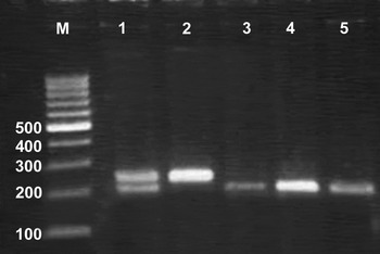
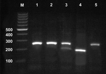
The method of Flekac and colleagues was used to determine the genotype distribution of the p.Ala16Val (C>T) polymorphism in the SOD2 gene and the −21A>T polymorphism in the CAT gene (encoding catalase).Reference Flekac, Lee, Hilgertova, Lacinova and Jarolimkova 13 Briefly, the DNA sequence surrounding the polymorphism was amplified by polymerase chain reaction, 25 µl reaction products were incubated with 1 µl restriction enzyme and 2.5 µl 10× restriction digest buffer, and the size of resulting DNA fragments was estimated by agarose gel electrophoresis. For genotyping the SOD2 p.Ala16Val polymorphism, the forward primer was 5′-GCTGTGCTTTCTCGTCTTCAG-3′, the reverse primer was 5′-TGGTACTTCTCCTCGGTGACG-3′, and polymerase chain reaction products were incubated with the BsaWI (Fermentas, Waltham, Massachusetts, USA) at 60 °C for 16 hours. A fragment of 267 bp was obtained for the C allele and fragments of 183 bp and 84 bp were obtained for the T allele. For genotyping the −21A>T exchange polymorphism in the CAT gene, the forward primer was 5′-AATCAGAAGGCAGTCCTCCC-3′, the reverse primer was 5′-TCGGGGAGCACAGAGTGTAC-3′ and polymerase chain reaction products were incubated with Hinf I (Fermentas) at 37 °C for 16 hours. Fragments of 203 bp and 47 bp were obtained for the A allele and a fragment of 250 bp was obtained for the T allele.
For genotyping the −277A>G polymorphism in the NOS2 (iNOS) gene (encoding inducible nitric oxide synthase), the polymerase chain reaction was performed using 5′-TGGACTCCTGTCACAAGCTGGAAA-3′ as the forward primer and 5′-CTCAgTTTTCgACTCgCTACAA-3′ as the reverse primary. The products of 446 bp was subjected to DNA sequencing.Reference Holla, Stejskalova, Znojil and Vasku 14
Statistical analysis
Study data were stored in a database and analysed using IBM SPSS Statistics software version 20.0 (Armonk, New York, USA). Relationships between variables were assessed using Pearson's correlation analysis. The distribution of genotypes and allele frequencies in patient and control groups was assessed by chi-square analysis. A p value of less than 0.05 was considered statistically significant.
Results
There were 72 patients (34 men, 38 women) in the tympanosclerosis group, with a mean age of 31.08 ± 12.02 years. There were 90 patients (49 men, 41 women) in the chronic otitis media group, with a mean age of 33.02 ± 8.38 years. There were 188 healthy volunteers (97 men, 91 women) in the control group, with a mean age of 34.41 ± 6.68 years. There were no significant differences among groups regarding age and sex (p > 0.05).
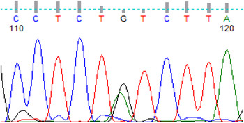
The DNA sequence of the NOS2 −277A>G polymorphism is shown in Figure 1. The distribution of GG, GA and AA genotypes was compared between the groups (Table I). The GG genotype frequency was 35 per cent, 30 per cent and 23 per cent in the tympanosclerosis, chronic otitis media and control groups, respectively. There was no significant difference in GG genotype frequency between the tympanosclerosis and chronic otitis media groups or between the chronic otitis media and control groups (p > 0.05). However, there was a significant difference in GG genotype frequency between the tympanosclerosis and control groups (p < 0.05).
Image showing heterozygosity of the NOS2 −277A>G polymorphism, as indicated by the superimposed peaks for the adenine (A) and guanine (G) nucleotides in the DNA sequence.

NOS2 −277A>G Polymorphism: genotype and allele frequency

*AA = wild type; GA = heterozygous; GG = polymorphic allele; CI = confidence interval. †Tympanosclerosis group vs control group. ‡Tympanosclerosis group vs chronic otitis media group. **Chronic otitis media group vs control group.
Agarose gel electrophoresis was used for genotyping the p.Ala16Val SOD2 (also known as Mn-SOD) polymorphism (Figure 2). The frequency of the CC genotype was 29 per cent, 23 per cent and 33 per cent in the tympanosclerosis, chronic otitis media and control groups, respectively. The frequencies of the CC, CT and TT genotypes are shown in Table II. There was no significant difference in CC genotype distribution between the tympanosclerosis and chronic otitis media groups, the tympanosclerosis and control groups, or the chronic otitis media and control groups (p > 0.05).
Agarose gel electrophoresis image showing the genotypes of the SOD2 p.Ala16Val polymorphism. Lanes: M, 100 bp DNA size marker; column 1, 267 bp product of the C allele and 183 bp and 84 bp products of the T allele; column 2, 267 bp product of the C allele; columns 3–5, 183 bp and 84 bp products of the T allele.

SOD2 p.Ala16Val Polymorphism: genotype and allele frequency

*TT = wild type; TC = heterozygous; CC = polymorphic allele; CI = confidence interval. †Tympanosclerosis group vs control group. ‡Tympanosclerosis group vs chronic otitis media group. **Chronic otitis media group vs control group.
The genotype of the CAT −21A>T exchange polymorphism was determined by agarose gel electrophoresis (Figure 3). The distribution of the TT, TA and AA genotypes was determined for all study groups (Table III). The frequency of the TT genotype was 22 per cent, 17 per cent and 8 per cent in the tympanosclerosis, chronic otitis media and control groups, respectively. There was no significant difference in TT genotype distribution between the tympanosclerosis and chronic otitis media groups (p > 0.05). However, significant differences in TT genotype distribution were observed between the tympanosclerosis and control groups and between the chronic otitis media and control groups (p < 0.05).
Agarose gel electrophoresis image showing the genotypes of the −21A>T exchange polymorphism in the CAT gene. Lanes: M, 100 bp DNA size marker; columns 1, 2 and 5, 250 bp product of the T allele; column 3: 203 bp and 47 bp products of the A allele and 250 bp product of the T allele; column 4, 203 bp and 47 bp products of the A allele.

CAT −21A>T Polymorphism: genotype and allele frequency

*AA = wild type; AT = heterozygous; TT = polymorphic allele; CI = confidence interval. †Tympanosclerosis group vs control group. ‡Tympanosclerosis group vs chronic otitis media group. **Chronic otitis media group vs control group.
There were no significant differences in allele distribution for the NOS2, CAT and SOD2 genes among groups (p > 0.05).
Discussion
Although tympanosclerosis has been widely studied, its aetiology and pathogenesis are not fully understood. Immunological reactions, genetic predisposition, infection, trauma and free oxygen radicals are possible causes of tympanosclerosis.Reference Tos 15 Oxidative stress is thought to contribute to the pathogenesis of tympanosclerosis. This study found that allelic variants of two important antioxidant genes, NOS2 and CAT, were differentially expressed by tympanosclerosis patients compared with the healthy age-matched controls.
In otitis media, serum antioxidant enzyme levels are reported to be low and the oxidative stress index high.Reference Baysal, Aksoy, Kara, Taysi, Taskin and Bilinc 16 , Reference Yilmaz, Kocan, Besler, Yilmaz and Gursel 17 Garcia Callejo et al. reported that in patients with chronic otitis media with effusion, malondialdehyde levels were approximately 10 times higher in effusion fluid than in middle-ear fluid.Reference Garcia Callejo, Estors Ferrero, Morant Ventura, Segarra Cortes and Velert Vila 18 Karlıdağ et al. reported that tympanosclerosis patients had higher malondialdehyde and nitric oxide levels in the eardrum and middle-ear materials and higher plasma malondialdehyde levels compared with patients without tympanosclerosis, whereas the average superoxide dismutase and catalase activity was lower in the patients with tympanosclerosis than in those without.Reference Karlıdağ, Ilhan, Kaygusuz, Keles and Yalcin 9 There was a significant difference in catalase levels between groups but not in superoxide dismutase levels. In addition, Forseni et al. reported higher B lymphocyte and macrophage numbers and higher levels of interleukin-6 in patients with otitis media with effusion, but higher inducible nitric oxide synthase levels in tympanosclerosis patients.Reference Forseni, Hansson, Bagger-Sjöback and Hultcrantz 19 Atmaca et al. administered saline to the right middle ear and nitric oxide to the left middle ear of rats via myringotomy.Reference Atmaca, Erol, Çoban, Yildiz, Çengel and Koyuncu 20 Histopathological examination of the middle-ear bullae did not identify significant differences in tympanosclerosis formation. These authors concluded that nitric oxide administration was not sufficient to cause tympanosclerosis, but that it may cause tympanosclerosis through an inflammatory reaction with a group of cytokines.
In humans, the NOS2 gene is located at chromosome 17q11.2-q12, and has been associated with a number of diseases.Reference Burgner, Xu and Rockett 21 , Reference Burgner, Usen and Rockett 22 This study revealed a significant increase in tympanosclerosis compared with chronic otitis media in individuals with the AA genotype of the NOS2 −277A>G polymorphism. Effects of the −277A>G polymorphism on NOS2 gene regulation and transcription are unknown because no study so far has reported the genotype. However, inducible nitric oxide synthase is already thought to contribute to tympanosclerosis formation. As well as its roles in vasodilatation and destroying pathogens, nitric oxide is reported to take part in middle-ear inflammatory response, as well as in the development of tympanosclerosis. Inducible nitric oxide synthase is also reported to participate in tympanosclerosis formation.Reference Karlıdağ, Ilhan, Kaygusuz, Keles and Yalcin 9 , Reference Forseni, Hansson, Bagger-Sjöback and Hultcrantz 19 , Reference Forseni, Bagger-Sjöback and Hultcrantz 23 Nitric oxide is thought to be secreted (dependent on cytokines) and to reduce osteoclast activity and osteoblast secretion, leading to tympanosclerosis formation.Reference Pirodda, Ferri, Bruzzi, Marini and Faggioli 24
The CAT gene is located at chromosome 11p 13.31. It encodes a ubiquitous enzyme that is found in all known organisms; in mammals, it is most abundant in the liver, kidneys and erythrocytes. The −21A>T CAT variant represents an A to T substitution within the promoter region, just proximal to the transcriptional start site.Reference Quan, Korneluk, Tropak and Gravel 25 Only a few studies have investigated this polymorphism. This study hypothesised that the T allele may reduce the antioxidant capacity of catalase and may therefore be a risk factor for diseases associated with oxidative stress, including tympanosclerosis.
The SOD2 gene is located at chromosome 6q25.3 (Online Mendelian Inheritance in Man entry 147460). A cytosine (C) to thymine (T) transition at position 677 of the SOD2 gene leads to a change in the structural conformation of the mitochondrial targeting sequence in the encoded superoxide dismutase enzyme.Reference Forsberg, de Faire and Morgenstern 26 The valine (Val) isoform (encoded by the T allele) has 30–40 per cent lower enzyme activity and thus reduces the capacity of cells to mitigate oxidative stress.Reference Brose, Avramopoulos and Smith 27
It is likely that high inducible nitric oxide synthase levels and low catalase levels play a role in the tympanosclerosis formation by increasing the production of nitric oxide and other free oxygen radicals. However, this study found no significant between-group differences in the CC genotype frequency for the SOD2 p.Ala16Val polymorphism.
-
• Oxidative stress contributes to tympanosclerosis aetiopathogenesis
-
• Genetic polymorphisms affecting inducible nitric oxide synthetase, superoxide dismutase and catalase were investigated in chronic otitis media patients with and without tympanosclerosis
-
• The GG genotype frequency of the NOS2 −277A>G polymorphism was significantly different in tympanosclerosis patients
-
• The TT genotype frequency of the CAT −21A>T polymorphism was significantly different in tympanosclerosis and chronic otitis media patients
-
• Genetic predisposition may influence tympanosclerosis aetiopathogenesis
In this study, there was a significant increase in the GG genotype of the NOS2 −277A>G polymorphism in tympanosclerosis patients, and a significant increase in the TT genotype of the −21A>T polymorphism in the CAT gene in chronic otitis media patients with and without tympanosclerosis compared with healthy participants. These results indicate that antioxidants controlled by several enzymes may contribute to tympanosclerosis formation, and that antioxidant levels may be linked to genetic polymorphisms.
Acknowledgements
This study was supported by Firat University Scientific Research Projects Office (FUBAP Number 1965).