Introduction
Prostate cancer is the most common malignancy in men, with a reported 47,000 cases diagnosed annually in the UK. The rate of incidence is increasing, and Masson et al.Reference Masson, Persad and Bahl1 highlighted that this is partly because of the increasing use of the prostate-specific antigen test in both symptomatic and asymptomatic patients. Radiotherapy is utilised in all stages of prostate cancer, from early localised disease to locally advanced and metastatic stages,Reference Khoo2 the clinical efficacy of radiotherapy is proven in the literature with high rates of response with no evidence of disease at 5 years of 80Reference Al-Mamgani, Heemsbergen and Levendag3 and 79%,Reference Dearnaley, Sydes and Graham4 respectively.
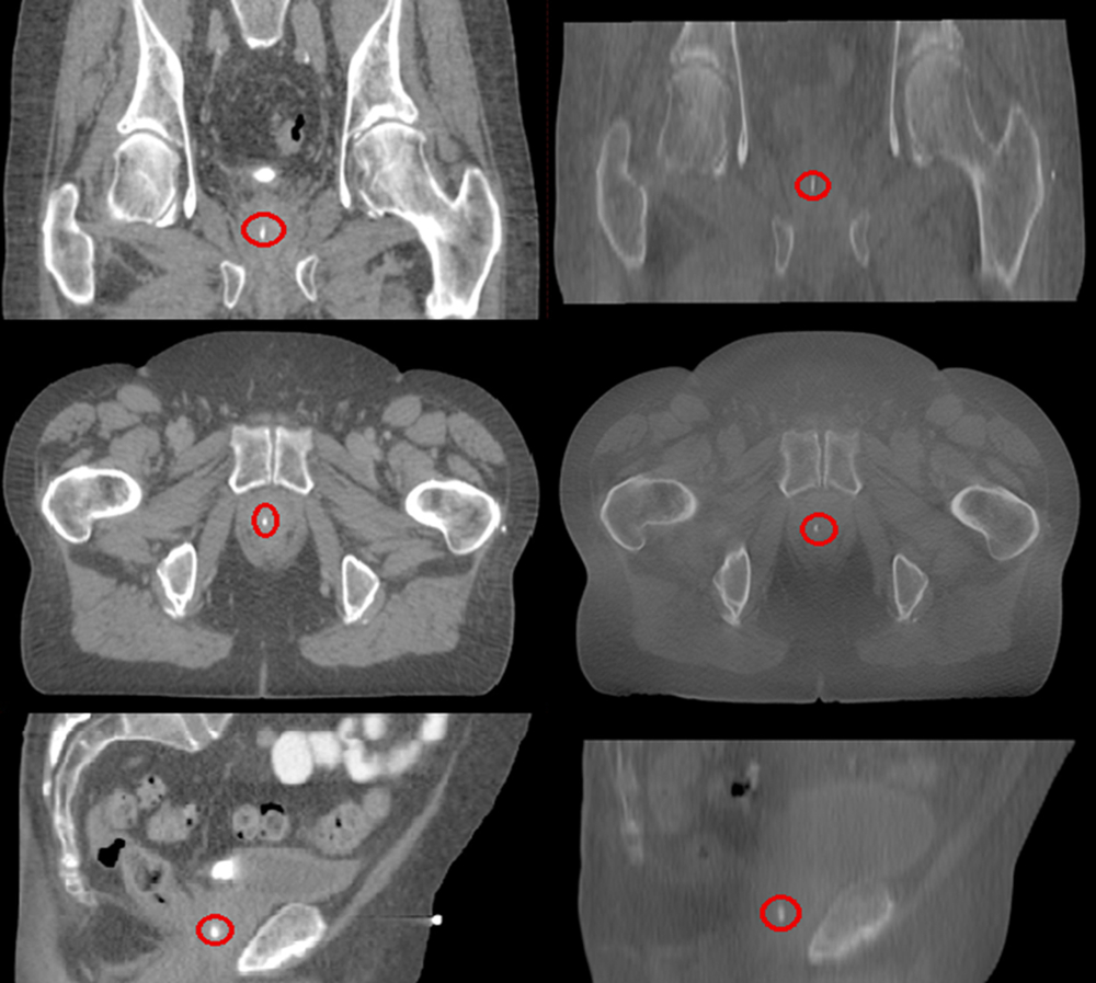
Prostate calcifications are a naturally occurring product associated with benign prostatic hyperplasia; however, any significant link between prostate calcifications and adenocarcinoma of the prostate is still to be identified.Reference Yang, Huang and Wang5 Prostate calcifications have been reported in nearly 90% of 298 prostatectomy patients according to findings in a pathological study carried out by Suh et al.Reference Suh, Gardner and Kee6 These common calcifications (Figure 1) that are clearly visible on a cone beam computed tomography (CBCT) scan and are found almost exclusively within the gland; however, they can occur in the seminal vesicles and ejaculatory duct; however, these are particularly rare. Another study highlighted the potential use of these naturally occurring prostate calcifications as surrogates to use to effectively localise the prostate with CBCT for radiotherapy treatment.Reference Zeng, McGowan and Larsen7 Zeng et al.Reference Zeng, McGowan and Larsen7 discovered that well-defined prostate calcifications located either inside the prostate or near its borders were identified in 35% of patients who were treated with radiotherapy.
Planning CT (left) and CBCT slices (right) highlighting prostatic calcifications. CT, computed tomography; CBCT, cone beam computed tomography.

It has been suggested that prostate calcifications are reliable markers for prostate localisation in a sub-analysis of data from four patients. These findings prompted Hanna et al.Reference Hanna, Pimenta and Marta8 to assess prostate calcification displacement during a course of radiotherapy and retrospectively analysed data from 10 patients, and the study concluded that centrally located prostate calcifications can be used as naturally occurring fiducial markers. Similar deductions were drawn by Sbai et al. who assessed 183 CBCT images from 9 patients and their respective displacements and also highlighted the potential use of using prostate calcifications in image guidance.Reference Sbai, Thariat and Tachfouti9
This study aimed to assess the clinical feasibility of employing an automatic match during CBCT imaging using prostatic calcifications within the 95% isodose as the region of interest. The study also looked to analyse the differences between the treated soft tissue matches versus the automatic calcification matches, and finally determine the levels of interobserver variability between the two radiographers who matched the images.
Method
Aim of the study:
(1) Assess if performing an automatic calcification match is clinically feasible for use in the verification of internal position of the prostate.
The study questions were:
(1) Is there a difference between standard manual soft tissue anatomy matching versus auto calcification matching within the 95% isodose?
(2) Is using auto matching clinically feasible when setting prostatic calcifications as a region of interest within the 95% isodose?
(3) As the matching process becomes more automated what is the effect on interobserver variability between the two matching radiographers?
The selection criteria included any patient referred for radiotherapy treatment to the prostate with known prostate calcifications inclusive of patients with seminal vesicle involvement. The rationale behind this was that regardless of clinically positive seminal vesicle involvement, the matching technique remains the same. Trust consent was sought at the study design phase and approval was given to commence the study. To ensure no selection bias, the patients were selected consecutively as they progressed through the radiotherapy pathway. A sample of 34 patients was chosen as it provides a large enough sample size to provide an accurate representation of the population as it is approximately 10% of the author’s departmental annual prostate throughput.
Thirty-four consecutive patients were selected with known prostatic calcifications undergoing a course of 74 Gy in 37 fractions of curative radiotherapy with daily CBCT imaging. All patients with prostatic calcifications that were visible on both the planning CT and CBCT images were considered as eligible for the study. The CBCT images were assessed offline using those slices acquired on the 5th fraction of the radiotherapy course using prostatic calcifications as naturally occurring fiducial markers set as the region of interest.
The images were analysed using slices from the 5th fraction as it allowed eligible patients time to familiarise themselves with the routine preparation before treatment and to give the radiographers the opportunity to provide any necessary intervention. Non-compliant bladder and rectal preparation led to withdrawal from the study as excessive rectal distension, and reduced bladder filling has a potentially negative impact on match quality.
The images were analysed offline at separate times by two experienced treatment radiographers in one session who are competent at matching CBCT images of the prostate, the subsequent image values were input onto an excel spread sheet. When reviewing the CBCT images, the ARIA (Varian, Palo Alto, USA) offline review software application was used. The region of interest was set inside the 95% isodose around either a single, defined large calcification or a cluster of >3 calcifications located in close proximity within the prostate gland depending on what was visible on the CBCT scan to a size of 1 cm3. The anatomy match was performed automatically using the auto match functionality within the Varian (Varian, Palo Alto, USA) offline review software application. After the auto match was completed by the application, the radiographer then scrolled through the slices and performed the necessary final alterations to the match to ensure clinically acceptable volumetric coverage of the 95% isodose. When matching the images, there was a strict adherence to the local standard operating procedure that requires the entire prostate be included within the 95% isodose with a high conformity index to the initial planning CT scan.
Results
In total, 34 patients were included in this study, and the age range was between 62 and 82 years with the median age being 76 years. The mode of the cohort was 76 years. The cohort histology consisted of 29 patients with disease confined solely within the capsule of the prostate, whereas in five patients, the disease had invaded the seminal vesicles. The mean age within the cohort was 75 years regardless of whether the disease was confined solely to the prostate or was inclusive of the seminal vesicles.
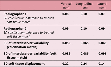
The standard deviation (SD) values of the difference between treated soft tissue match (baseline) versus automatic calcification matches performed by radiographer 1 were 0·08 cm in the vertical axis, 0·10 cm in the longitudinal axis and 0·07 cm in the lateral axis.
The SD values for radiographer 2 when assessing the difference between the treated CBCT soft tissue match versus automatic calcification matches were 0·09 cm in the vertical axis, 0·10 cm in the longitudinal axis and 0·09 cm in the lateral axis.
The prostate automatic calcification matches were performed offline by selecting the region of interest set inside the 95% isodose around either a single, defined large calcification or a cluster of smaller calcifications to a size of 1 cm3 within the Aria offline review application. To avoid being influenced by the final treated position, the two radiographers performed matches starting from the CBCT acquisition position and set the region of interest for automatic matching from this point.
Discussion
During the data collection phase, no patients within the cohort were excluded from the study due to non-compliance of bladder and bowel preparation. All of the patients included within the cohort had no issues with bladder and bowel preparation to a point that would negatively impact on the matching process and ultimately compromise volumetric coverage. The CBCT images taken on the 5th fraction were analysed for each of the patients within the cohort. These images were matched and analysed using the Varian ARIA offline review software package in one session at separate times with the data input onto an excel spread sheet.
When composing the research aim at the study design stage, the authors expected the SD when comparing the soft tissue anatomy matches to the calcification matches to be no greater than 2 mm in any axis. Upon reviewing the CBCT images, the deviations fluctuated around 1 mm in all three axes for all of the matches carried out by the two reviewing radiographers (Table 1). Both the reviewing radiographers matched the CBCT slices from 34 separate treatment sessions taken on fraction 5 from patients within the cohort. These images were reviewed in a single sitting session with the radiographers carrying out analysis separately to avoid any matching bias.
Results of radiographer matches

SD, standard deviation.
In terms of mean displacement, the longitudinal axis observed the largest deviation in all the images matched and this finding was also highlighted by Sbai et al.Reference Sbai, Thariat and Tachfouti9 The interobserver variability observed between the two radiographers was 0·055, 0·065 and 0·045 cm in the vertical, longitudinal and lateral axes, respectively. The clarity of the calcifications on the CBCT images with their large size might go some way to explaining the small amount of interobserver variability displayed by the two matching radiographers. Using calcifications provides a clear starting point for the radiographers to match to within the treatment volume before commencing a check of volumetric coverage, if the matching process begins in the same place, it can allow for a standardisation of matching technique between radiographers.
The rationale for utilising the automatic match functionality within the Varian offline review software application is that the matching process by the radiographer begins at a more advanced stage as a basic anatomy match has already been performed. Whereas a standard manual match starts with the images displaced and requires the radiographer to scout through the slices first to analyse the amount of anatomical and positional displacement, in a sense the manual matching speed is dictated by where the deviation between the images is first detected by the software application. If there is a larger positional displacement, the longer the matching process will take before the radiographer reaches the final checking phase of the match and commencing the treatment. This speed can also be influenced by the current skill level and experience of the radiographer, the complexity of internal anatomy and assessment of bladder/rectal filling which requires decision-making and other technical aspects such as the pixel size and clarity of the images the screen monitors offer the radiographer to perform their duties. These variables can have an influence on the speed and accuracy of a match and cannot be fully mitigated against, and this further provides the rationale of streamlining the matching process. The patient preparation in terms of drinking required fluid levels and bowel emptying within the cohort was of a satisfactory standard so this made matching less difficult as there was reduced ambiguity between the anatomical structures on the CBCT images, which can be hindered by excessive rectal filling and inadequate bladder volume.
The automatic function allows the radiographer to select the region of interest and the algorithm within the software application will perform this task and then the radiographer can perform the necessary minor adjustments to ensure adequate dose coverage. This type of matching allows the process to become more automated and can help to speed up the matching process while not affecting quality as proven by the SD difference between the auto calcification matches compared to the initial soft tissue matches that were subsequently treated on. A potential drawback of setting a very specific region of interest is the time taken to set these parameters up on the first fraction before the parameters are saved, this time ensures accuracy of setting to a cluster or large calcification for subsequent fractions. Time must also be sought to analyse which patients are clinically appropriate to use this auto matching function, a triaging process with patients with large calcifications will be required so that these smaller regions of interest are set correctly.
In terms of the image matching process, the matching mind set shifts slightly using more automated methods, and the current manual matching process is to scout, match and final check, in comparison to the automatic process which is to auto function, adjust and final check. From a radiographer’s perspective, the auto matching protocol can standardise a departmental process of matching as the calcification is already outlined as a region of interest. Once the algorithm within the offline review application has produced a match to review, the radiographers are in a position to analyse the slices and assess volumetric coverage. Instead of a manual match that can suffer from differences in matching speeds between operators and levels of clinical experience with slightly altered matching styles, by adopting an automatic match, the radiographers perform matches in the same standardised way. Further investigation is needed to determine if setting the region of interest around calcifications reduces interobserver variability as radiographers can use it as a reference point to commence the matching process.
From a radiographer’s perspective, the educational implications of employing a slightly altered matching technique need to be considered but should not prove to be an insurmountable barrier to trialling prostatic calcifications for image guidance in a clinically appropriate patient cohort. The radiographers will require a short educational session detailing the rationale of the automatic matching process and how it fits into existing protocols alongside reviewing the clinical evidence to highlight its potential efficacy in this patient group.Reference Hanna, Pimenta and Marta8, Reference Sbai, Thariat and Tachfouti9
In order to determine efficacy of using prostatic calcifications for automatic matching further investigation is required within the literature. Consistent placement of high quality matches is imperative in radiotherapy as well as carrying out matching tasks efficiently due to increasing workloads.
Conclusion
This study compared the accuracy of CBCT image matching using soft tissue anatomy and prostatic calcification using the automatic matching functionality; it also compared the interobserver variability of two radiographers deemed competent to match prostate CBCT images. Upon reviewing the results of the cohort in their entirety, the deviations from the initial soft tissue anatomy match fluctuated around 1 mm in all three axes for all of the matches carried out by the two reviewing radiographers. This highlights the efficacy of employing auto matching and the potential for further investigation regarding setting the region of interest within the matching volume to a large calcification or cluster. The purpose of using the automatic match is to create a standardised approach to the matching process and to improve levels of efficiency when it is common practice to treat upwards of 40 patients per day.
From the results of this study and when considering the limited conclusions drawn currently within the literature, prostatic calcifications appear to be suitable to use as a region of interest for automatic matching; however, this area of image guidance requires further clinical investigation to bring about a constructive debate within the literature.
Author ORCIDs
Paul O'Connor 0000-0002-6612-0713
Acknowledgements
None.