Introduction
Major depressive disorder (MDD) and bipolar disorder (BD) are severe psychiatric diseases with overlapping symptomatology (Niu et al., Reference Niu, Wang, Jia, Wang, Zhong, Lin and Huang2017). Despite the high global lifetime prevalence of MDD (13.2%) (Hasin, Goodwin, Stinson, & Grant, Reference Hasin, Goodwin, Stinson and Grant2005) and bipolar spectrum disorder (2.4%) (Merikangas et al., Reference Merikangas, Akiskal, Angst, Greenberg, Hirschfeld, Petukhova and Kessler2007), the underlying pathophysiology is poorly understood (Niu et al., Reference Niu, Wang, Jia, Wang, Zhong, Lin and Huang2017). BD is characterized mainly by recurrent mood exacerbations of opposite polarity, ranging from major depressive to manic episodes (Gitlin, Reference Gitlin2006), with depression the most frequent presentation (MacDonald et al., Reference MacDonald, Krishnan, Cervenka, Hu, Guadagno and Trakadis2019). Current diagnostic systems, which are limited by the lack of reliable biomarkers, split BD and MDD into two independent diagnostic entities based on symptoms (Parker, Reference Parker2014). However, genetic studies have established that BD and MDD share a similar polygenic component (Schulze et al., Reference Schulze, Akula, Breuer, Steele, Nalls, Singleton and McMahon2014). A large survey in the USA detected MDDs spanning the spectrum from MDD as least severe, BD-II as more severe and BD-I as most severe (Moreno et al., Reference Moreno, Hasin, Arango, Oquendo, Vieta, Liu and Blanco2012). These results suggest that dimensional approaches may contribute to describing mood disorders more accurately (Moreno et al., Reference Moreno, Hasin, Arango, Oquendo, Vieta, Liu and Blanco2012).
Activated peripheral inflammation plays a key role in the pathophysiology of BD and MDD (Bai et al., Reference Bai, Chen, Hsu, Huang, Tu, Chang and Tsai2020; Solmi et al., Reference Solmi, Suresh Sharma, Osimo, Fornaro, Bortolato, Croatto and Carvalho2021), although the severity of such inflammation in each disorder is unclear (Bulut, Yorguner, & Çarkaxhiu Bulut, Reference Bulut, Yorguner and Çarkaxhiu Bulut2021). Many patients with BD remain misdiagnosed with MDD for many years, leading to inadequate treatment, high healthcare costs, and poor prognosis involving, for example, mania and increased suicidality (Han, De Berardis, Fornaro, & Kim, Reference Han, De Berardis, Fornaro and Kim2019). Therefore, analyzing levels of inflammatory factors in BD and MDD may help differentiate the two disorders and improve treatment (Felger, Reference Felger2017; Rosenblat et al., Reference Rosenblat, Kakar, Berk, Kessing, Vinberg, Baune and McIntyre2016).
Structural abnormalities in the brain have recently been identified as promising endophenotypes for affective disorders, so their analysis may improve the treatment and even prevention of these conditions (Opel et al., Reference Opel, Goltermann, Hermesdorf, Berger, Baune and Dannlowski2020). Since changes in gray matter volume (GMV) and cortical thickness (CT) can reflect degeneration, loss of neuroplasticity, or neurotoxicity (Lan et al., Reference Lan, Chhetry, Oquendo, Sublette, Sullivan, Mann and Parsey2014), volume-based gray matter measures such as voxel-based morphometry (VBM) and CT-based measures such as surface-based morphometry (SBM) are widely used for structural analysis of the brain (Kong et al., Reference Kong, Herold, Zöllner, Salat, Lässer, Schmid and Schröder2015). For instance, previous work found that GMV in the medial orbital frontal cortex (mOFC) was reduced to a similar extent in BD and MDD patients (Wise et al., Reference Wise, Radua, Via, Cardoner, Abe, Adams and Arnone2017). In a VBM-based study, both MDD and BD patients showed smaller GMV in the left anterior cingulate cortex (ACC) and right hippocampus than healthy controls (HCs), while GMV in the left superior frontal gyrus (SFG) and left ACC was lower in MDD than in BD patients (Chen et al., Reference Chen, Wang, Niu, Zhong, Hu, Chen and Huang2018b). In another study, BD patients showed significantly smaller CT in the frontal lobe than controls or patients with MDD, while both patient groups showed significantly smaller CT in the left inferior temporal cortex than controls (Niu et al., Reference Niu, Wang, Jia, Wang, Zhong, Lin and Huang2017). In contrast, Fung et al. (Fung et al. Reference Fung, Deng, Zhao, Li, Qu, Li and Chan2015) found no CT differences between MDD or BD patients. These findings suggest that the two mood disorders feature common and unique alterations in GMV and CT, for which the underlying neuropathology needs to be elucidated.
Clinical and experimental studies suggest that structural changes in the brains of patients with mood disorders are due to chronic low-grade inflammation (Leonard, Reference Leonard2018). The inflammatory response involves cytokine cascades, pathways of cellular immunity, increased levels of C-reactive proteins, and altered levels of complement components (Berk et al., Reference Berk, Kapczinski, Andreazza, Dean, Giorlando, Maes and Malhi2011). BD patients are known to present more severe dysregulation of interleukin (IL)-6, tumor necrosis factor-α, and C-reactive protein than MDD patients (Bai et al., Reference Bai, Chen, Hsu, Huang, Tu, Chang and Tsai2020; Chang et al., Reference Chang, Wang, Lee, Lee, Chen, Huang and Chen2017), but whether plasma levels of complement factors differ between the two types of patients is unclear. The complement cascade, a key component of the inflammatory system, is a first-line defense against pathogens and waste materials. It also serves as a bridge between innate and acquired immune responses (Woo, Pouget, Zai, & Kennedy, Reference Woo, Pouget, Zai and Kennedy2020). There are three major pathways of complement activation: classical, mannan-binding lectin, and alternative (Pillai et al., Reference Pillai, Bruno, Nierenberg, Pandya, Feng, Reichert and Pomara2019). All pathways converge on the generation of C3 convertase, which cleaves complement C3 into C3a and C3b (Pillai et al., Reference Pillai, Bruno, Nierenberg, Pandya, Feng, Reichert and Pomara2019). Upon antigen-antibody binding, C1q is the primary initiator of the classical pathway, and it contributes to antigen recognition in adaptive and innate immunity (Reid, Reference Reid2018). The complement components C1q, iC3b, C3, and C4 are involved in classical pathways (Kopczynska et al., Reference Kopczynska, Zelek, Touchard, Gaughran, Di Forti, Mondelli and Morgan2019). C3b, generated by the classical or lectin pathways, activates or reinforces the alternative pathway, which involves the proteins properdin, factor B, factor H, and iC3b (Kopczynska et al., Reference Kopczynska, Zelek, Touchard, Gaughran, Di Forti, Mondelli and Morgan2019). MDD patients show increased serum levels of C1q, C3, C4, and factor H (Shin et al., Reference Shin, Ham, Ko, Pae, Park, Steffens and Han2019; Yao & Li, Reference Yao and Li2020). The corresponding serum levels in BD patients are unclear: for example, different studies have reported increased or decreased C3 levels (Reginia et al., Reference Reginia, Kucharska-Mazur, Jabłoński, Budkowska, Dołȩgowska, Sagan and Samochowiec2018; Yang et al., Reference Yang, Tao, Xiao, Li, Tang and Liu2018). A comprehensive investigation of the complement cascades in BD and MDD is needed.
Changes in complement activity could underpin aberrant neurodevelopment or neuropathology by triggering excessive synaptic pruning, neurogenesis, and cell death (Gallego, Blanco, Morell, Lencz, & Malhotra, Reference Gallego, Blanco, Morell, Lencz and Malhotra2021). Increased C4 gene expression was found in the dorsolateral prefrontal cortex and in the parietal cortex of schizophrenic patients (Rey et al., Reference Rey, Suaud-Chagny, Bohec, Dorey, d'Amato, Tamouza and Leboyer2020), which implicates excessive complement activity in the development of schizophrenia and may help explain the reduced numbers of synapses in the brains of patients (Sekar et al., Reference Sekar, Bialas, de Rivera, Davis, Hammond, Kamitaki and McCarroll2016). An analysis of 33 003 brains of healthy individuals and genetic data in the UK Biobank linked higher expression of the isoform C4A expression was significantly associated with reduced surface area for the transverse temporal sulcus (O'Connell et al., Reference O'Connell, Sønderby, Frei, van der Meer, Athanasiu, Smeland and Djurovic2021). Animal studies have consistently shown that activation of C1q, C4, and C3 contribute to synapse loss in Alzheimer's disease, leading to gray matter loss and brain atrophy (Wu et al., Reference Wu, Dejanovic, Gandham, Gogineni, Edmonds, Schauer and Hanson2019). However, few studies have explored the association between complement components and brain structure in mood disorders. Levels of C3 mRNA were significantly higher in post-mortem prefrontal cortex of people who committed suicide than in the corresponding brain region of HCs (Crider et al., Reference Crider, Feng, Pandya, Davis, Nair, Ahmed and Pillai2018). In addition, complement components have been found to enhance pro-inflammatory signaling, leading to increased IL-1β production (Zhang et al., Reference Zhang, Kimura, Fang, Zhou, Sfyroera, Lambris and Song2007). These findings justify further exploration of the relationship between complement cascades and brain structure in mood disorders.
The present study builds upon the abovementioned evidence of shared and unique alterations in inflammatory biomarkers and brain structures in BD and MDD to comprehensively investigate complement cascade factors of all three complement pathways, as well as to explore correlations of their levels with changes in brain structure. We hypothesized that both disorders would be associated with increased levels of complement factors, with BD showing greater increases. We also hypothesized that the disorders would show shared and unique alterations in GMV and CT. Finally, we hypothesized that the disorders would share associations between complement factors and structural alterations in the brain, which may help clarify how the two mood disorders arise through shared pathways (Lanz et al., Reference Lanz, Reinhart, Sheehan, Rizzo, Bove, James and Kleiman2019).
Methods
Participants
Patients with BD and MDD were recruited from the inpatient and outpatient departments of West China Hospital of Sichuan University. BD or MDD was diagnosed using the structured clinical interview for DSM-IV Axis-I disorders (Patient Version) (First, Spitzer, Gibbon, & Williams, Reference First, Spitzer, Gibbon and Williams2002). All subjects were between 16 and 55 years old and were right-handed Han Chinese who were physically and neurologically healthy. Their demographic characteristics were recorded, including disease duration, treatment history, illness stage, age, sex, education level, and body mass index (BMI). Manic and depressive symptoms were assessed using the Young Mania Rating Scale (Young, Biggs, Ziegler, & Meyer, Reference Young, Biggs, Ziegler and Meyer1978) and the Hamilton Rating Scale for Depression (Hamilton, Reference Hamilton1960).
Patients with concurrent neurological illness, intellectual disability, cardiovascular disease, schizophrenia, anxiety disorder, alcohol or drug abuse, and history of loss of consciousness were excluded from the study. None of the patients had received electroconvulsive therapy before participation.
For comparison, demographically matched HCs were recruited from the local community through advertising. HCs were included if they (1) were 16–55 years old; (2) did not meet the diagnostic criteria of the structured clinical interview for DSM-IV (Non-patient Version) (First et al., Reference First, Spitzer, Gibbon and Williams2002); (3) had no current or past significant medical or physical disease, such as diabetes, thyroid diseases, hypercholesteremia, liver disease, epilepsy, stroke, or systemic lupus erythematosus; and (4) had no history of psychiatric illness among first-degree relatives.
In order to exclude individuals with any intellectual disability, we assessed the intelligence quotient (IQ), verbal IQ, and performance IQ of all participants using the seven-subtest short form (information, arithmetic, digital symbol, digital span test, block design, picture completion, and similarities) on the revised Wechsler Adult Intelligence Scale in Chinese (Wechsler, Reference Wechsler1981). The IQ scores of all participants were over 70.
All participants provided written informed consent after the study procedures were explained. The study was approved by the Institutional Review Board of West China Hospital of Sichuan University. Additional details about inclusion and exclusion criteria are provided in the Supplementary Materials.
Measurement of complement component levels
Blood samples without fasting were collected by venipuncture between 4:00 p.m. and 4:30 p.m. using ethylenediaminetetraacetic acid as an anti-coagulant. Peripheral blood mononuclear cells were removed by refrigerated centrifugation at 1000 g for 10 min, and the separated plasma was immediately divided into 0.5-mL aliquots and stored at −80 °C. Plasma factors were evaluated using MILLIPLEX® MAP kits (Merck KGaA, Darmstadt, Germany). The human complement panel 2-immunology multiplex assay (HCMP2MAG-19 K, Merck Millipore, Billerica, MA, USA) was used to measure the levels of C1q, C3, C3b/iC3b, C4, factor B, properdin and factor H, according to the manufacturer's instructions. Data were analyzed using a FLEXMAP 3D® instrument (Luminex, Merck Millipore) operated with xPONENT® software (version 4.0, Luminex). Standard curves were generated using standard samples in the multiplex assay kit. Median fluorescent intensity data were analyzed using a weighted 5-parameter logistic method to calculate the factor concentrations.
The manufacturer-specified sensitivities of the assays of complement components were as follows: C1q, 0.048 ng/mL; C3, 0.120 ng/mL; C3b or iC3b, 3.639 ng/mL; C4, 0.191 ng/mL; factor B, 0.024 ng/mL; factor H, 0.135 ng/mL; and properdin, 0.0032 ng/mL. The intra-assay coefficient of variation was 10% for the panel.
Image acquisition
Magnetic resonance imaging (MRI) was performed on a 3.0-T MR scanner (Achieva; Philips, Amsterdam, The Netherlands) using an eight-channel phased-array head coil. High-resolution T1 images were acquired using a 3D magnetization-prepared rapid gradient-echo sequence with the following parameters: repetition time, 8.37 ms; echo time, 3.88 ms; flip angle, 7°; in-plane matrix resolution, 256 × 256; field of view, 24 × 24 cm2; and number of slices, 188. During MRI, none of the participants showed more than 2 mm displacement in the x, y, or z direction or more than 2° angular motion.
VBM and SBM
VBM and SBM analyses were conducted using the Computational Anatomy Toolbox 12 (CAT12, version 12.5, Jena University Hospital, Jena, Germany, http://dbm.neuro.uni-jena.de/cat/), which is an extension of Statistical Parametric Mapping software (University College London, London, UK, http://www.fil.ion.ucl.ac.uk/spm/software/spm12/), according to the developers' instructions (http://dbm.neuro.uni-jena.de/cat12/CAT12-Manual.pdf).
T1 MRI scans of all subjects were registered to the Montreal Neurological Institute space. Whole-brain structural data were then segmented into white matter, gray matter, and cerebrospinal fluid, and bias was corrected in order to remove intensity non-uniformities. The images of segmented gray matter were preserved to allow assessment of volume changes based on spatial registration (Hong et al., Reference Hong, Zhao, Wang, Li, Tang, Li and Chan2020). The total intracranial volume (TIV) of each subject was calculated and used as a covariate in subsequent statistical analyses.
Normalized gray matter images were smoothed using a Gaussian filter with a full-width half-maximum (FWHM) of 8 mm. The CT of the left and right hemispheres was automatically estimated by CAT12 using the projection-based thickness method, and the corresponding images were smoothed with a 15-mm FWHM Gaussian kernel (Zhang et al., Reference Zhang, Wu, Su, Yao, Wang, Li and Du2017b).
Statistical analysis
Statistical analyses were carried out using SPSS 24.0 (IBM, Chicago, IL, USA). Differences in demographic and clinical data were assessed for significance using one-way analysis of variance for continuous variables and Fisher's chi-squared test for categorical variables. Data normality was evaluated by visual inspection and the Kolmogorov–Smirnov test. The concentrations of all complement components were log10-transformed prior to analysis in order to normalize their distributions. Complement factors were compared using analysis of covariance (ANCOVA) while controlling for age, sex, education level, and BMI. Clinical parameters between the two patient groups were compared using the two-samples t test. To explore correlations of complement components with clinical parameters, partial correlation analysis was conducted after controlling for demographic parameters. To explore effects of drugs on complement factors, we performed simple comparisons between drug-treated or untreated groups (online Supplementary Methods). Differences associated with p < 0.05 were considered statistically significant.
Image analysis
Voxel-wise GMV and vertex-wise CT differences among the three groups were investigated using ANCOVA, with age, sex, education level and BMI treated as covariates. For GMV analysis, all voxels with GMV probability value < 0.1 (absolute threshold; range, 0–1) were excluded in order to avoid possible edge effects around the margin between different tissue types. While controlling for age, sex, education level, and BMI, statistical maps were created to identify potential linear correlations between plasma levels of complement components and whole-brain GMV or CT in BD patients, MDD patients and HCs considered separately as three groups or together as one large group (Tseng et al., Reference Tseng, Chang, Wei, Lu, Hsieh, Yang and Chen2021).
We also conducted linear modeling to test whether observed relationships differed among the groups. Group and complement factor level were entered as regressors of interest, and contrasts for group by complement factor level interaction (Group × complement factor > 0 and Group × complement factor < 0) were generated, while age, sex, education and BMI were entered as regressors of non-interest (Tseng et al., Reference Tseng, Chang, Wei, Lu, Hsieh, Yang and Chen2021). TIV was also controlled as a covariate in GMV analysis. Significance was defined as p < 0.05 after correcting for family-wise error rate at the cluster level with conservative voxel-wise p < 0.0005.
To further analyze the relationship between GMV or CT and other variables, GMV and CT were calculated for various brain regions defined according to the anatomical automatic labeling atlas (AAL) or the Desikan–Killiany (DK40) cortical atlas. These regions were matched to brain areas that differed significantly between patients and controls or between the two types of patients, and the regions were separately matched to neuroanatomical alterations that correlated significantly with levels of complement components in whole-brain analysis. The values were then subjected to partial correlation analysis using SPSS 24.0, with the significance level defined as p < 0.05. During this partial correlation analysis outside SPM, we did not correct for multiple comparisons because the brain areas had survived multiple-comparison corrections in whole-brain analysis.
Results
Demographic and clinical characteristics of patients and HCs
A total of 140 subjects, including 52 BD patients, 35 MDD patients, and 53 age- and sex-matched controls were enrolled in the study (Table 1). Across the three groups, there were no significant differences in sex or smoking status. All three groups had similar estimated average IQ scores. Between BD and MDD patients, there were no significant differences in age at onset, number of depressive episodes, general functioning, or anti-depressant usage. Both patient groups had a lower education level than HCs, while BD patients had a higher BMI than MDD patients and HCs. The BD group had longer illness duration than the MDD group. Although BD patients had lower Hamilton depression total scores than the MDD group, they were treated more often with mood stabilizers and/or antipsychotics.
Demographic and clinical characteristics of BD, MDD and HC

1.BD, bipolar disorder; MDD, major depressive disorder; HC, healthy controls; WAIS, Wechsler Adult Intelligence Scale Vocabulary Score (37 items); IQ, intelligence quotient; VIQ, verbal intelligence quotient; PIQ, performance intelligence quotient; TMT-A, Trail Making Test Parts A; TMT-B, Trail Making Test Parts B; DSST, digit symbol substitution test; C1q, Complement component 1q; C3, Complement component 3; C4, Complement component 4; Factor B, Complement factor B; Factor H, Complement factor B; HAMD, Hamilton depression rating scale (17 items); YMRS, Young Mania Rating Scale (11 items); GAF, Global Assessment of Functioning; n, number; s.d., standard variance; M, male; F, female. For the comparison of complement factors, age, gender, education years and BMI were co-variated out.
2.*p < 0.05, **p < 0.01, ***p < 0.001. Bold represents statistically significant results (p < 0.05).
Levels of complement components
Levels of C1q were under the limit of detection in 3 subjects (2 BD patients, 1 HC). Levels of C3, C4 and factor H were under the limit of detection in 2 subjects (1 BD patient, 1 HC). Levels of factor B were under the limit of detection in 6 subjects (5 BD patients, 1 HC). Levels of properdin were under the limit of detection in 4 subjects (3 BD patients, 1 HC). C3b was not detected in more than half of subjects, so it was excluded from the analysis.
The log10-transformed concentrations of all complement components were higher in the BD group than in HCs. The same trend was also observed in the MDD group, except for C3. Levels of C3, C4, and factor H were higher in patients with BD than in those with MDD, after controlling for age, sex, education level, and BMI (Table 1). No other significant correlations were found between levels of complement components and clinical parameters in either patient group (online Supplementary Table S1). Within each group of patients, levels of complement components did not differ significantly between patients who received certain drug treatments or not (online Supplementary Table S3a–S3d).
Altered GMV and CT in BD and MDD patients
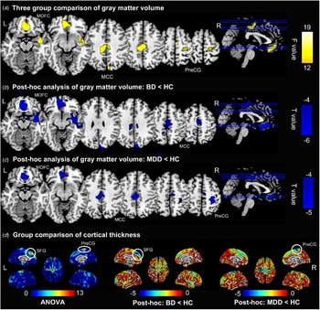
Voxel-based comparison of GMV in the mOFC, middle cingulum, and right precentral gyrus using age, sex, education years, and intracranial volume as covariates indicated significant differences among the three groups (Table 2 and Fig. 1a). Post-hoc analysis identified brain volume deficits in the mOFC and middle cingulum common to both patient groups (Table 2 and Figs 1b–c), and MDD patients additionally showed lower GMV in the left precentral gyrus than HCs (Table 2 and Fig. 1c).
GMV and CT differences between participants with BD and MDD, compared to HCs. (a). Three group comparisons: The GMV of the middle cingulum, mOFC, right superior temporal gyrus, right pre/post central gyrus and right occipital gyrus differed significantly among the three groups (color in yellow); (b). Compared with controls, BD patients showed decreased GMV in middle cingulum, right superior temporal gyrus, medial frontal orbital cortex and right superior occipital cortex (color in blue); (c). Compared with controls, the MDD patients showed decreased GMV in right pre/postcentral gyrus, middle cingulum and mOFC (color in blue); (d). The figure in the left showed three group comparison results of the CT, including bilateral precentral gyrus and left SFG (color in red); the figure in the middle showed that CT in the left SFG was decreased in bipolar patients group compared with controls (cold color); the figure in the right showed that compared with controls, MDD patients showed decreased CT in bilateral precentral gyrus (cold color). (All the results were co-variated out for age, gender and education years, and the TIV was additionally co-variated out for GMV analysis, voxel threshold significance was set at p < 0.0005, cluster level was set at p < 0.05, family wise error corrected). BD, bipolar disorder; MDD, major depressive disorder; HC, healthy control.

Significant difference of GMV and CT between BD, MDD, and HC groups

1. BD, bipolar disorder; MDD, major depressive disorder; HC, healthy controls; BA, Brodmann area; MNI, Montreal Neurological Institute.
2. Age, sex and education years were co-variated out in GMV and CT analysis. The intracranial volume was additionally controlled in GMV analysis. Voxel-wise threshold was set at p < 0.0005. Significance level was set at p < 0.05, cluster wise family wise error corrected. *p < 0.05, **p < 0.01, ***p < 0.001. Bold represents statistically significant results.
Vertex-based comparisons of CT in the left precentral gyrus and left SFG revealed significant differences among the three groups after controlling for age, sex, and education level (Table 2 and Fig. 1d). CT of the left precentral gyrus was significantly smaller in the MDD group than in HCs, while CT in the left SFG was significantly smaller in the BD group than in HCs (Table 2 and Fig. 1d). However, no significant differences were observed in GMV or CT between BD and MDD patients after controlling for individual variations in intracranial volume (for GMV), age, sex, and education level.
Correlation of plasma levels of complement components with whole-brain GMV or CT
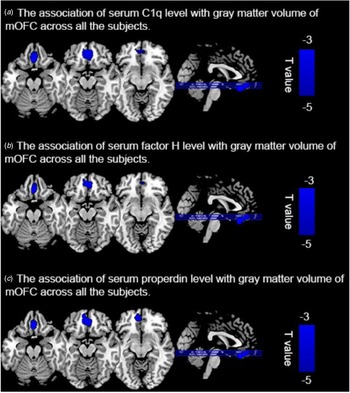
Whole-brain correlation analysis across all subjects after controlling for age, sex, education level, BMI, and TIV revealed a negative association between GMV in the mOFC and log10-transformed levels of C1q, factor H, and properdin (Table 3 and Fig. 2). However, whole-brain correlation analysis did not reveal significant correlations of CT with plasma levels of complement components, whether among all subjects or between different groups. Nevertheless, CT of several brain areas correlated significantly with several complement factors in SPSS (online Supplementary Table S2).
(a) Figure shows significant negative correlation between log10-transformed C1q and volume of medial orbital prefrontal cortex (cold color) across all the subjects. (b) Figure shows significant negative correlation of the log10-transformed factor H and GMV in medial orbital prefrontal cortex (cold color) across all the subjects. (c). Figure shows significant negative correlation of the log10-transformed properdin and GMV in medial orbital prefrontal cortex (cold color) across all the subjects. For the correlation analysis, age, gender, education years, TIV and body mass index were set as the covariates. Significance was set at a threshold with a peak-level uncorrected p < 0.0005, with a family-wise error rate (FWE)-corrected cluster level of p < 0.05). MOFC: medial orbital frontal cortex.

Whole brain-based negative correlation analysis of GMV with complement component factors

1. Age, sex, education years and body mass index were controlled. BA, Brodmann area; MNI, Montreal Neurological Institute.
2. Voxel-wise threshold was set at p < 0.0005. Significance level was set at p < 0.05, cluster wise family wise error corrected. *p < 0.05, **p < 0.01, ***p < 0.001. Bold represents statistically significant results. Bold represents statistically significant results.
Discussion
Consistent with previous findings, we found that complement component levels were significantly higher in both patient groups than in HCs, suggesting that both BD and MDD are associated with increased levels of inflammatory biomarkers (Reginia et al., Reference Reginia, Kucharska-Mazur, Jabłoński, Budkowska, Dołȩgowska, Sagan and Samochowiec2018; Wei et al., Reference Wei, Liu, Zhao, Yang, Ni, Wang and Ma2018). The log10-transformed levels of C3, C4, and factor H were significantly higher in BD than MDD patients, even after controlling for age, sex, years of education, and BMI. These results suggest that patients with BD might involve more severe inflammatory response than MDD. Group comparison of brain structures identified structural alterations that were shared and different between BD and MDD patients. Compared to HCs, both patient groups showed reduced GMV in the middle cingulum and mOFC. However, MDD patients showed lower GMV in the right precentral gyrus and smaller CT in the left precentral gyrus than HCs, while the BD group showed smaller CT in the left SFG than HCs. Finally, we found that plasma levels of C1q, factor H, and properdin were negatively associated with GMV in the bilateral mOFC across all subjects, which further supports the idea that BD and MDD share elements of their etiology (Lanz et al., Reference Lanz, Reinhart, Sheehan, Rizzo, Bove, James and Kleiman2019). To the best of our knowledge, this is the first HC-controlled study examining relationships between brain structure and complement components in mood disorders.
Our study associated BD and MDD with increased levels of complement components involved in the classical pathway (C1q and C4) and alternative pathway (factor B, factor H and properdin). C1q initiates the classical pathway upon antigen-antibody binding (Akcan, Karabulut, İsmail Küçükali, Çakır, & Tüzün, Reference Akcan, Karabulut, İsmail Küçükali, Çakır and Tüzün2018). Patients with chronic BD show significant elevation of C1q mRNA expression in peripheral blood (Akcan et al., Reference Akcan, Karabulut, İsmail Küçükali, Çakır and Tüzün2018). C3, C4 and factor B, whose levels in BD are much better studied than levels of C1q, are elevated in the peripheral blood of patients with manic or psychotic BD (Fontana et al., Reference Fontana, Storck, Angst, Dubs, Abegg and Grob1980; Wadee et al., Reference Wadee, Kuschke, Wood, Berk, Ichim and Maes2002). Patients with chronic BD show elevated levels of mRNAs encoding C4 and factor B in peripheral blood mononuclear cells (Akcan et al., Reference Akcan, Karabulut, İsmail Küçükali, Çakır and Tüzün2018). Our observation of increased C1q and factor H in MDD patients is consistent with previous work (Shin et al., Reference Shin, Ham, Ko, Pae, Park, Steffens and Han2019; Yao & Li, Reference Yao and Li2020). Other studies have shown that depressed subjects have significantly higher plasma levels of C4 than normal controls (Kronfol & House, Reference Kronfol and House1989; Maes et al., Reference Maes, Delange, Ranjan, Meltzer, Desnyder, Cooremans and Scharpé1997). Consistent with our observation that properdin, a key regulator of the alternative complement pathway (Chen, Cortes, & Ferreira, Reference Chen, Cortes and Ferreira2018a), is increased in patients with BD or MDD, previous studies have reported dysregulation of the alternative pathway in neurological and neuropsychiatric disorders such as schizophrenia and epilepsy (Kopczynska et al., Reference Kopczynska, Zelek, Vespa, Touchard, Wardle, Loveless and Morgan2018; Zhang, Lv, Fan, Tang, & Yi, Reference Zhang, Lv, Fan, Tang and Yi2017a). Taken together, our results are consistent with the idea that inflammatory markers are associated with mood disorders such as BD and MDD, without being specific to a particular disorder (Swardfager, Rosenblat, Benlamri, & McIntyre, Reference Swardfager, Rosenblat, Benlamri and McIntyre2016).
We found that plasma levels of C3 were significantly higher in BD patients, but not MDD patients, than in HCs. Our results are consistent with other studies in which no difference in C3 was found between MDD and controls (Berk, Wadee, Kuschke, & O'Neill-Kerr, Reference Berk, Wadee, Kuschke and O'Neill-Kerr1997). In fact, we found that BD patients had higher levels of C3, C4, and factor H than MDD patients. While we are unaware of previous studies directly comparing levels of these complement components between the two patient groups, our results are consistent with a previous report that BD patients have higher levels of pro-inflammatory cytokines than MDD patients (Bai et al., Reference Bai, Chen, Hsu, Huang, Tu, Chang and Tsai2020). Nevertheless, our results should be verified in future comparisons of complement components between the two disorders. If verified, our findings suggest that BD involves more severe inflammatory response than MDD.
Our findings of shared structural alterations in the brains of BD and MDD patients are consistent with the results of a recent meta-analysis, where the volume of the ventromedial prefrontal cortex was found to be lower in both patient groups, suggesting a volume deficit common to the two disorders (Wise et al., Reference Wise, Radua, Via, Cardoner, Abe, Adams and Arnone2017). A previous study also reported that middle cingulum volume was significantly smaller in MDD patients who did not respond to treatment than in HCs or in patients who did respond to treatment (Batail et al., Reference Batail, Coloigner, Soulas, Robert, Barillot and Drapier2020). Although no such reduction has been reported for BD patients, our results suggest that altered middle cingulate gyrus volume may underlie brain pathology in both disorders.
We identified structural impairments that may be specific to BD or MDD. Our MDD patients had significantly smaller GMV in the precentral and postcentral gyri, as well as smaller CT in the left precentral gyrus than HCs. As both the precentral and postcentral gyri are key parts of the sensorimotor network (Chenji et al., Reference Chenji, Jha, Lee, Brown, Seres, Mah and Kalra2016), our results confirm the potential role of the precentral region in MDD pathogenesis. Our BD patients had significantly smaller CT in the left SFG than HCs did. The SFG is the gyral-based representative of the dorsolateral prefrontal cortex (Niu et al., Reference Niu, Wang, Jia, Wang, Zhong, Lin and Huang2017) and is involved in self-awareness, complex cognitive behavior, executive function and emotional regulation (Yang et al., Reference Yang, Liu, Jiang, Yu, Ding, Lu and Lv2019). Our results are consistent with a previous whole-brain meta-analysis showing widespread cortical thinning in the SFG of BD patients, suggesting that structural defects in the SFG may reflect the main emotional regulatory and processing symptoms of BD (Hanford, Nazarov, Hall, & Sassi, Reference Hanford, Nazarov, Hall and Sassi2016).
In both patient groups, we found that structural defects of mOFC were associated with elevated levels of C1q, factor H, and properdin, which may provide insight into the shared neuro-inflammatory pathogenesis of mood disorders (Lanz et al., Reference Lanz, Reinhart, Sheehan, Rizzo, Bove, James and Kleiman2019). The mOFC is part of the medial prefrontal cortex (mPFC), which is a stress-sensitive brain area (McEwen & Morrison, Reference McEwen and Morrison2013) that is involved in the regulation of emotions, cognition, and innate immunological responses to stress (Belleau, Treadway, & Pizzagalli, Reference Belleau, Treadway and Pizzagalli2019; Savitz, Price, & Drevets, Reference Savitz, Price and Drevets2014). Human and rodent studies have shown that in mood disorders, repeated environmental stress can activate innate immune receptors, such as Toll-like receptors, through exogenous or endogenous ligands, thereby triggering inflammation and attenuating neuronal response, while inducing dendritic atrophy and activating microglia in the mPFC (Nie et al., Reference Nie, Kitaoka, Tanaka, Segi-Nishida, Imoto, Ogawa and Furuyashiki2018). More severe mPFC thinning in mood disorders has been associated with higher levels of peripheral pro-inflammatory cytokines, greater expression of inflammation-related genes, and reduced expression of neuroprotective genes (Belleau et al., Reference Belleau, Treadway and Pizzagalli2019). Given that C1q initiates the classical complement pathway, while factor H and properdin are key regulators of the alternative pathway (Chen et al., Reference Chen, Cortes and Ferreira2018a; Noris & Remuzzi, Reference Noris and Remuzzi2013), the present study suggests that both classical and alternative complement pathways contribute to mOFC structural defects in BD and MDD.
Limitations
Our study has some limitations, including its cross-sectional design. The single assessment of peripheral blood samples could not provide evidence of causality, leaving open the question of whether the observed alterations in complement levels and brain structure are a cause or consequence of BD and MDD. The second limitation is the unbalanced sample size in our study, which may decrease statistical power. Third, we did not correct for the seven complement components in the partial correlation analysis that we conducted outside SPM.
Fourth, the association between elevated complement components and mOFC structural changes needs to be carefully interpreted. In the present study, patients were excluded if they had severe physical illnesses such as systemic lupus erythematosus, and blood samples were collected from all subjects at the same time to avoid potential confounding due to variation in inflammation state. However, other factors affecting complement levels were not taken into account, such as concurrent immunological conditions (infections, atopy, autoimmune disorders) or sleep level (Nilsson & Ekdahl, Reference Nilsson and Ekdahl2012). Future studies should address potential confounding from pre-existing inflammatory conditions.
Fifth, our BD patients did not show severe symptoms, despite showing increased levels of inflammatory markers. This may limit the generalizability of our findings to different BD subpopulations.
Sixth, all our BD patients were receiving medication at the time of the study. Traditional mood stabilizers such as lithium and valproate can substantially reduce neuroinflammation (Muneer, Reference Muneer2016), which may have affected our results. However, we believe that medication was not a strong confounder because we did not observe significant differences in complement factors between patients treated with drugs or not. In addition, BD patients showed high levels of inflammatory markers despite receiving treatment. Nevertheless, the potential influence of medication history should be rigorously assessed in future studies of complement components in mood disorders.
Finally, the patients in our study with BD had disease considerably longer than patients with depression. Nevertheless, we did not find any significant correlation between illness duration and levels of complement components. Future studies may wish to enroll only first-episode patients in order to exclude potential influence from illness duration on inflammatory response.
Conclusion and outlook
To the best of our knowledge, our study is the first to analyze the association between brain structures and complement cascades in BD patients, MDD patients, and HCs. Our results identify alterations in complement proteins and brain structures that are shared and unique between BD and MDD. Our findings underscore the need for combining dimensional and categorical approaches when describing mood disorders. Our work also reveals aspects of neuro-inflammatory pathogenesis that may be shared among mood disorders, which may help develop novel therapeutic strategies (Felger, Reference Felger2017).
Supplementary material
The supplementary material for this article can be found at https://doi.org/10.1017/S0033291722003282
Acknowledgments
The authors would like to thank all their coworkers at the Affiliated Mental Health Centre & Hangzhou Seventh People's Hospital, West China Hospital, the State Key Laboratory of Biotherapy in West China Hospital and Suzhou Psychiatry Hospital for their contributions to this research study.
Author contributions
H.Y., P.N., J.C., and T.L. developed the study, had full access to all data, and take responsibility for data integrity and accuracy. H.Y., P.N., Q.W., and W.G. drafted the manuscript. W.W., J.W., X.D., W.D., and X.M. collected the data, and Y.T., L.Z., M.L., and X.L. performed all data analyses. All authors agree to be accountable for all aspects of the work, ensuring that questions related to the accuracy or integrity of the data and results are appropriately investigated and resolved. All authors critically revised and approved the final version of the manuscript.
Financial support
This work was supported by the National Natural Science Foundation of China Key Project (81630030 and 81920108018 to T.L.), National Natural Science Foundation of China (81871054 and 81501159 to P.N., 82101598 to H.Y.), Special Foundation for Brain Research from Science and Technology Program of Guangdong (2018B030334001), 2021 Project for Hangzhou Medical Disciplines of Excellence & Key Project for Hangzhou Medical Disciplines, 1.3.5 Project for Disciplines of Excellence at West China Hospital of Sichuan University (ZY2016103, ZY2016203, and ZYGD20004 to T.L.), and Introductory Project of the Suzhou Clinical Expert Team (SZYJTD201715 to X.D. and T.L.).
Conflict of interest
The authors declare no conflicts of interest.
Ethical standards
The study was approved by the Ethics Committee of West China Hospital, Sichuan University, and complied with the principles of the Declaration of Helsinki. All participants received a complete description of the study and provided written informed consent. A copy of the written consent is available for review by the Editor of this journal.
Consent for publication
Not applicable.
Availability of data and materials
All data used in the current study are available from the corresponding author on reasonable request.