Introduction
In recent years, the impact of parasitic diseases in commercial poultry has decreased due to poultry industry modernization and biosecurity improvement (Puttalakshmamma et al., Reference Puttalakshmamma, Ananda, Prathiush, Mamatha and Suguna2008). Ascaridia galli (Nematoda: Ascaroidea) is the most common and important helminthic parasite found in poultry. Females lay smooth and thick-walled eggs, which are eliminated in faeces and subsequently survive for long periods in the environment. Poultry become infected by ingesting embryonated eggs from a contaminated environment (Tonner-Klank et al., Reference Tonner-Klank, Moller, Forslund and Dalsgaard2007). Ascaridia galli infections cause direct losses in poultry, especially growth-rate reductions, leading to weight loss, reducing feed conversion rate and causing damage to intestinal mucosa (Ramadan & Znada, Reference Ramadan and Znada1991). For the control of this and other poultry nematodes, anthelmintics are usually used, such as hygromycin B, piperazine, thiabendazole, fenbendazole and levamisole (Reid & McDougald, Reference Reid, McDougald, Calnek, Barnes, Beard, McDougald and Saif1997; Ruff, Reference Ruff1999). However, the literature reports that there are some disadvantages in the utilization of chemical compounds, such as resistance development, relatively high costs, potential ecological impacts and residual effects of chemical compounds (Craig, Reference Craig1993; Waller, Reference Waller1994; Sangster, Reference Sangster1999; Jackson & Miller, Reference Jackson and Miller2006). In this context, biological control with nematophagous fungi is suggested, which could help combat environmental decontamination by helminth eggs and infective larvae (Larsen, Reference Larsen1999; Araújo et al., Reference Araújo, Braga, Araujo, Silva and Tavela2008).
Ovicidal nematophagous fungi secrete extracellular enzymes that play an important role, contributing greatly to the reduction of infection and to the destruction of helminth eggs (type 3 effect) present in faeces and the environment (Segers et al., Reference Segers, Butt, Kerry, Beckett and Peberdy1996; Braga et al., Reference Braga, Carvalho, Silva, Araujo, Araújo, Freitas, Geniêr and Queiroz2010b). Crude extracts of Pochonia chlamydosporia fungus have been shown to have ovicidal enzymic activity against cyathostomin eggs in horses and against Ancylostoma sp. in dogs (Braga et al., Reference Braga, Araújo, Carvalho, Silva, Araujo, Freitas, Geniêr, Ferreira and Queiroz2010a, Reference Braga, Carvalho, Silva, Araujo, Araújo, Freitas, Geniêr and Queirozb). However, there is no report demonstrating a type 3 effect or crude extract activity against gastrointestinal nematode eggs in poultry. This information in future could help to decrease the recurrence of infection in these animals by decreasing gastrointestinal parasite eggs in the environment.
The present study aimed to evaluate the activity of P. chlamydosporia (VC1 and VC4) fungus in solid medium and its crude extract against eggs of A. galli.
Materials and methods
Fungal isolates
Two isolates of P. chlamydosporia (VC1 and VC4) fungus from the mycology collection of ‘Laboratório de Parasitologia do Departamento de Veterinária da Universidade Federal de Viçosa, Minas Gerais, Brasil’ (Parasitology Laboratory of Veterinary Department, Viçosa Federal University, Minas Gerais State, Brazil) were maintained in assay tubes at 4°C containing 2% corn-meal-agar (2% CMA) in a dark environment for 10 days. Culture plates of 4 mm in diameter were extracted from fungal cultures and maintained in test tubes containing 2% CMA and transferred to Petri dishes of 9.0 cm in diameter containing 20 ml of 2% potato dextrose agar, maintained at 26°C in a dark environment for 10 days. After growth of the isolates, new culture plates of 4 mm diameter were transferred to Petri dishes of 9.0 cm in diameter containing 20 ml of 2% water-agar (2% WA) and kept at 26°C in a dark environment for 10 days.
Production of crude extracts from fungal isolates
Two isolates of P. chlamydosporia (VC1 and VC4) fungus were cultured in Erlenmeyer flasks (250 ml) containing 50 ml of liquid minimal medium (0.3 g/l NaCl, 0.3 g/l MgSO4.7H2O, 0.3 g/l K2HPO4 and 0.2 g/l yeast extract; Merck, Germany) and supplemented with 0.2% gelatin. Gelatin was filtered through Millipore filters (45 μm pore size) and aseptically added to the autoclaved medium. Samples containing the isolate were incubated in a shaker at 120 rpm at 28°C in a dark environment. After 5 days, the supernatant fraction was collected and filtered using Whatman filter paper no. 1 at 4°C, according to Esteves et al. (Reference Esteves, Peteira, Atkins, Magan and Kerry2009).
Collection of Ascaridia eggs
Eggs of A. galli were recovered from adult worms following the biopsy of poultry. Egg viability was analysed under a light microscope using a 10 × objective lens according to Urquhart et al. (Reference Urquhart, Armour, Duncan, Dunn and Jennings1998). After that, all eggs were washed ten times in distilled water and, at each time, centrifuged at 1000 rpm for 5 min. The supernatant fraction was discarded after each centrifugation. Eggs were incubated at 25°C for 14 days with a solution containing 0.005% streptomycin sulphate and 0.1% chloramphenicol, as described by Araújo et al. (Reference Araújo, Santos and Ferraz1995).
Experimental assays
Two in vitro assays, A and B, were performed in distinct steps during an interval of 5 days. In assay A, the ovicidal activity of P. chlamydosporia (VC1 or VC4) against A. galli eggs on solid medium in Petri dishes was evaluated. In assay B, the action of crude extracts obtained from VC1 or VC4 against A. galli eggs was evaluated after interaction for 24 h. The Petri dishes of assays A and B were sealed with Rolapack film and put in a BOD incubator at 26°C in a dark environment.
Assay A
One thousand A. galli eggs were placed on to Petri dishes of 9.0 cm in diameter containing 2% WA with one grown fungal isolate (VC1 or VC4) for 10 days, constituting the treated group. The control group consisted of 1000 eggs without fungi. Six repetitions were made to both the treated and control group. At 1, 3 and 5 days post-inoculation, 100 eggs were removed from each plate containing fungal isolates and from the control (without fungi) as described by Araújo et al. (Reference Araújo, Santos and Ferraz1995). These eggs were placed on glass slides with a drop of 1% blue Amam and evaluated under light microscopy (40 × objective lens) according to Lysek et al. (Reference Lysek, Fassatiová, Pineda and Hernández1982), being: type 1 effect, physiological and biochemical effect with no morphological damage to eggshell and with hyphal adherence; type 2 effect, lytic effect with morphological changes in eggshells and embryos without hyphal penetration; and type 3 effect, lytic effect with morphological change in eggshells and embryos with hyphal penetration and internal egg colonization.
Assay B
Five hundred eggs of A. galli were placed on to Petri dishes of 4.5 cm in diameter with 5 ml of P. chlamydosporia (VC1 or VC4) crude extract, being the treated group. The control group consisted of 500 eggs in 10 ml of distilled water in each plate without fungal extract. Petri dishes of the control group and of the treated group containing VC1 or VC4 crude extract were sealed with Rolapack film and put in a BOD incubator at 26°C in a dark environment for 24 h. After this period, the eggs were counted from each Petri dish of the treated and control groups, according to the modified methodology of Mukhtar & Pervaz (Reference Mukhtar and Pervaz2003) and Braga et al. (Reference Braga, Araújo, Carvalho, Silva, Araujo, Freitas, Geniêr, Ferreira and Queiroz2010a).
Data analysis
Data from each interval (assay A) were analysed using Friedman's non-parametric test at the 1% probability level (Ayres et al., Reference Ayres, Ayres, Ayres and Santos2003). In assay B, the number of observed eggs in Petri dishes from the treated group compared with the control group was assessed by Tukey's test at 1% probability. BioStat 3.0 software (Ayres et al., Reference Ayres, Ayres, Ayres and Santos2003) was used for data analysis. Subsequently, the percentage reduction in eggs was calculated according to the following equation:

Results and discussion
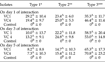
Pochonia chlamydosporia isolates VC1 and VC4 were capable of destroying A. galli eggs during experimental assay A at the interaction intervals of 1, 3 and 5 days. All tested fungal isolates induced percentage reductions in egg number (type 1, 2 and 3 effects) by 3 days post-inoculation. In the control group, nematophagous fungi were not present during the experiment (table 1).
Percentages and standard deviations of the ovicidal activity (type 1, 2 and 3 effects) of Pochonia chlamydosporia (VC1 and VC4) fungus and the control group without fungi against eggs of Ascaridia galli at 1, 3 and 5 days of interaction.
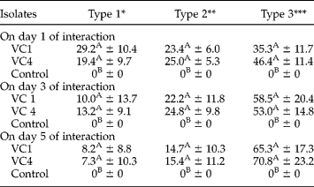
Percentages followed by same letter in the same column are not significantly different (P>0.01) by Friedman test. *Physiological, biochemical effect without morphological damage to eggshell, with hyphae adhered to the shell. **Lytic effect with morphological alteration of embryo and eggshell, without hyphal penetration through the eggshell. ***Lytic effect with morphological alteration of embryo and eggshell, besides hyphal penetration and internal colonization.
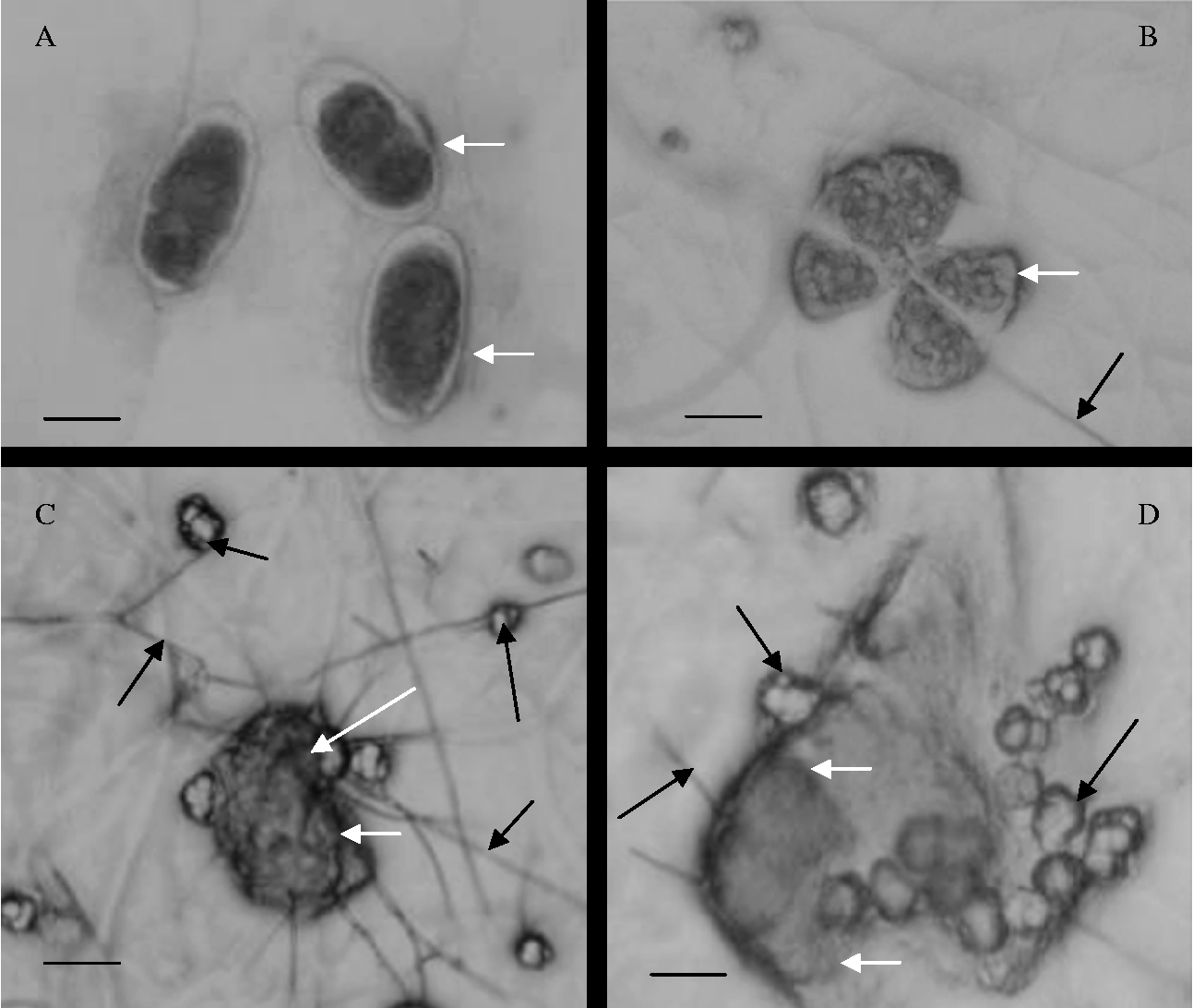
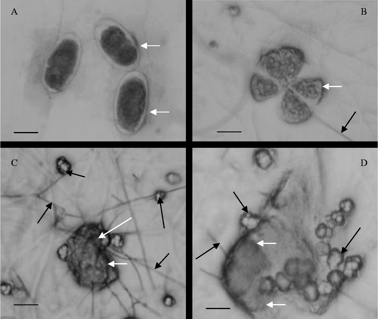
Using 40 × objective light microscopy, A. galli eggs were observed in Petri dishes of the control group (without fungus) (fig. 1A). In fig. 1B–D, vegetative structures (hyphae) of isolates (VC1 and VC4) were observed colonizing A. galli eggs and subsequently causing their rupture, characterizing a type 3 effect. Furthermore, we observed the production of P. chlamydosporia chlamydospores in Petri dishes of the fungal isolates (fig. 1B–D). In the present study, isolate VC1 demonstrated ovicidal activity (egg destruction), with percentage reductions of 35.3, 58.5 and 65.3% on the first, third and fifth days of interaction, respectively. Likewise, isolate VC4 induced percentage reductions of 46.4%, 53.0% and 70.8% for a type 3 effect for the same periods studied. Moreover, comparing the ovicidal activity of VC1 to VC4 against A. galli eggs in Petri dishes during this assay, there was no difference (P>0.01) among the percentage reductions for a type 3 effect. On the other hand, the same happened to type 1 and 2 effects, since these isolates showed no difference (P>0.01) in their action.
(A) Eggs of Ascaridia galli in Petri dishes of the control group (white arrows). (B–D) Destroyed A. galli egg (white arrows) after interaction with the hyphae and chlamydospores of Pochonia chlamydosporia fungus (black arrows) at 1, 3 and 5 days, respectively. Scale bars: 36 μm (A), 40 μm (B, C) and 25 μm (D).
In assay B after 24 h of interaction, crude extracts of isolates VC1 and VC4 induced reductions of 64.1% and 56.5%, respectively, in the number of A. galli eggs compared with controls.
Some control alternatives against A. galli using plants have been tested. In a recent study, Brito et al. (Reference Brito, Fernandes, Fernandes, Ferreira, Rolim and Filho2009) concluded that aqueous and ethanolic extracts of Morinda citrifolia fruit showed satisfactory in vitro anthelmintic activity against A. galli. However, the authors emphasize the need for more studies with higher concentrations of this fruit, since the results showed a low efficiency of action of the fruit extract (aqueous and ethanolic) against A. galli. Moreover, the present study also showed that at the end of 24 h, crude extracts (VC1 and VC4) of P. chlamydosporia reduced the number of A. galli eggs by 64.1% and 56.5%, respectively. In addition, isolates VC1 and VC4 were effective at destroying eggs in 2% WA medium in the studied intervals (1, 3 and 5 days of interaction). Thus, these results suggest that P. chlamydosporia may be used successfully for the biological control of gastrointestinal nematodiosis, including the control of A. galli, since these and the eggs of other nematode parasites are present in the environment (Tonner-Klank et al., Reference Tonner-Klank, Moller, Forslund and Dalsgaard2007).
In relation to their commercial applicability, even for environmental biological control, García et al. (Reference García, Bulnes, Melchor, Veja, Miranda, Montes, Hidalgo and Marrero2004) and Khan et al. (Reference Khan, Williams and Nevalainen2004) state that P. chlamydosporia fungus is harmless to animals, humans and the environment. It also produces chlamydospores (resistant structures) that enable passage through the gastrointestinal tract of animals. In a recent study, Braga et al. (Reference Braga, Araújo, Carvalho, Silva, Araujo, Freitas, Geniêr, Ferreira and Queiroz2010a) reported the passage of P. chlamydosporia through the gastrointestinal tract of horses to show its viability in colonizing faeces and destroying eggs of Oxyuris equi. In this assay, P. chlamydosporia (VC1 and VC4) chlamydospore production was observed in Petri dishes of the treated group, confirming previously cited studies.
During the experimental assays, the ovicidal activity (assay A) and the activity of P. chlamydosporia crude extract (assay B) were demonstrated by a reduction in A. galli eggs. Results from these assays using P. chlamydosporia fungus are consistent with the results of Braga et al. (Reference Braga, Araújo, Carvalho, Silva, Araujo, Freitas, Geniêr, Ferreira and Queiroz2010a), who observed its action in destroying cyathostomin eggs from horses, and with those of Esteves et al. (Reference Esteves, Peteira, Atkins, Magan and Kerry2009) who, working with the same fungus grown in liquid medium supplemented with 0.2% gelatin, demonstrated its action in destroying eggs of Meloidogyne spp., a plant parasitic nematode.
In another study, Braga et al. (Reference Braga, Araújo, Campos, Carvalho, Silva, Tavela and Maciel2007) demonstrated the efficacy of P. chlamydosporia isolates VC1 and VC4 against eggs of Ascaris lumbricoides, a common gastrointestinal nematode of humans, at intervals of 7, 10 and 14 days. At the end of the experiment, these authors reported results above 26% for ovicidal activity (type 3 effect) for both tested isolates. When this effect was compared with the reported results of the present study at the end of 5 days, it was noted that ovicidal activity was higher against A. galli eggs, the percentage reductions of which were 65.3% and 70.8% for VC1 and VC4, respectively. Moreover, Khan et al. (Reference Khan, Williams and Nevalainen2004) mentioned that nematophagous fungi produce enzymes that disintegrate different egg layers; this could be the cause of different percentage reductions for P. chlamydosporia ovicidal activity against different genera of helminth eggs.
After incubation for 24 h, it was observed that A. galli eggs were destroyed, with 64.1% and 56.5% reductions for VC1 and VC4, respectively. These results are consistent with the study of Mukhtar & Pervaz (Reference Mukhtar and Pervaz2003), suggesting that this occurrence can be attributed to different culture conditions. These authors reported that the action of a fungal filtrate of P. chlamydosporia against eggs of Meloidogyne javanica, a nematode parasite of plants, caused a reduction of 94.0% in egg eclosion after the same interval studied (24 h). In the present study, we used different P. chlamydosporia concentrations to estimate fungal filtrate toxicity. However, there may be differences in enzyme production from several P. chlamydosporia isolates (Esteves et al., Reference Esteves, Peteira, Atkins, Magan and Kerry2009). Moreover, these authors report that culture conditions seem to have an important role; therefore conditions should always be standardized so that significant comparisons can be made. According to Cayrol et al. (Reference Cayrol, Djian and Pijarowski1989), culture conditions can affect the toxic properties of fungi. Webb et al. (Reference Webb, Ghafoor and Heale1972) and Segers et al. (Reference Segers, Butt, Kerry and Peberdy1994) cited by Mukhtar & Pervaz (Reference Mukhtar and Pervaz2003) state that the nematicidal action of P. chlamydosporia filtrate against M. javanica juveniles can be attributed to the production of certain enzymes and toxins.
According to Ruff (Reference Ruff1999), the production of broilers in confinement tends to favour the growth of short-cycle parasites such as Eimeria sp., A. galli, Heterakis gallinarum, Capilaria sp., and their direct transmission. In addition, poultry raised in systems that provide greater soil contact have higher parasitosis rates. So, according to the percentage reductions shown in the present study, it could be suggested that P. chlamydosporia be applied as an alternative for the biological control of eggs eliminated in faeces, intervening directly in this parasite's biological cycle. Furthermore, resistance of A. galli eggs in the environment shows the importance of finding alternatives that can contribute to a reduction in recurrent infections.
This is the first report describing the interaction and the crude extract activity of P. chlamydosporia against eggs of A. galli. Results from the present study showed that P. chlamydosporia (VC1 and VC4) is effective at destroying these eggs and therefore can be used in the biological control of nematodes. However, the authors suggest the characterization of the substances produced by P. chlamydosporia (VC1 and VC4) in future studies.
Acknowledgements
The authors would like to thank the Conselho Nacional de Desenvolvimento Científico e Tecnológico (CNPq), Coordenação de Aperfeiçoamento de Pessoal de Nível Superior (CAPES) and Fundação de Amparo à Pesquisa do Estado de Minas Gerais (FAPEMIG) for the financial support and grant concession. J.V.A. received a scholarship from CNPq.