Introduction
Anterior cricoid split is a procedure performed for grades 2 and 3 subglottic stenosis, which can be carried out endoscopically or as an open procedure. The open procedure was originally described by Cotton and Seid as an alternative to tracheostomy for patients in whom extubation failed because of subglottic stenosis.Reference Cotton and Seid1
In our unit, anterior cricoid split is performed endoscopically as we believe it to be superior to the open method. To facilitate this, a bespoke sickle knife was developed in conjunction with the company Single Use Surgical (Cardiff, Wales, UK).
This paper describes a case series of patients who underwent endoscopic anterior cricoid split and balloon dilatation for extubation failure and mature stenosis. The study aimed to assess the success rate of endoscopic cricoid split in our unit and to review the indications for the procedure.
Materials and methods
Ethical considerations
No ethics approval was sought for this study.
Study design
A retrospective case note review was performed of all paediatric patients who underwent endoscopic cricoid split in a tertiary referral paediatric ENT unit between August 2012 and March 2015. The degree of subglottic stenosis was graded according to the Cotton–Myer grading system.
Surgical procedure
Endoscopic anterior cricoid split is performed with the patient spontaneously ventilating, or apnoeic if this is not possible. A microlaryngoscopy and bronchoscopy is performed to: confirm the diagnosis, grade the stenosis and assess the remainder of the airway.
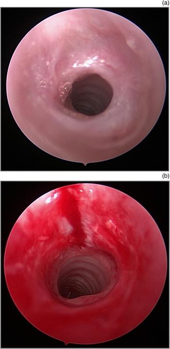
The assistant holds the endoscope whilst the surgeon uses a bespoke sickle knife (Single Use Surgical) (Figure 1) to perform the anterior cricoid split (Figure 2). The operating surgeon places their other hand externally on the anterior neck to stabilise the cricoid and to judge the depth of the split. The sickle knife divides the first two tracheal rings of the cricoid and lower thyroid cartilage. The endoscope allows the surgeon to identify the subglottis, so that the upper limit of the incision is just below the anterior commissure of the vocal folds.
Purpose-built sickle knife.

(a) Endoscopic view of grade 3 subglottic stenosis, and (b) view following endoscopic cricoid split and balloon dilatation.

The subglottis is subsequently dilated with an age-appropriate oesophageal balloon (Boston Scientific, Marlborough, Massachusetts, USA) and dilated for two sets of 90 seconds each. The balloon diameter was chosen equal to the expected cricoid diameter plus 1 mm, using the Great Ormond Street Hospital chart for sizing paediatric airways.Reference Tweedie, Skilbeck, Cochrane, Cooke and Wyatt2
The patient is intubated with an age-appropriate endotracheal tube and ventilated on the intensive care unit for approximately 5 days, after which they are extubated (steroids are administered prior to extubation).
Results
Nine patients underwent an endoscopic cricoid split procedure between August 2012 and March 2015, five for failure to extubate and four for treatment of an established subglottic stenosis (Table 1). Three of the infants were born at term and six were born pre-term, with a mean age at operation of 30 weeks (range, 11–104 weeks). The mean duration of ventilation in the pre-term neonates was 51 days (range, 17–133 days). All of the patients had multiple intubations. Four (67%) of the children born pre-term had chronic lung disease.
Demographics and clinical outcome of patients undergoing endoscopic cricoid split

O2 = oxygen; pre-op = pre-operatively; ETT = endotracheal tube; post-op = post-operatively; CGA = corrected gestational age; CLD = chronic lung disease; CPAP = continuous positive airway pressure; CHARGE = coloboma, heart defects, atresia choanae, growth retardation, genital abnormalities and ear abnormalities syndrome
The mean age of the patients at operation in those born pre-term was 18 weeks (range, 11–30 weeks) or 5 weeks (range, 1–16 weeks) corrected gestational age, whilst the mean age for those born at term was 53 weeks (range, 11–104 weeks). The mean body weight at operation was 2.9 kg (range, 1.8–3.7 kg) in the pre-term neonates and 7.2 kg (range, 3.6–10.3 kg) in the term neonates.
The mean number of days that the patients were intubated post-operation was 5.6 days (range, 4–9 days). All five patients intubated pre-operatively were extubated.
One child had coloboma, heart defects, atresia choanae, growth retardation, genital abnormalities, and ear abnormalities (‘CHARGE’) syndrome. This led to multi-level airway obstruction associated with bilateral choanal atresia, laryngomalacia and subglottic stenosis.
Seven patients required repeat balloon dilatations post-operation: three patients required one dilatation and four patients required two dilatations. Those who underwent cricoid split for mature subglottic stenosis had not undergone previous airway procedures.
The operative time for the endoscopic cricoid split was approximately 30 minutes; this was comparatively shorter than the time for the open approach of approximately 80 minutes.
Mean length of follow up was 10 months (range, 4–22 months).
Complications
There was one failure of the endoscopic cricoid split. The child was a pre-term infant, who had been intubated for several weeks, but had been extubated for several days prior to undergoing the cricoid split procedure. The patient was extubated after the operation, but had to be re-intubated because of stridor and poor pulmonary reserve associated with chronic lung disease. Following a repeat balloon dilatation and failed extubation, the child underwent tracheostomy. Unfortunately, the child died as a result of his chronic lung disease.
There was one direct complication, which happened when the senior surgeon was operating without assistance. An inadvertent vocal fold injury occurred when the cricoid split procedure was performed without a hand stabilising the cricoid externally on the neck. The vocal fold healed well, with no further treatment required. The procedure is now always performed with two operators, one holding the endoscope and one performing the cricoid split bi-manually. There were no cases of post-operative surgical emphysema.
Discussion
Every patient who was intubated pre-operatively was extubated following their endoscopic cricoid split procedure. Only one patient was re-intubated as a result of poor pulmonary reserve secondary to chronic lung disease, and eventually underwent tracheostomy, but this patient had been extubated prior to the endoscopic cricoid split procedure.
It is evident from our series that there are two main indications for an endoscopic cricoid split procedure: extubation failure in the neonate; and established subglottic stenosis, which may occur in the older child.
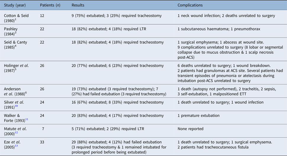
The extubation rate of the endoscopic cricoid split procedure compares favourably with the open method rates of 67–88 per cent quoted in the literature.Reference Cotton and Seid1, Reference Pashley3–Reference Anderson, Tom, Wetmore, Handler and Potsic6, Reference Silver, Myer and Cotton10–Reference Eze, Wyatt and Hartley13 Rotenberg and Berkowitz reported decreased extubation rates with an open cricoid split procedure from 71 per cent in the period 1989–2006 to 41 per cent between 1996 and 2005, which they attributed to an increase in the duration of pre-operative intubation in increasingly premature neonates with a multitude of co-morbidities.Reference Rotenberg and Berkowitz7 They stated that tracheostomy may be a more suitable primary treatment for these neonates and that parents should be counselled accordingly.
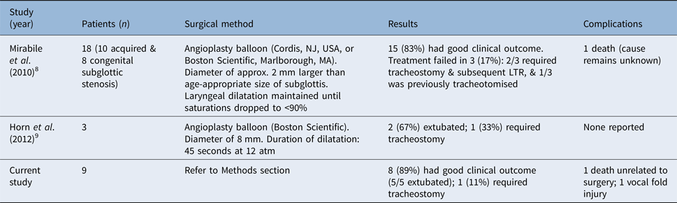
Summaries of the published studies on open and endoscopic anterior cricoid split procedures are provided in Tables 2 and 3.Reference Cotton and Seid1, Reference Pashley3–Reference Anderson, Tom, Wetmore, Handler and Potsic6, Reference Mirabile, Serio, Baggi and Couloigner8–Reference Eze, Wyatt and Hartley13
Summary of published studies of open anterior cricoid split

LTR = laryngotracheal reconstruction; ACS = anterior cricoid split; ETT = endotracheal tube
Summary of published studies of endoscopic anterior cricoid split

LTR = laryngotracheal reconstruction
Seven patients required repeat balloon dilatations post-operatively, ranging from one to two procedures. Our rate of repeat balloon dilatations is lower than the rates quoted by Mirabile et al.Reference Mirabile, Serio, Baggi and Couloigner8 In their series of 18 patients, they reported that 83 per cent required dilatations, ranging from 1 to 7 procedures.
In 2010, Mirabile et al. were the first to describe the combination of endoscopic cricoid split with balloon dilatation as a treatment for mature subglottic stenosis.Reference Mirabile, Serio, Baggi and Couloigner8 In their series, six of eight patients with congenital or acquired stenosis avoided tracheostomy. This is in agreement with our study, as we demonstrated successful outcomes in children with mature stenosis, and all of the children avoided tracheostomy. Our results are better than those of Horn et al., who reported that two of three children avoided tracheostomy.Reference Horn, Maguire, Simons and Mehta9
• The extubation rates for endoscopic cricoid split are at least as good as those for the open procedure
• Endoscopic cricoid split is safe and efficient for managing subglottic stenosis, whether for extubation failure or mature stenosis
• Shorter operative time and scar avoidance make the endoscopic method an attractive alternative to open method for extubation failure in a neonate
• The endoscopic method may also reduce the need for tracheostomy in early subglottic stenosis
We have demonstrated that endoscopic cricoid split is a safe and efficient procedure for the treatment of extubation failure and mature subglottic stenosis. The operative time was shorter when using the endoscope compared to the open procedure; in addition, an external scar and drain could be avoided.
Competing interests
None declared