Transposition of the great arteries combined with totally anomalous pulmonary venous connection is extremely rare outside of heterotaxy syndrome. Since the first description in 1964,Reference Whitaker, Watson and Keates 1 few cases have been reported,Reference Sapsford, Aberdeen and Watson 2 – Reference Mykychak, Fedevych, Maksymenko and Yemets 11 most have been treated by an atrial switch operation. We report the successful treatment of a neonate with transposition of the great arteries, intact ventricular septum and obstructed sub-diaphragmatic totally anomalous pulmonary venous connection, repaired by arterial switch operation and connection of the pulmonary venous return to the left atrium.
Case presentation
A female neonate was delivered at term via urgent caesarean section due to reduced foetal movements and podalic presentation. The birth weight was 2850 g and the Apgar scores were 8 and 9. Central cyanosis was noted within a few hours that was unresponsive to oxygen administration but partially resolved with prostaglandin infusion.
The infant was irritable and tachypneic but not dysmorphic. The heart rate was 160 beats per minute, oxygen saturation on room air was 85% and blood pressure was 80/50 mmHg and equal in all extremities. S2 was loud, and a soft blowing ejection murmur was audible at the upper left sternal border. The exam was otherwise normal.
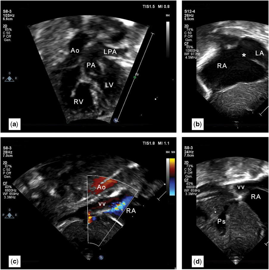
The electrocardiogram (EKG) demonstrated sinus rhythm with right ventricular hypertrophy. The initial echocardiographic diagnosis was levocardia, transposition of the great arteries {S,D,D} (D-transposition of the great arteries) (Fig 1a), large secundum atrial septal defect with right-to-left shunt and a patent arterial duct (Fig 1b). The right heart chambers appeared dilated.
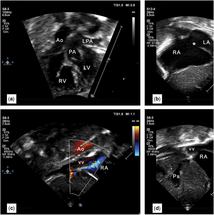
Echocardiographic 2D images. ( a ) In this subxiphoid view, D-transposition of the great arteries with intact ventricular septum is clearly demonstrated. ( b ) There is a large secundum atrial septal defect (asterisk) with right-to-left shunt. RA is enlarged. ( c ) Subxiphoid short-axis imaging with colour Doppler mapping shows a large vertical vein coursing inferiorly from behind the heart through the diaphragm; this is anterior to the aorta (Ao). Flow is towards the probe (red) in both vessels. ( d ) Colour aliasing indicates an obstruction of the vertical vein at the site of entry into the PS, which is dilated. Ao = aorta; LPA = left pulmonary artery; LV = left ventricle; PA = pulmonary artery; PS = portal sinus; RA = right atrium; RV = right ventricle; VV = vertical vein.
Given the unrestrictive atrial communication, balloon atrial septostomy was considered unnecessary. The patient was scheduled for an arterial switch operation.
Prompted by the right-to-left atrial shunt, a repeat echocardiogram documented the absence of pulmonary venous return in the left atrium. A large vascular channel was interposed between the descending aorta and inferior caval vein, with phasic venous flow directed towards the abdomen consistent with sub-diaphragmatic totally anomalous pulmonary venous connection (Fig 1c). The venous channel showed a connection to the portal sinus (Fig 1d) with a restrictive venous duct characterised by a high-velocity, continuous, and non-phasic venous flow profile, consistent with an obstructed anastomosis.
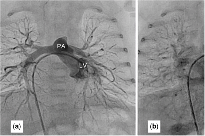
Given the extreme rarity of this association, it was decided to further investigate the anatomy, to best plan the surgical repair. Cardiac catheterisation and angiography confirmed the diagnosis and demonstrated usual coronary anatomy for D-transposition of the great arteries (Fig 2).
Cardiac catheterisation. Catheter course: femoral vein – inferior vena cava – right atrium – left atrium – left ventricle. ( a ) The left ventricular angiogram shows an intact ventricular septum and the PA arising from the LV, thus demonstrating the D-transposition of the great arteries. ( b ) Levophase of the LV angiogram showing all pulmonary veins draining into a large VV, with an inferior course through the diaphragm and a restrictive connection to the portal sinus. LV = left ventricle; PA = pulmonary artery; VV = vertical vein

The surgical repair consisted of anastomosis of the pulmonary venous confluence to the left atrium, arterial switch operation and atrial septal defect closure with a 3.5-mm-fenestrated bovine pericardial patch. Inhaled nitric oxide administration was started due to pulmonary artery hypertension and sternal closure was delayed.
Inhaled nitric oxide therapy was stopped as pulmonary artery pressure normalised, and the chest was closed on the 4th post-operative day. The patient was extubated the following day. Recovery was complicated by a right chylothorax that resolved with thoracic drainage, octreotide infusion, intravenous furosemide and a medium chain triglyceride diet. The patient was discharged on the 14th post-operative day in good clinical condition.
The patient is now doing well at 1-year follow-up. The last echocardiography documented mild neopulmonary valve stenosis with preserved biventricular systolic function, normal chamber dimensions and unobstructed pulmonary veins.
Discussion
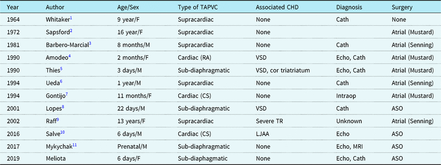
D-transposition of the great arteries with totally anomalous pulmonary venous connection is an extremely uncommon association, except as part of heterotaxy syndrome.Reference Delisle, Ando and Calder 12 To the best of our knowledge, in the setting of a definite visceroatrial situs (solitus or inversus), only 11 in vivo cases of D-transposition of the great arteries with totally anomalous pulmonary venous connection have been reported (Table 1). Among post-mortemReference Delisle, Ando and Calder 12 and in vivo reports, sub-diaphragmatic drainage was the rarest form of totally anomalous pulmonary venous connection, with only three cases before ours.Reference Thies, Matthies, Minami, Pott, Meyer and Körfer 5 , Reference Lopes, Tavares, Mailho, Almeida and Mangione 8 , Reference Mykychak, Fedevych, Maksymenko and Yemets 11 Mykychak described the only case with the same anatomy as our report (intact ventricular septum and no other associated CHDs).Reference Mykychak, Fedevych, Maksymenko and Yemets 11
Transposition of the great arteries and totally anomalous pulmonary venous connection: in vivo cases reported in the literature

ASO = arterial switch operation; Cath = cardiac catheterisation; CS = coronary sinus; Eco = echocardiography; Intraop = intraoperative; LJAA = left juxtaposition of atrial appendages; RA = right atrium; TAPVC = total anomalous pulmonary venous connection; TR = tricuspid regurgitation; VSD = ventricular septal defect
As in Lopes et al,Reference Lopes, Tavares, Mailho, Almeida and Mangione 8 our first echocardiogram did not detect the totally anomalous pulmonary venous connection. While in that report totally anomalous pulmonary venous connection was diagnosed during cardiac catheterisation, in our case a repeat echocardiogram revealed the totally anomalous pulmonary venous connection before catheterisation. The clues to the diagnosis were an unusual aliased flow pattern below the diaphragm and the unidirectional right-to-left atrial shunt with right heart dilation.
These two defects together partly correct the abnormal haemodynamics caused by each one alone. Totally anomalous pulmonary venous connection directs pulmonary venous blood to the right heart from which it is pumped to the aorta due to D-transposition of the great arteries, increasing systemic arterial saturation. This combination can produce fewer symptoms, milder cyanosis, and result in later diagnosis than the isolated defects.
Since the first successful repair in 1972,Reference Sapsford, Aberdeen and Watson 2 most reported cases have been treated by a modified atrial switch operation (Table 1), at least in part due to late diagnosis as discussed above. Some have even proposed that D-transposition of the great arteries with totally anomalous pulmonary venous connection precludes neonatal arterial switch operation because the left ventricle is not prepared to handle the systemic circulation, because of reduced left heart flow during foetal life.Reference Gontijo, Fantini, Barbosa, Gomes, Gutierrez and Vrandecic 7 This seems unlikely because pulmonary blood flow in utero accounts for only about 10% of combined ventricular output and three patients diagnosed as neonates underwent an arterial switch operation with satisfactory results.
Conclusion
Although extremely rare, transposition of the great arteries can be associated with anomalous pulmonary venous drainage even with normal atrial situs. A systematic, segmental approach to echocardiographic examination is essential to avoid overlooking important diagnoses. In D-transposition of the great arteries, special attention should be given to unusual indirect findings, such as a continuous right-to-left atrial shunt and disproportionately dilated right chambers, as they can be hints for an associated major anomaly. If diagnosed within the first weeks of life, arterial switch operation should be preferred over an atrial switch operation, as it avoids late complications related to a systemic right ventricle.
Acknowledgements
We would like to acknowledge Prof. Stephen P. Sanders (Boston Children’s Hospital, Boston, MA, USA) for his review of the report and valuable suggestions.
Financial Support
This work received no specific grant from any funding agency, commercial or not-for-profit sectors.
Conflicts of Interest
None.
Ethical Standards
No human or animal experimentation was used.