Glioblastoma is an aggressive malignant primary brain tumour which grows extremely fast and invades adjacent tissues. The self-renewing capacity and invasive nature of these cells have devastating consequences. Ageing contributes significantly to glioblastoma initiation and progression (Refs Reference Ladomersky1, Reference Kim2). On a different front, two main sources of neural stem cells (NSC) exist in the adult brain; these include the sub-granular zone of the hippocampus (SGZ) and the subventricular zone (SVZ) of the lateral ventricle. A recent pioneering study by Lee and colleagues (Ref. Reference Lee3) on human isocitrate dehydrogenase (IDH)-wildtype glioblastoma, which accounts for 90% of the cases and with frequent occurrence in older individuals, reported that glioblastoma originates from NSC located in SVZ. These cells contain somatic mutations that are implicated in gliomagenesis.
The physiology of adult NSC is under the control of a complex repertoire of signals residing in the SVZ niche. SVZ-located NSC, in contrast to those of SGZ, are in direct contact with soluble factors of the cerebrospinal fluid (CSF) via NSC processes that penetrate between the overlying ciliated ependyma. Thus, factors in the CSF influence the proliferation rate of NSC (Ref. Reference Codega4). Following the integration and decoding of the numerous CSF signals, NSC either remains in the quiescent state, undergo self-renewal or differentiate. Self-renewal allows NSC to retain their number, whereas differentiation promotes the generation of new cell types, including astrocytes, neurons and oligodendrocytes.
Uncontrolled proliferation of NSC may increase their susceptibility to malignant transformation and trigger glioblastoma initiation. The current knowledge regarding the physiology of NSC is poorly understood. Recently, Gengatharan et al. (Ref. Reference Gengatharan5) estimated the cell proliferation rate of adult NSC in freely-behaving mice, using the most advanced in vivo imaging techniques. This study reported that NSC primarily exists in the quiescent state, a mitotic-dormant state, with less than 10% regularly undergoing mitosis. The proliferation rate of these cells is under the control of melatonin, a master circadian hormone. In response to darkness, pineal melatonin is synthesised and released from the pineal gland into both the blood and the third ventricular CSF. The authors report a significant difference in NSC division rate throughout a 24-hour period, with 70% of the mitoses occurring during the day. In addition to in situ hybridisation of melatonin receptors (MT1 and MT2) on NSC in the SVZ, pharmacological and genetic manipulations of melatonin receptors confirmed their role in melatonin regulation of cell division. Activation of melatonin receptors by this ligand decreased NSC division. Conversely, blockade of the receptors with luzindole or CRISPR technology, to selectively repress melatonin receptor expression, caused an increase in cell division. Furthermore, boosting endogenous melatonin synthesis by keeping mice in constant darkness for 3 to 7 days induced a significant reduction in NSC division. In contrast, suppressing melatonin synthesis by retaining the mice in continuous light for 3 or 7 days resulted in a marked rise in NSC division.
Regarding the role of melatonin in NSC physiology, the study by Gengatharan et al. is the most comprehensive study to date. This relates to the use of the most advanced in vivo imaging approach in freely behaving mice where the NSC population was labelled with fluorescent tags and with an NSC-specific promoter. The inhibitory effect of melatonin on SVZ-NSC proliferation in vivo, and the mounting evidence that glioblastoma originates from SVZ, bolsters the correlation between melatonin and glioblastoma. In line with these observations, in vitro studies reported a therapeutic role of melatonin in reducing tumour cell proliferation, self-renewal and clonogenic ability, especially in glioblastoma cells (Ref. Reference Fernandez-Gil6). Furthermore, an in vivo study showed that constant light exposure for 5 weeks, a strategy that inhibits circadian-dependent melatonin synthesis, enhanced glioma tumour growth in rats (Ref. Reference Guerrero-Vargas7).
Nestin is one of the markers expressed by neural stem/progenitor cells (NSPCs) of SVZ. Wu and colleagues carried out a systematic meta-analysis and proposed a positive correlation between nestin expression and the overall survival in glioma patients, especially at the early stage of disease (Ref. Reference Wu8). Interestingly, Jang et al., using a rat model of chemical glioma, showed that over-expression of nestin cells is considered as an early stage of the neoplastic process. Furthermore, they found a cluster of nestin cells in the wall of SVZ whose size grew markedly by age (Ref. Reference Jang9). Since there is a significant decline in melatonin synthesis in rats, similar to human, we suggest that the expansion in the size of nestin cluster cells in SVZ might be caused by a reduction of melatonin associated with ageing.
Since the introduction of artificial electrical light sources, human lifestyles have changed dramatically with marked changes in sleeping habits which impacts human health. The exposure of humans to blue light wavelengths inhibits the synthesis and release of melatonin from the pineal gland and shortens the duration of nocturnal melatonin secretion (Refs Reference Gooley10, Reference Gooley11). This occurs during night shift work, indoor lightning when blue-enriched light-emitting diodes are used, and viewing screens of electrical devices such as smartphones, TVs, or tablets before sleeping. Moreover, excessive exposure to light-at-night reportedly increases the incidence of a variety of different cancers (Ref. Reference Garcia-Saenz12). We suggest that melatonin suppression due to this lifestyle, which results in a reduced total melatonin production, may increase cancer incidence by itself or synergises with other environmental factors such as ingestion or inhalation of toxins.
Increased age is a significant factor in decreasing endogenous melatonin levels and in altering the expression of melatonin receptors. Melatonin levels are higher in children than in adults. After adulthood, endogenous melatonin levels fade such that a nocturnal rise in blood melatonin levels may be barely discernible in elderly individuals (Ref. Reference Grivas and Savvidou13). The rapidity with which melatonin levels drop during ageing varies widely among individuals and may relate to their general health (Ref. Reference Karasek14). Gengatharan et al. did not quantify the expression levels of MT1 and MT2 receptors, however, in situ hybridisation showed a stronger expression for MT2 in the SVZ. When they used an in vivo treatment strategy with luzindole, a non-selective receptor antagonist, to block both MT2 and MT1 melatonin receptors, this was associated with a significant promotion in cell division. Consistent with this finding, a recent in vitro study by Kinker et al. demonstrated that MT1 activation reduced the proliferation of glioma and medulloblastoma cell lines, while MT2 had the opposite effects (Ref. Reference Kinker15).
It is noteworthy that there is a sex-specific difference in glioblastoma incidence, as it is 60% higher in men than women (Ref. Reference Carrano16). Interestingly, a slightly higher amplitude of melatonin rhythm has been reported in women compared to that in men (Ref. Reference Cain17). The higher melatonin in women and the lower glioblastoma incidence might justify considering melatonin as a significant factor in glioblastoma inhibition.
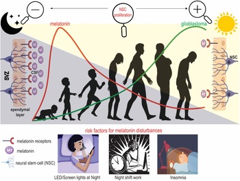
In conclusion, the recent study by Gengatharan et al. not only presented a better view of SVZ NSC division in vivo, it also identified a day/night difference in mitotic activity that is mediated by melatonin and its receptors. This clearly introduces melatonin as a key regulator of neural mitosis, since higher levels of melatonin and increased function of melatonin receptors are associated with lower NSC division. Other studies have reported that endogenous melatonin levels and its receptors drop significantly during ageing, which highlights the possibility that initiation or progression of glioblastomas, originating from SVZ-NSC, may accelerate as a result of the age-associated loss of melatonin. Moreover, a greater understanding of the potential negative consequences of risk factors arising from modern lifestyles, including artificial light-at-night, night shift work, sleep disturbances and insomnia is required. This information may lead to the definition of a mechanistic pathway allowing for the design of new therapeutic strategies to reduce/treat glioblastoma. Figure 1 depicts how these variables come together to initiate glioblastoma over time.
The proposed association of melatonin with the incidence of glioblastoma. In human newborns, pineal melatonin synthesis begins at 4–6 months of age and thereafter melatonin is produced in a light-dependent circadian rhythm. Maximum levels of melatonin are attained during early childhood between 5–10 years old and after adulthood, they exhibit a gradual decline into old age. Circadian melatonin is released directly into the third cerebral ventricle from where it circulates throughout the cerebrospinal fluid (CSF). Cilia of the ependymal cells and processes of NSC in the SVZ are in direct contact with circadian and non-circadian CSF melatonin to decode and integrate the signals in order to determine the fate of NSC. Among other factors, the mitotic activity of NSC is under the control of physiological melatonin levels, which seem to fine-tune cell division of NSC. Elevated melatonin levels during darkness and longer night periods cause a reduction of NSC mitotic activity, while light exposure or longer day length reduces the regulatory effects of melatonin on NSC and causes an increased NSC division. Melatonin synthesis perturbations due especially to exposure of blue wavelengths during the normal period of darkness depress the normal circadian melatonin rhythm and may stimulate glioblastoma incidence; likewise, this also occurs in aged individuals, where the circadian melatonin rhythm is severely dampened.

Authors contributions
MG, KZ, RR and SR contributed to writing-original draft, and writing-review & editing. All authors read and approved the final version of the manuscript.
Conflicts of interest
The authors declare no conflict of interest.