Beta-thalassemia major, associated with mutations in the globin gene, is the most common hereditary haemoglobinopathy. This disease causes severe chronic haemolytic anaemia and is seen in early childhood. Reference Flint, Harding, Boyce and Clegg1,Reference Olivieri2 Regular blood transfusion is the main treatment for these patients. However, these transfusions can lead to cardiac iron overload and cause severe cardiac complications such as heart failure and malignant atrial and ventricular arrhythmias. Reference Flint, Harding, Boyce and Clegg1,Reference Kyriacou, Michaelides, Senkus and Simamonian3 Despite advances in chelating therapy, cardiac complications are still the main cause of morbidity and mortality in β-thalassemia major patients. Reference Modell, Khan and Darlison4
Iron overload is associated with an increase in the risk of atrial and ventricular arrhythmias. Reference Russo, Rago and Pannone5,Reference Russo, Rago and Pannone6 Twelve-lead surface electrocardiography remains the most commonly used and easily accessible tool for diagnosis. Previously, electrocardiographic markers representing atrial depolarisation and ventricular repolarisation such as P wave dispersion, QT dispersion and corrected QT dispersion, T peak to end interval, which is the measurement of the interval between the peak and the end of the T-wave, T peak to end/QT ratio and T peak to end dispersion have emerged as markers of transmural dispersion of repolarisation, can predict the development of atrial and ventricular arrhythmias, respectively Reference Dilaveris, Gialafos and Sideris7,Reference Gupta, Patel and Patel8
Cardiovascular MRI is being used as a non-invasive method to evaluate the cardiac iron overload in β-thalassemia major patients. Myocardial iron deposition can be assessed using spin echo (T2) and gradient echo (T2*) techniques. Reference Anderson, Holden and Davis9 Cardiac T2* MRI is a more sensitive and specific method than either plasma ferritin levels or hepatic T2* measurement. Reference Westwood, Wonke and Maceira10 However, data regarding the relationship of electrocardiographic markers with cardiac T2* MRI are scarce. Reference Kayrak, Acar and Gul11 Therefore, we aimed to investigate electrocardiographic parameters and their relationship with cardiac T2* MRI value in patients with β-thalassemia major.
Materials and methods
Study population
In this prospective study, conducted from March 2018 to March 2019, 52 patients with β-thalassemia major who were admitted to our paediatric haematology outpatient clinic and 52 age- and gender-matched healthy patients were assessed. The study was approved by the local ethical committee. The β-thalassemia patients were being regularly transfused (every 3–4 weeks) and were receiving chronic chelation therapy (deferoxamine or deferiprone and combination treatment). Exclusion criteria were chronic inflammation, thyroid disorders, diabetes mellitus, use of antiarrhythmic drugs, use of QT-prolonging drugs, and congenital or acquired arrhythmia syndromes. Demographic, clinical, echocardiographic, and electrocardiographic parameters were carefully recorded.
Electrocardiography and echocardiography
The standard 12 lead electrocardiography were recorded for each patient with a paper speed of 25 mm/s at an amplitude of 10 mm/mV. Electrocardiography measurements QT, T peak to end, and P wave intervals were made by one cardiologist who was blind to patients’ data. An average value from three readings was calculated for each lead. P wave duration was measured as the time between the beginning of the P wave (the point from the isoelectric line of the first visible upward slope for positive waves, or the first visible downward slope for negative waves) and the end of the P wave (the point of return to the isoelectric line). The difference between the maximum and minimum of the P wave duration was defined as the P wave dispersion. P wave dispersion was not measured if fewer than nine leads were eligible for analysis. Reference Karadeniz, Özdemir and Demirol12
The QT interval was measured from the beginning of the QRS complex to the end of the T wave and corrected for heart rate using the Bazett formula (QTc = QT/√R − R interval). Reference Bazett13 The QT dispersion was calculated for at least 8 of the 12 leads and usually included leads I, II, and V5. The differences between the maximum and minimum QT and QTc intervals were defined as the QT and QTc dispersions, respectively. Reference Karadeniz, Özdemir and Demirol12
The T peak to end interval was defined as the interval from the peak of T wave to the end of T wave. We defined T peak to end as the intersection point of the tangent to the downward slope of the T wave with the isoelectric line. Measurements of the T peak to end interval were taken from precordial leads. The T peak to end/QT ratio was calculated from these measurements. Reference Karadeniz, Özdemir and Demirol12
The same paediatric cardiology specialist who took the electrocardiography measurements performed the echocardiographic examination using a Philips Ultrasound System (model iE33, Philips Medical Systems, Andover) and an S3–1 probe. Conventional echocardiographic measurements were made according to the standards of the American Echocardiography Society. Reference Rudski, Lai and Afilalo14 The dimensions of the left ventricle in the parasternal long axis on M mode tracing, the ejection fraction, and fractional shortening were analysed using the Teichholz formula. Reference Teichholz, Cohen, Sonnenblick and Gorlin15
Both electrocardiographic and echocardiographic examinations were performed while β-thalassemia patients were in a stable haemodynamic state, 5–7 days after the last red blood cell transfusion.
Cardiac magnetic imaging
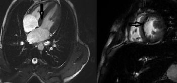
Cardiac iron loading was evaluated with cardiac T2* MRI. Magnetic resonance images were acquired for all patients with a 1.5 tesla (T) magnet GE Optima MR 360 (General Electric Company, Chicago). All magnetic resonance image examinations were performed in a supine position. Total shooting time was approximately 10 minutes for each patient. The multi-TE GRE sequence was used to measure cardiac iron accumulation. To evaluate cardiac function, short axis multi-slice cine images were taken to scan the entire left ventricle. Parameters used for magnetic resonance examinations were as follows: functional cine short axis: TR = 4.4 ms, TE = 1.896 ms, FOV = 380 mm, matrix = 224 × 256, FA = 60, NEX = 1, section thickness = 8 mm, number of sections = 10; cardiac iron (R2): TR = 21.9 ms, TE = 1.52–18, 544 ms, 8 different TE values, FOV = 380 mm, matrix = 224 × 160, FA = 20, NEX = 1, section thickness = 8 mm, section number = 5. Evaluation was performed on a separate workstation (Advantage Workstation Volume Share 5; General Electric Healthcare, New York) using special software (Cardiac VX) (Fig 1).
Cardiovascular magnetic resonance T2* images showing the apical four chamber and short-axis view of the heart at the same echo time (7 ms). Dark signal indicating myocardial siderosis (black arrow).

Statistical analysis
Data were analysed using the statistical programme SPSS Statistics 25.0 (IBM Corp., Armonk, New York, USA). Descriptive statistics are given as number of units, percentage, mean ± SD. Normal distribution of continuous variables was evaluated using the Shapiro–Wilk test for normality and Q–Q plots. The homogeneity of group variances was evaluated using the Levene’s test. To compare two groups under the assumption of normally distributed continuous variables, independent samples t tests were used for parametric statistical tests, and one-way analysis of variance was used for comparison of more than two groups. Median (Q1–Q3) values are given as descriptive statistics in the analysis of the variables that are not distributed normally. Non-parametric statistical tests were used for comparison of more than two groups, the Mann Whitney U was used to compare two groups, and the Kruskal–Wallis H test was used to compare more than two groups. Relationships between variables were evaluated with Spearman’s correlation analysis. The relationships between variables with categorical structure were evaluated using the continuity correction chi-square test in 2 × 2 tables. p Values less than 0.05 were considered statistically significant.
Results
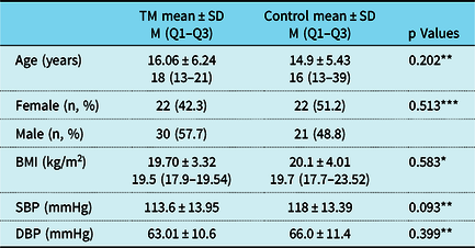
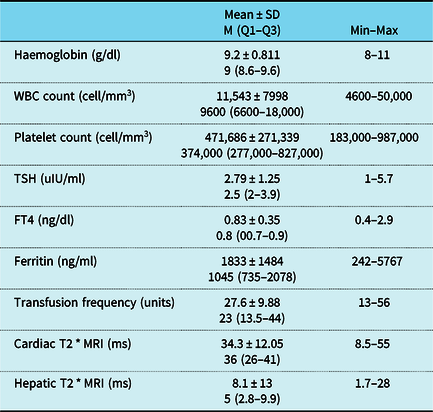
The mean age of the patients in the study was 16.06 ± 6.24 years. Of these patients, 30 were male and 22 were female. Comparisons of the demographic parameters of the patients and control group are shown in Table 1. The groups were similar in terms of age, gender, body mass index, and blood pressure levels. Laboratory characteristics of the study population are given in Table 2.
Clinical and demographic characteristics of the study population

DBP = diastolic blood pressure; SBP = systolic blood pressure; TM = thalassemia major.
*Independent sample t test.
**Mann Whitney U testing.
***Continuity correction test.
Laboratory characteristics of the study population

FT4 = free T4; TSH = thyroid stimulating hormone.
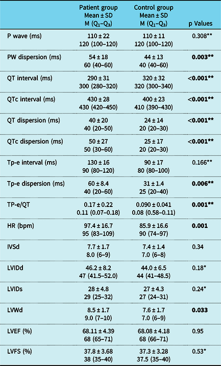
Electrocardiographic and echocardiographic parameters of the study group and the healthy controls are listed in Table 3. Mean heart rate significantly increased in the patient group compared to the controls (97.4 ± 16.7 versus 85.9 ± 16.6 bpm, p = 0.001). The atrial depolarisation parameter P wave dispersion was significantly higher in β-thalassemia major patients compared to controls (54 ± 18 ms versus 44 ± 13 ms, p = 0.003). Interestingly, we did not find any difference in P wave measurements between groups.
Comparisons of the electrocardiographic and echocardiographic parameters of the study group and healthy controls

bpm = beat per minute; FS = fractional shortening; HR = heart rate; IVSd = interventricular septum thickness in diastole (mm); LVEF = left ventricular ejection fraction; LVIDs and LVIDd = LV internal dimensions in systole and diastole; LVWd = left ventricular posterior wall thickness in diastole (mm); QTc = corrected QT interval; TM = thalassemia major
Significant values of p values are indicated in bold.
*Independent sample t test.
**Mann Whitney U test.
With regard to QT parameters, the QTc interval, dispersions of QT and QTc were significantly prolonged in the patient group compared to controls (all p < 0.001). However, we found higher QT intervals in controls compared to β-thalassemia major patients. When we compared the echocardiographic parameters of the patients and the controls, all conventional echocardiographic parameters were similar between groups except the thickness of the left ventricular posterior wall in diastole. Left ventricular posterior wall thickness in diastole was higher in β-thalassemia patients compared to controls (8.5 ± 1.7 mm versus 7.6 ± 1.7 mm, p = 0.033). T peak to end dispersion and T peak to end/QT ratio were significantly higher in β-thalassemia major patients compared to controls (p = 0.006 and 0.001, respectively). No significant difference was found for the T peak to end interval (p = 0.166).
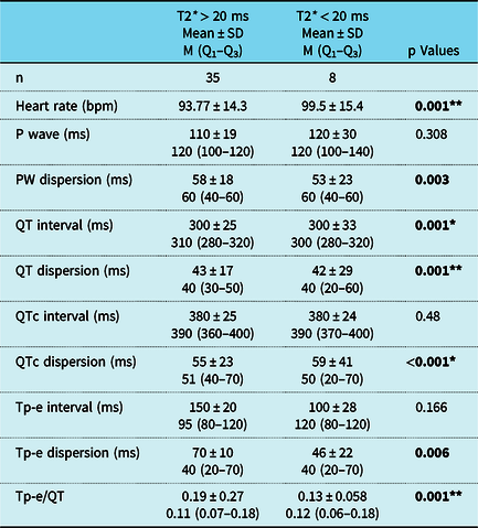
Electrocardiographic findings of β-thalassemia major patients according to cardiac T2* scores are listed in Table 4. Interestingly, we found prolonged P wave, QT, and T peak to end dispersions, T peak to end intervals, and T peak to end/QT ratios in patients with T2* greater than 20 ms.
Electrocardiographic parameters of β-thalassemia major patients according to cardiac T2* scores

Significant values of p values are indicated in bold.
*Independent sample t test.
**Mann Whitney U test.
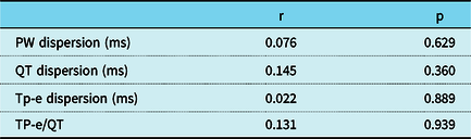
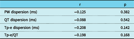
Correlations of the electrocardiographic parameters with cardiac T2* and with plasma ferritin levels are shown in Tables 5 and 6, respectively. We did not observe a significant correlation between cardiac T2* MRI values and plasma ferritin levels.
Correlation and statistical significance of the ECG parameters to cardiac T2*

Correlation and statistical significance of the ECG parameters to ferritin levels

Discussion
In the current study, we found that the atrial depolarisation parameter P wave dispersion and ventricular repolarisation parameters including QT, QTc, T peak to end intervals, and T peak to end dispersions were significantly prolonged, and T peak to end /QT ratio was significantly higher in patients with β-thalassemia major compared to healthy controls. Neither atrial depolarisation parameters nor ventricular repolarisation parameters were correlated with cardiac T2* MRI values. To our knowledge, ours is the first study of children that investigated electrocardiographic markers of important atrial and ventricular arrhythmic events and their correlation with cardiac iron overload.
Heart failure and cardiac arrhythmias are the leading causes of morbidity and mortality among transfusion-dependent β-thalassemia major patients, second only to iron overload. Reference Murphy and Oudit16 Therefore, early identification of these abnormalities is crucial for long-term survival in these patients. To date, different echocardiographic and electrocardiographic parameters have been investigated as markers for early detection of cardiac involvement in β-thalassemia major patients. Reference Teichholz, Cohen, Sonnenblick and Gorlin15,Reference Ulger, Aydinok, Levent, Gurses and Ozyurek17 Theories have been put forward to explain the mechanisms of the iron toxicity: one is that excessive intracellular iron may interfere with the electrical function of the heart, another proposes the generation of free radicals and selective Na channel dysfunction, while another involves apoptosis and fibrosis. Reference Schellhammer, Engle and Hagstrom18,Reference Kuryshev, Brittenham and Fujioka19
P wave dispersion has been reported as an important non-invasive electrocardiographic marker for assessing the homogeneous distribution of sinus node impulses within the atrial myocardium. Its predictive value in atrial arrhythmias, especially in atrial flutter and fibrillation, has been shown in cases without underlying heart disease and under different clinical conditions. Reference Karadeniz, Özdemir and Demirol12,Reference Ozer, Aytemir and Atalar20 Previous studies showed prolonged P wave dispersion in β-thalassemia major patients. However, some of these studies did not evaluate the cardiac iron deposit with cardiac T2* MRI. Russo et al. showed a correlation between P wave dispersion and myocardial iron overload by assessing the cardiac T2* MRI in β-thalassemia major patients with normal cardiac functions. Reference Russo, Rago and Pannone6 In contrast to Russo et al. Reference Russo, Rago and Pannone6 , Acar et al. Reference Acar, Kayrak, Gul, Abdulhalikov, Özbek and Uçar21 did not find prolonged P wave dispersion or a correlation with cardiac T2* MRI. In our study, although we found prolonged P wave dispersion in β-thalassemia major patients, similar to Acar et al., we did not find any association between P wave dispersion and cardiac T2* MRI.
QT parameters including QT, QTc intervals, QT and QTc dispersions, and QT variability index have been evaluated in β-thalassemia major patients before. However, the results of these studies were conflicting. Some of them found prolonged QT, QTc, and QT dispersion and corrected QT dispersion in β-thalassemia major patients compared to controls Reference Russo, Rago and Pannone5,Reference Gupta, Patel and Patel8 while others did not. Reference Magrì, Sciomer and Fedele22,Reference Garadah, Kassab, Mahdi, Abu-Taleb and Jamsheer23 In our study, we found prolonged QTc, QT, and QT dispersion in β-thalassemia major patients compared to controls. The relationship between QT parameters and iron overload from serum ferritin levels was reported by Ulger et al., who did not compare the QT parameters with cardiac T2* MRI. Reference Ulger, Aydinok, Levent, Gurses and Ozyurek17 Consistent with their study, we did not find any correlation between QT parameters and either serum ferritin or liver iron concentrations.
Previous studies that investigated the relationship between QT parameters and cardiac iron overload measured by cardiac T2* MRI did not find any difference in QT parameters between β-thalassemia major patients who had T2* MRI measures less than or greater than 20 ms. Reference Kayrak, Acar and Gul11 Furthermore, no difference was found in QT and QTc dispersions between β-thalassemia major patients and controls. Reference Kayrak, Acar and Gul11 Similar to Kayrak et al. Reference Teichholz, Cohen, Sonnenblick and Gorlin15 , we did not find any relationship between QT parameters and cardiac T2* MRI measures and serum ferritin levels. However, contrary to Kayrak et al., we found prolonged QT and QTc dispersions compared to controls.
Recent studies have suggested that the interval between the peak and the end of the T wave (the T peak to end interval) and T peak to end dispersion can be a novel electrocardiographic marker of increased dispersion of ventricular transmyocardial repolarisation. Reference Gupta, Patel and Patel8,Reference Kors, Ritsema and Van Herpen24 Some clinical conditions such as long QT syndrome, Brugada syndrome, and hypertrophic cardiomyopathy may lead to an increased heterogeneity of transmural dispersion of repolarisation (represented by prolonged T peak to end intervals and T peak to end dispersions). This dispersion of repolarisation becomes the substrate for reentry causing malignant ventricular arrhythmias and even sudden cardiac death. Reference Castro Hevia, Antzelevitch and Tornés Bárzaga25,Reference Shimizu, Ino and Okeie26 Besides T peak to end and T peak to end dispersion, the T peak to end/QT ratio has emerged as a more accurate non-invasive electrocardiographic marker of arrhythmogenesis due to its independence from heart rate and body weight. Reference Gupta, Patel and Patel8 In β-thalassemia major patients, data regarding the T peak to end interval, T peak to end dispersion, and the T peak to end/QT ratio are very scarce. Kayrak et al. showed prolonged T peak to end intervals and T peak to end dispersion except for T peak to end/QT ratio in β-thalassemia major patients. They also found a correlation between T peak to end interval and cardiac T2* MRI values. However, they did not find any difference in transmyocardial parameters in patients with cardiac T2* MRI measures shorter than 20 ms or longer than 20 ms. Reference Teichholz, Cohen, Sonnenblick and Gorlin15
To date, no study has investigated both atrial depolarisation and ventricular repolarisation parameters and their relationship with serum ferritin level and cardiac iron load. For the first time, we investigated the correlation of these parameters. In our study, we found prolonged T peak to end interval, T peak to end dispersion, and increased T peak to end/QT ratio in patients with β-thalassemia major. However, in contrast with Kayrak et al. Reference Kayrak, Acar and Gul11 , we could not find any correlation between electrocardiographic parameters and cardiac iron load (cardiac T2* MRI) or between electrocardiographic parameters and serum ferritin levels. Interestingly, we found prolonged P waves, QT and T peak to end dispersions, T peak to end intervals, and increased T peak to end/QT ratio in patients with T2* greater than 20 ms.
Our results showed that iron overload toxicity alone does not influence atrial and ventricular conduction. Iron overload may increase fibrosis and oxidative stress in atrial and ventricular myocardium that may be associated with a tendency for arrhythmias. Additionally, our results demonstrated that cardiac arrhythmias were not related to iron overload in haemochromatosis alone. In light of results from previous studies, our results indicate that iron levels may be necessary but not sufficient in the development of cardiac arrhythmias in the conditions of iron overload.
For practical usage of the electrocardiographic parameters described in this study, we recommend longitudinal long-term follow-up of these parameters in patients with β-thalassemia major. For this reason, all individuals with β-thalassemia major should be evaluated with 12-lead electrocardiography periodically according to their clinical situation and cardiac imaging findings (echocardiography and cardiac magnetic resonance). In the coming years, we anticipate advances in mobile applications for assessing ECG and rhythm disturbances, allowing these parameters to be calculated automatically. Such applications may provide useful information for risk assessment to predict malignant ventricular arrhythmias.
The current study has some limitations that should be considered. First, the foremost limitation of our study was a small sample size. Second, we did not investigate non-invasive markers of fibrosis in β-thalassemia major patients. Third, due to the short follow-up period, we may not have been able to detect a correlation between electrocardiographic parameters and cardiac T2* MRI values. Lastly, we could not prospectively follow up with patients with β-thalassemia major to check atrial and ventricular arrhythmias with 24-hour electrocardiography monitoring, and therefore, we could not evaluate future arrhythmic events.
Conclusions
Our study demonstrated that atrial depolarisation and ventricular repolarisation parameters are affected in β-thalassemia major patients and that these parameters are not correlated with cardiac iron load. Due to long life expectancy, children with β-thalassemia major may be considered at risk of developing arrhythmias. Therefore, careful evaluation of these parameters is necessary in children with β-thalassemia major. More long-term prospective electrophysiological studies are needed to further demonstrate the clinical and prognostic implications of these parameters.
Acknowledgments
None.
Authorship contributions
Concept – T.D. and B.T.G.; Design – T.D.; Supervision – C.K.; Funding – Z.Ö.S.; Materials – Z.Ö.S., T.D., and B.T.G.; Data collection and/or processing – T.D. and C.K.; Analysis and/or interpretation – C.K., T.D., Z.Ö.S., and B.T.G.; Literature search – T.D., C.K., Z.Ö.S., and B.T.G.; Writing – T.D., C.K., Z.Ö.S., and B.T.G.; Critical review – C.K., T.D., B.T.G, and Z.Ö.S.
Financial support
This research received no specific grant from any funding agency, commercial, or not-for-profit sectors.
Conflicts of interest
None.
Ethical standards
The authors assert that all procedures contributing to this work comply with the Helsinki declaration of 1975, as revised in 2008, and have been approved by the Ethical Review Committee at the Tepecik Training and Research Hospital, Izmir.