Tetralogy of Fallot with pulmonary atresia and diminutive or absent intrapericardial pulmonary arteries is a rare and challenging congenital abnormality, with a high morbidity and need for multiple interventions. Since the 1970s, multiple institutions have devoted considerable effort developing methods for effective palliation.Reference Malhotra and Hanley1–Reference Lofland3 The greatest advances have been in the surgical technique of unifocalisation of major systemic-to-pulmonary collateral arteries, and in catheter-based methods for maintaining patency and size of both major collateral arteries and intrapericardial pulmonary arteries. Despite these advances, considerable controversy still exists as to the indications for unifocalisation, as opposed to the need for recruitment of all branches of the pulmonary arterial tree when they are all, or mostly, already in continuity with the intrapericardial pulmonary arteries. This controversy is exemplified by two recent papers with seemingly opposing views on the approach to this lesion.Reference Brizard, Liava’a and d’Udekem4, Reference Davies, Mussa and Davies5
Our approach, probably not unlike that of many institutions, is to pursue a scheme for management governed by the findings in each individual patient, seeking to maximise the ultimate size and distribution of the intrapericardial pulmonary arteries, irrespective of their initial size and appearance. With the belief that flow encourages growth, we establish antegrade flow in the intrapericardial pulmonary arteries as a first step in palliation. We unifocalise major systemic-to-pulmonary collateral arteries only when the specific collateral artery supplies a pulmonary segment that is truly isolated from the intrapericardial pulmonary arterial tree, or when there is true congenital absence of the intrapericardial pulmonary arteries. In instances of dual supply, we occlude the collateral artery, with the belief that its competitive flow may inhibit growth and development of the associated intrapericardial pulmonary arterial tree. Given that, in general, the pulmonary arterial tree is considerably hypoplastic at the first stage of palliation, we usually leave open the ventricular septal defect. After considerable catheter-based intervention on the arterial tree, we assess the general appearance of the arterial tree, as opposed to using a formal index based on cross-sectional areas or diameters, determining, in this manner, the time to close the ventricular septal defect. The shunt fraction is generally an invalid parameter for this determination, as relative obstruction at the source of antegrade flow, be it through an aorto-pulmonary window, a systemic-to-pulmonary shunt, or a conduit placed from the right ventricle to the pulmonary arteries, is common at the time of assessment for closure of the ventricular septal defect. We make the final decision in terms of closure by measuring right ventricular pressures at the time of operation. In this study, we describe our surgical methods and results in managing this complex abnormality.
Population and methods
We carried out a retrospective study of our database and medical records. We obtained the Institutional Review Board approval from both the Arnold Palmer Hospital for Children and Miami Children’s Hospital, with these two institutions comprising our joint programme. We searched our databases for all patients with diagnosis including keywords “pulmonary atresia” and “tetralogy of Fallot” (or “ventricular septal defect”), and “major aortopulmonary collateral arteries” between September, 1988 and August, 2009. We excluded patients who were not predominantly managed in our institutions over their lifetime. We obtained demographics, records of all catheterisations and operations, and the most recent clinical evaluation, including mortality, open or closed state of the ventricular septal defect, right ventricular function as assessed by echocardiography, and clinical functional state. The latter was graded as none, mild, moderate, or severely decreased exercise tolerance as described by the cardiologist in the clinic note. The last checking of the database and charts was complete to January, 2010.
Statistical methods
We expressed data as mean and standard deviation or median and 95% confidence limits. We used chi-square and Fisher’s exact tests to compare patients who were unifocalised with those not undergoing unifocalisation. We performed survival analysis using the Kaplan–Meier (product–limit) method.
Initial assessment
At our institutions, we perform catheterisation and, in some cases undertake magnetic resonance imaging, in all patients diagnosed echocardiographically as having tetralogy of Fallot with pulmonary atresia and diminutive pulmonary arteries and/or evidence of major systemic-to-pulmonary collateral arteries. Initial management is then determined by the presence and size of the intrapericardial pulmonary arteries, and by the extent of dual supply. In the absence of intrapericardial pulmonary arteries, or if there are more than one or two minor lung segments without dual supply, the first procedure is usually unifocalisation and establishment of continuity from the right ventricle to the pulmonary arteries with or without closure of the ventricular septal defect. If there is nearly complete dual supply, and the right and left pulmonary arteries are less than 2.5 millimetres in diameter, the first procedure is usually the creation of an aortopulmonary window, and either catheter or surgical ligation of major collateral arteries. If there is complete dual supply, and the pulmonary arteries are greater than 2.5 millimetres in diameter, the first procedure is most likely the establishment of continuity from the right ventricle to the pulmonary arteries, closure of the ventricular septal defect, and ligation of major collateral arteries. We perform the first procedure as early in infancy as possible because stenosis within the major collateral arteries progresses with time, and we believe that early establishment of antegrade flow in the pulmonary arteries promotes their growth. In addition, we establish continuity from the right ventricle to the pulmonary arteries as early as it is feasible to allow optimal catheter access for interventional rehabilitation of the pulmonary vasculature.
Operative techniques for unifocalisation
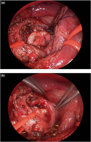
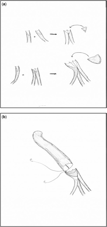
Our techniques for unifocalisation largely follow those developed by Hanley et al.Reference Reddy, Liddicoat and Hanley6 We prefer a midline approach and bilateral unifocalisation. We make a long midline incision and thoroughly mobilise the ascending aorta and superior caval vein. We incise the pleuropericardial reflection between the phrenic nerves and pulmonary hilum to maximise distal hilar or intrapulmonary exposure. We expose major collateral arteries from the arch or brachiocephalic arteries superiorly, and from the descending aorta through the transverse sinus (Fig 1). The latter dissection is facilitated by auto-retracting the ascending aorta leftward, the caval vein rightward, and the right atrial appendage inferiorly. As it is well known, the exact design of the unifocalisation differs according to the individualised anatomy. It requires, as suggested by Hanley et al,Reference Reddy, Liddicoat and Hanley6 both pre- and intraoperative creativity. In general, the goal is to unifocalise as much as possible to a central position, using autogenous tissue to make the connections. In absence of the intrapericardial pulmonary arteries, this can often be done by redefining a suitable major collateral artery as the central vessel. Otherwise, a suitable non-autogenous conduit must be used. In the latter situation, our preference is to use a roll created from a pulmonary homograft by suturing longitudinally around a dilator of 5–8 millimetres in diameter. We use interrupted sutures so the graft can be opened at any point, and so it can also be trimmed in length to avoid any tension on any of the major collateral arteries. The graft can be prepared before making a skin incision. Recognising that the use of such a tube will create a circumferential non-autologous suture line, we attempt to amalgamate two or more major collateral arteries to each other first, so that the circumference of the final connection to the non-autogenous tube is as large as possible (Fig 2). In this manner, the future management of the final circumferential connection is not fundamentally different from that of the distal connection of a conduit placed from the right ventricle to the pulmonary arteries. We find that we can perform about half to three quarters of this reconstruction before there is any decrease in the saturation of oxygen, at which point we begin cardiopulmonary support. We complete the remaining unifocalisation with mild hypothermia and a beating heart. Our decision regarding closure of the ventricular septal defect is based partially on the pre-operative anatomy, and partially on our subjective assessment of the completed unifocalisation. If the collateral arteries are large, and without peripheral stenosis, and we anticipate perfusing 15 or more lung segments, we attempt closure of the ventricular septal defect as a primary procedure. We have not commonly used an intraoperative physiological test. If the ventricular septal defect is to be closed, we arrest the heart at this point, close the defect, and restore continuity from the right ventricle to the pulmonary arteries using a valved aortic homograft or, occasionally, a gluteraldehyde-treated valved bovine jugular venous graft. If the ventricular septal defect is to be left open, we prefer to use a humble-sized polytetrafluoroethylene graft with a small homograft hood, thus avoiding pulmonary over-circulation, and reducing the risk of formation of aneurysms or pseodoaneurysms due to persistence of systemic pressure in the right ventricle. Occasionally, we have used a transjunctional patch having found suitable anatomy of the infundibulum and the pulmonary trunk.
Panel a shows exposure of major collateral arteries arising from the arch or great vessels superiorly and from the descending aorta through the transverse sinus. The left bronchus is retracted cephalic, and the superior caval vein rightward by vessel loops. Panel b shows how a second collateral is dissected off the bronchus.

Panel a shows an illustration of autogenous connection between two or more major collateral arteries. Panel b shows connection to a homograft tube when a central autogenous connection cannot be made. The homograft patch in panel a can be replaced with a beveled connection to the end of the tube.

Results
Initial assessment
We included 38 patients in our analysis, of whom 18 were male and 20 female. Follow-up was complete for 89% of the cohort. All patients had one predominant malalignment ventricular septal defect, with two patients having additional small muscular ventricular septal defects. In 24% of patients, we were unable to find discernable intrapericardial pulmonary arteries during operation, while 42% of the patients had predominantly dual arterial supply. The median number of major collateral arteries was 3, with a range from 2 to 6.
Operative sequence
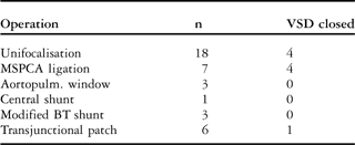
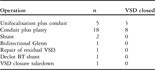
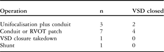
With a median duration of follow-up of 9 years, the median number of catheterisations was 5, with a range from 1 to 15, and the median number of operations was 2.2, with a range from 1 to 6. We undertook unifocalisation in 26 of 38 patients (68%). This was achieved in two patients with one collateral artery, 14 with two collateral arteries, six with three arteries, and four with four collateral arteries. The remaining patients underwent procedures to recruit the native pulmonary vasculature. The mean age at the time of first operation was 5.9 plus or minus 11.2 months. The types of first operations performed, as well as the patency of the ventricular septal defect, are shown in Table 1. After the first operation, two patients died, two were lost to follow-up, and five are currently at risk for a second operation. Thus, 29 patients underwent a second operation at a mean age of 2.7 plus or minus 3.7 years, with a mean interval between the first and second operations of 2.2 plus or minus 3.5 years. The types of second operation are shown in Table 2. After the second operation, four patients died, one was lost to follow-up, and 18 are currently at risk for a third operation. Thus, 12 patients underwent a third operation. The types of third operation are shown in Table 3. After the third operation, two patients died, one was lost to follow-up, and six are currently at risk for a fourth operation. In one patient, we performed fourth, fifth, and sixth operations for unifocalisation; closure of the ventricular septal defect and placement of a conduit, and repair of a residual ventricular septal defect, respectively.
Types of first operation performed and patency of the VSD.
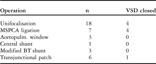
Aortopulm. = aortopulmonary; BT = Blalock–Taussig; MSPCA = major systemic-to-pulmonary collateral artery; VSD = ventricular septal defect
Types of second operation performed and patency of VSD.
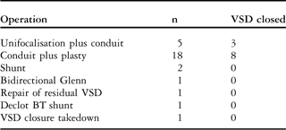
BT = Blalock–Taussig; VSD = ventricular septal defect
Types of third operation performed and patency of VSD.
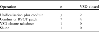
RVOT = right ventricular outflow tract; VSD = ventricular septal defect
Mortality
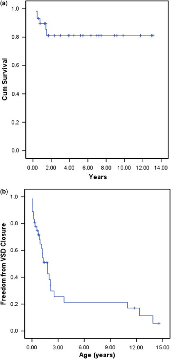
Of 38 patients, eight have died, while we are unaware of the situation in four patients lost to follow-up. Of 23 patients who underwent unifocalisation, and whose state is currently known, 18 (78%) are alive. Of 11 patients who did not have unifocalisation and whose state is currently known, 8 (73%) are alive (p > 0.1). The overall Kaplan–Meier survival curve is shown in Figure 3a.
Kaplan–Meier curves for (a) freedom from death, and (b) freedom from closure of the ventricular septal defect (VSD).

Closure of the ventricular septal defect
We closed the defect in 26 of 38 patients at a median age of 2.39 years, with 95% confidence limits from 0.93 to 3.86 years. Of 26 patients known to be alive, 23 have had the defect closed. Of the eight patients known to have died, the defect had been closed in three. Of those patients unifocalised, the defect has been closed in 68%, while in those not undergoing unifocalisation, closure was achieved in 69% (p > 0.1). The Kaplan–Meier curve for freedom from closure of the ventricular septal defect is shown in Figure 3b.
Functional state
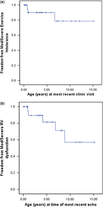
The Kaplan–Meier freedom from greater than or equal to moderately decreased exercise tolerance is shown in Figure 4a.
Kaplan–Meier curves (a) for freedom from moderately (Mod) or severely decreased exercise tolerance, and (b) from right ventricular (RV) dysfunction.

Right ventricular function by echocardiography
The overall Kaplan–Meier freedom from moderate or greater dysfunction of the right ventricle by echocardiography is shown in Figure 4b. For patients who have had closure of their ventricular septal defect, the cumulative hazard for moderate or greater dysfunction of the right ventricle continuously increases out to at least 10 years beyond the time of complete repair.
Discussion
By analysing the records of 38 patients with tetralogy of Fallot with pulmonary atresia and systemic-to-pulmonary collateral arteries managed within our joint programme, and followed for up to 15 years, we have demonstrated a freedom from death after 10 years of 80%, with the ventricular septal defect closed in four-fifths of the patients. Most patients had no or mild exercise intolerance at their most recent clinical evaluation.
Our results are in keeping with those of several other reports published in the past year.Reference Davies, Mussa and Davies5, Reference Farouk, Zahka and Siwik7–Reference Carotti, Di Donato, Squitieri, Guccione and Catena9 Like these other contemporary reports of the intermediate outcomes for patients with this lesion, our results add to the likelihood that we are improving longevity and intermediate quality of life when compared to the known natural history.Reference Bull, Somerville, Ty and Spiegelhalter10, Reference Leonard, Derrick, O’Sullivan and Wren11 The contemporary cumulative reported experience with well over 1000 patients with this abnormality relegates, in our opinion, the option of no intervention to historical interest only.
A more important problem is to determine the optimal therapeutic strategy yielding the best outcomes over the long term. The criteria for incorporating collateral vessels into the pulmonary circulation still remain the source of considerable debate. At the extremes, the unit at the Royal Children’s Hospital, Melbourne, has argued for repair without unifocalisation in all cases, whereas the group at Birmingham Children’s Hospital has asserted that unifocalisation is essential, irrespective of the morphology of the intrapericardial pulmonary arteries.Reference Brizard, Liava’a and d’Udekem4, Reference Davies, Mussa and Davies5, Reference d’Udekem, Alphonso and Nørgaard12–Reference Brawn, Jones, Davies and Barron14 A careful study of their reports, however, reveals a plethora of explicit references to “beliefs” and “preferences” not always deducible from direct evidence. Both groups have achieved currently acceptable outcomes in the intermediate term. Most other groups, including our own, take more moderate, individualised, yet far from identical, approaches to these patients.
The underlying reason for such variety in approach is the lack of solid, evidence-based answers to several fundamental questions. First, what are the signalling pathways for the growth of the collateral arteries? How do they respond to somatic growth, and to increased circulatory volume, compared with normal pulmonary arteries? Second, do the collateral arteries have a continuing propensity for focal stenosis? Third, as the collateral arteries evolve, do they facilitate development of a normal pulmonary circulation and normal right ventricular function? In other words, what is their myotonic response to low pulmonary pressure with and without exercise? Does abnormal right ventriculo-arterial coupling contribute to right ventricular dysfunction? Does abnormal coupling of the collateral arteries with the intrapericardial pulmonary arteries contribute to progressive abnormalities in the newly created pulmonary vasculature? Fourth, what constitutes abnormal arborisation of the intrapericardial pulmonary arteries? How often is it present in patients with tetralogy with pulmonary atresia? How can we objectively assess it? Does it hibernate and can it be recruited, or might it be anatomically and permanently incomplete in this lesion? Fifth, what is meant by dual supply? Where are the points of transition, and what is their behaviour with time? How can dual supply be objectively and accurately assessed? Does intrapulmonary collateralisation occur within or across segments? Is collateralisation consistent with long-term normal pulmonary circulation? In our view, these questions must form the emerging studies of this lesion before, first, we can further improve outcomes in the intermediate term, and second, address the disturbing finding, revealed in our study as well as others, that right ventricular dysfunction is a continuing hazard that may compromise outcomes over the long-term.
Our present small and retrospective study does not address any of these questions, but serves only to add to the body of evidence that an individualised approach, using unifocalisation as well as aggressive attempts to recruit the available intrapulmonary vasculature, achieves outcomes in the intermediate term superior to the natural history of the lesions, and comparable with those of other studies. In our study, we explicitly omitted the important role played by catheter-based intervention. In our joint programme, this contribution has been critical in achieving our current outcomes, and will be detailed in a subsequent study.
Acknowledgement
This study was supported in part by the Orlando Health Foundation, Orlando, Florida, United States of America.