Introduction
Fertilization is a process of cellular fusion (Ohta, Reference Ohta1991), encompassing the contact between the spermatozoon and the oocyte up to the union of the nuclei of both cells (Schatten, Reference Schatten1999; Moore, Reference Moore2001). This phenomenon promotes the activation of the female gamete, which triggers the end of meiosis, previously stuck at metaphase II (Moore, Reference Moore2001). Therefore, a series of events is initiated that include: depolarization of the plasma membrane of eggs, spermatozoon penetration, cortical alveoli breakdown, formation of the perivitelline space and completion of meiosis (Iwamatsu, Reference Iwamatsu1992; Ohta & Nashirozawa, Reference Ohta and Nashirozawa1996).
The spermatozoon penetrates through an opening, the micropyle, located on the egg's animal pole in teleosteans (Kudo, Reference Kudo1980; Kobayashi & Yamamoto, Reference Kobayashi and Yamamoto1981; Hart, Reference Hart1990; Iwamatsu, Reference Iwamatsu, Tarín and Cano2000). The first spermatozoon that reaches the micropyle and penetrates it will interact with the microvilli found on the oocyte plasmatic membrane (Hart & Donovan, Reference Hart and Donovan1983; Iwamatsu, Reference Iwamatsu, Tarín and Cano2000).
As fertilization in fish is generally monospermatic (Kobayashi & Yamamoto, Reference Kobayashi and Yamamoto1981), after the first spermatozoon penetration, mechanisms that prevent polyspermy should take place. These include mechanical barriers, such as the closure of the internal opening of the micropyle (Brummett & Dumont, Reference Brummett and Dumont1979; Kobayashi & Yamamoto, Reference Kobayashi and Yamamoto1981; Hart & Donovan, Reference Hart and Donovan1983, Kudo et al., Reference Kudo, Linhart and Billard1994), formation of the fertilization cone (Kudo, Reference Kudo1980; Iwamatsu et al., Reference Iwamatsu, Onitake, Yoshimoto and Hiramoto1991; Linhart & Kudo, Reference Linhart and Kudo1997; Freire-Brasil et al., Reference Freire-Brasil, Nakaghi, Santos, Grassiotto and Foresti2002) and activation of the cortical reaction in order to eliminate any supernumerary spermatozoa (Iwamatsu & Ohta, Reference Iwamatsu and Ohta1981; Iwamatsu et al., Reference Iwamatsu, Ishijima, and Nakashima1993).
Fertilization events have been studied in several fish species, such as Fundulus heteroclitus (Brummett & Dumont, Reference Brummett and Dumont1979), Cyprinus carpio (Kudo, Reference Kudo1980), Oryzias latipes (Iwamatsu & Ohta, Reference Iwamatsu and Ohta1981; Iwamatsu et al., Reference Iwamatsu, Onitake, Yoshimoto and Hiramoto1991), Oncorhynchus keta (Kobayashi & Yamamoto, Reference Kobayashi and Yamamoto1987), Oreochromis niloticus and O. aureus (Bern & Avtalion, Reference Bern and Avtalion1990), Rhodeus ocellatus ocellatus (Ohta, Reference Ohta1991; Ohta & Nashirozawa, Reference Ohta and Nashirozawa1996), Silurus glanis (Kudo et al., Reference Kudo, Linhart and Billard1994), and Polyodon sphatula (Linhart & Kudo, Reference Linhart and Kudo1997). However, fertilization studies in Brazilian fish species have received little attention so far. Reports on fertilization events are available for Prochilodus lineatus, used as an experimental model (Freire-Brasil et al., Reference Freire-Brasil, Nakaghi, Santos, Grassiotto and Foresti2002) and, subsequently, other studies have been carried out in some native species at the Aquaculture Center – UNESP, as follows: Salminus brasiliensis (Nakaghi et al., Reference Nakaghi, Marques, Faustino, Ganeco and Senhorini2006), Pseudoplatystoma coruscans × P. fasciatum hybrids (Faustino et al., Reference Faustino, Nakaghi, Marques, Makino and Senhorini2007), P. coruscans (Marques et al., Reference Marques, Nakaghi, Faustino, Ganeco and Senhorini2008), Paulicea luetkeni and Brycon amazonicus (Nakaghi, unpublished data). Studies that involve native fish species are essential to provide both a better knowledge about their biology and reproductive features and a proper management in fish culture systems.
Brycon orbignyanus (piracanjuba) is a tropical fish from South America found in Paraná, Uruguay and Paraguay basins, as well as in Bolivia and the Amazon basin (Nakatani et al., Reference Nakatani, Agostinho, Baumgartner, Bialetzki, Sanches and Cavicchioli1999). This species is of great economic interest as it has high quality meat, fast growth, easy farming, besides being very popular as a sport fishing species because of its aggressive behaviour (Vaz et al., Reference Vaz, Torquato and Barbosa2000). Nevertheless, this species has become endangered as a consequence of constraints in the reproductive migration caused by the large number of dams along river channels and environmental degradation (Paiva, Reference Paiva1982; Vaz et al., Reference Vaz, Torquato and Barbosa2000). In addition, the spawning of B. orbignyanus in captivity has been hindered by the interruption of the gonadal cycle and successful reproduction can only be achieved artificially by hormonal induction. Therefore, studies on the reproductive biology of this species are important to assure its survival. Based on the scarcity of data about the fertilization events in South American fish and the necessity of conservation of B. orbignyanus in order to avoid its extinction, the goal of the present work was to characterize the first fertilization stages of this species under light and scanning electron microscopy.
Materials and Methods
Sampling
Samples were collected at Estação de Pesquisa e Desenvolvimento Ambiental de Volta Grande, Conceição das Alagoas, Minas Gerais (CEMIG – Companhia Energética de Minas Gerais) and at the Aquaculture Center of UNESP (CAUNESP). Spawning was induced in adult piracanjuba (Brycon orbignyanus) specimens between November and January – the reproductive season of this species. The females received three inoculations of Cyprinus carpio, common carp pituitary extract. Two doses (0.25 mg/kg and 0.5 mg/kg, respectively) were applied in a 4 h interval, and the third dose (5 mg/kg) was applied after 12 h. Ovulation occurred 4 h after the third application. The males received a dose of 5 mg/kg at the time of the third dose in females.
After extrusion, the oocytes were carefully mixed with sperm. Hydration occurred 1 minute later, when the oocytes were transferred into a 1600 l aquarium equipped with water exchange, temperature at 27 °C, and aeration system.
The samples were collected at the moment of oocyte extrusion, when oocytes and sperm were mixed (time 0), at 10, 20 and 30 s after mixing, every minute up to 10 min and then, at 15 and 20 min.
Light microscopy
The samples were fixed in Karnovsky's solution or 10% buffered formol for 24 h and washed in 70% alcohol. After washing, the samples were dehydrated for 5 min in a series of 80, 90 and 95% alcohol (absolute alcohol I, II and III), cleared in xylol I, II and III and then, embedded in paraplast for 20 min. Given the biological nature of the material, the microtomy technique was adapted to obtain cuts with a good quality for further observation. Therefore, the blocks used for microtomy had the cut sides immerse in a solution of glycerin and distilled water (1:1) for 18–24 h. Histological sections were 4 μm in thickness and stained with haematoxylin–eosin and Masson's Trichrome (Bancroft & Gamble, Reference Bancroft and Gamble2006). Analyses were performed using an Olympus BX 50 photomicroscope.
Scanning electron microscopy (SEM)
The samples were fixed in Karnovsky's solution for 24 h and washed up in 0.1 M cacodylate buffer, pH 7.2; post-fixed in 1% osmium tetroxide solution for 2 h and washed in the same buffer solution. After, they were dehydrated in a graded series of ethanol, at 30, 50, 70, 80, 90 and 95% concentrations and two baths at 100% (5 min each). The samples were then dried to the critical point in a Drier EMS 850 using liquid CO2 and metallized with gold–palladium ions in a Desk II Denton Vacuum. The material was electron-microphotographed under SEM (JEOL-JSM 5410).
Morphological analysis
Oocyte diameter was determined using a stereomicroscope equipped with a micrometer ocular lens. Twenty oocytes were measured three times for each sampling period. The values were statistically analyzed using Tukey's test at 5% confidence level (p < 0.05).
Results
As most of teleosteans, Brycon orbignyanus presents external fertilization. The oocytes were spherical, translucent, and greenish. The average diameter after extrusion was 1.3 ± 0.11 mm.
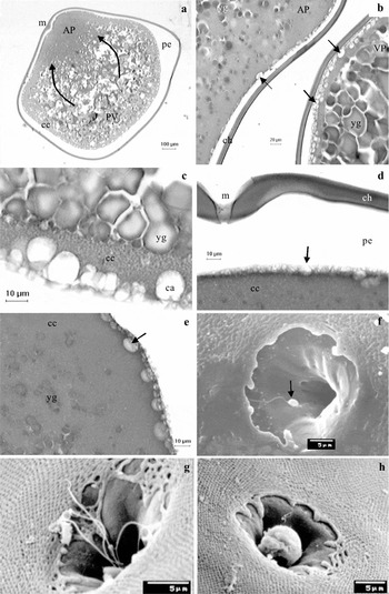
Within samples collected at extrusion, the oocytes presented a cytoplasmic movement towards the micropyle, which characterizes the animal pole (Fig. 1a). Under SEM, the oocyte surface, which corresponds to the chorion or radiate zone, had pores and a single micropyle (Fig. 1g, h).
Oocytes and eggs of B. orbignyanus. Light photomicrography (a–e); scanning electron microscopy (f–h). (a) Oocyte showing cytoplasmic movement (arrows) and consequent animal pole definition at time 0 (HE). (b) Egg showing cortical alveoli (arrows) in the animal and vegetative poles 10 s after fertilization (TM). (c) Oocyte showing the cortical alveoli (ca) in the vegetative pole at the moment of extrusion (HE). (d, e) Oocyte showing cortical alveoli (arrows) in the animal pole at the moment of extrusion (HE). (f) Oocyte evidencing spermatozoon (arrow) close to the micropyle opening at 1.5 min after fertilization. (g) Egg evidencing the micropyle region and showing spermatozoa tails at 1 min after fertilization. The presence of pores in the chorion can also be seen. (h) Egg evidencing the micropyle obstructed by the fertilization cone, resembling externally a spherical structure, at 1 min after fertilization. Animal pole (AP), chorion (ch), cortical alveoli (ca), cortical cytoplasm (cc), micropyle (m), perivitelline space (ps), vegetative pole (VP), yolk granule (yg).

The moment the oocytes and the sperm were mixed was referred to as time 0. At this stage, the animal and vegetative poles were formed. The vegetative pole was formed by the yolk whereas the animal pole was formed by the fused female and male pronuclei plus the displaced cytoplasm. The animal pole was basophilic and homogenous in appearance under light microscopy (Fig. 1a). The vegetative pole was acidophilic and had yolk globules (Fig. 1a), which were smaller when located closer to the animal pole (Fig. 1b). The larger globules, located in the cytoplasm and opposite to the animal pole, resulted from yolk globule coalescence (Fig. 1c).
In the cortical cytoplasm, several cortical alveoli were found aligned around the oocyte perimeter at the moment of fertilization, forming a thin basophilic layer (Fig. 1b, c). It was also observed that, after fertilizing B. orbignyanus oocytes, the number of cortical alveoli layers present in the perimeter of the egg decreased (Fig. 1a, d). These cortical alveoli were larger in size and more abundant at the vegetative pole (Fig. 1c). On the other hand, they were rarer at the animal pole (Fig. 1b, d).
As for the perivitelline space, the vegetative pole region was larger when compared to the animal pole. This feature was observed in B. orbignyanus oocytes from 10 s up to 5 min after fertilization.
It was also observed that, at time 0, the cortical cytoplasm of oocytes presented a higher concentration of cortical alveoli in the vegetative pole in relation to the animal pole (Fig. 1a, b), indicating that the cortical reaction in piracanjuba gets started in the animal pole and proceeds towards the vegetative pole. A few cortical alveoli were observed in the cytoplasm beneath the region close to the micropyle (Fig. 1e) and the cortical reaction was not promptly observed. Thus, the reaction was probably activated by spermatozoon penetration and/or by egg hydration at 1 min after fertilization. The cortical alveoli breakdown promoted the chorion elevation and the consequent enlargement of the perivitelline space, which increased significantly the diameter of the eggs (1.95 ± 0.08 mm) from 6 min on, due to the hydration level of these eggs.
After 10 s, spermatozoa were observed around the micropyle opening (Fig. 1f). After 1 min, several spermatozoa tails were found in the micropyle vestibule (Fig. 1g).
The penetration of the spermatozoon promoted the formation of the fertilization cone. This structure had a spherical shape at the external extremity (Fig. 1h), which obstructed the opening of the micropyle.
Discussion
The eggs of B. orbignyanus were spherical, translucent and greenish, similar to other Brycon species (Eckmann, Reference Eckmann1984; Andrade-Talmeli et al., Reference Andrade-Talmelli, Kavamoto, Romagosa and Fenerich-Verani2001; Romagosa et al., Reference Romagosa, Narahara and Fenerich-Verani2001; Reynalte-Tataje et al., Reference Reynalte-Tataje, Zaniboni-Filho and Esquivel2004). In the present study, the average egg diameter of B. orbignyanus was equal to 1.3 ± 0.11 mm after the extrusion, showing a normal size for this gender, on one occasion Vazzoler (Reference Vazzoler1996) reported an average diameter of 1.55 mm for mature oocytes of B. orbignyanus and Romagosa et al. (Reference Romagosa, Narahara and Fenerich-Verani2001) observed that B. cephalus oocytes presented an average diameter of 1.01 mm.
A cytoplasmic movement towards the micropyle region was observed in the oocyte samples collected during extrusion, which characterized the animal pole. This cytoplasmic motion is stimulated by the fertilization (Kimmel et al., Reference Kimmel, Ballard, Kimmel and Ullmann1995). Under SEM, the oocyte surface of B. orbignyanus, corresponding to the chorion or radiate zone, presented pores and a single micropyle, as previously described in this species by Ganeco & Nakaghi (Reference Ganeco and Nakaghi2003).
An accumulation of yolk granules was observed in the vegetative pole, which decreased in size close to the animal pole. The larger granules located in the cytoplasm opposite to the animal pole resulted from the coalescence of yolk granules. Kobayashi & Yamamoto (Reference Kobayashi and Yamamoto1985) observed that yolk coalescence in Oncorhynchus keta began at the vegetative pole, while the nuclear membrane was disintegrated.
In the cortical cytoplasm of B. orbignyanus, several cortical alveoli, aligned along the oocyte periphery, were observed at the moment of fertilization. The cortical cytoplasm surrounding the micropyle is composed of fat granules and cortical alveoli (Iwamatsu & Ohta, Reference Iwamatsu and Ohta1981; Iwamatsu, Reference Iwamatsu, Tarín and Cano2000). Bazzoli & Godinho (Reference Bazzoli and Godinho1994) analyzed mature oocytes of some freshwater teleosteans and classified the cortical alveoli of B. orbignyanus as discontinuous, bearing multiple layers of small vesicles, always aligned close to the radiate zone. In the present study, we noticed that the number of cortical alveoli layers present in the periphery of B. orbignyanus oocytes have decreased after fertilization.
The perivitelline space was larger at the vegetative pole than at the animal pole. Few cortical alveoli were also observed at the animal pole, mainly within the region close to the micropyle, in other fish species as well, such as Fundulus heteroclitus (Brummett & Dumont, Reference Brummett and Dumont1979), Oryzias latipes (Iwamatsu & Ohta, Reference Iwamatsu and Ohta1981), Oncorhynchus keta (Kobayashi & Yamamoto, Reference Kobayashi and Yamamoto1981), and Prochilodus lineatus (Freire-Brasil et al., Reference Freire-Brasil, Nakaghi, Santos, Grassiotto and Foresti2002). According to Ohta (Reference Ohta1985) and Hart (Reference Hart1990), the animal pole would present few cortical alveoli in order to delay the formation of the perivitelline space in this region thereby favouring the entrance of the spermatozoon.
The cortical alveoli breakdown and the consequent elevation of the chorion in fertilized eggs are known as the cortical reaction (Hart, Reference Hart1990). This was observed in B. orbignyanus eggs 10 s after fertilization and lasted up to 5 min. In this case, the reaction was probably activated by the penetration of the spermatozoon and/or by the hydration of the eggs 1 min after fertilization, similar to the results reported by Ohta et al. (Reference Ohta, Iwamatsu, Tanaka and Yashimoto1990) and by Kobayashi & Yamamoto (Reference Kobayashi and Yamamoto1981) in eggs of Rhodeus ocellatus ocellatus and Oncorhynchus keta, respectively.
This cortical reaction in B. orbignyanus started at the animal pole, similar to observations in Oryzias latipes (Iwamatsu & Ohta, Reference Iwamatsu and Ohta1981; Abraham et al., Reference Abraham, Gupta and Fluck1993), Polyodon sphatula (Linhart & Kudo, Reference Linhart and Kudo1997), Fundulus and Oncorhynchus (Hart, Reference Hart1990). However, in Rhodeus ocellatus ocellatus (Ohta et al., Reference Ohta, Iwamatsu, Tanaka and Yashimoto1990), the cortical reaction initiated at the vegetative pole towards the animal pole. It should be pointed out that the cortical reaction was not readily detected around the micropyle after fertilization, which agrees with the occurrence of few cortical alveoli in the cytoplasm immediately beneath the micropyle as a strategy to retard the formation of the perivitelline space and allow the entrance of the spermatozoon (Ohta, Reference Ohta1985; Hart, Reference Hart1990).
The formation of the perivitelline space in the studied species and the consequent increase caused the chorion to separate from the egg membrane. This event was also observed by Laale (Reference Laale1980). It should be highlighted that during the cortical alveoli breakdown, ions Ca2+ are absorbed from the medium and participate in the hardening of the chorion (Iwamatsu et al., Reference Iwamatsu, Ohta, Oshima and Sugiura1985; Iwamatsu, Reference Iwamatsu, Tarín and Cano2000), playing a key role in the mechanical protection of the developing embryo (Laale, Reference Laale1980; Lönning et al., Reference Lönning, Kjorsvik and Davenport1984).
As for the time spent by the fertilizing spermatozoon to reach the micropyle in the present study, spermatozoa were observed at the opening of the micropyle 10 s after sperm and oocytes were mixed up. In Fundulus heteroclitus, the presence of a fertilizing spermatozoon was observed from 3 s up to 3 min after mixing sperm and oocytes (Brummett & Dumont, Reference Brummett and Dumont1979). In Silurus glanis, the first spermatozoon reached the micropyle 20 s after fertilization (Kudo et al., Reference Kudo, Linhart and Billard1994). In Prochilodus lineatus, the spermatozoon passed through the micropyle between 10 and 20 s after fertilization (Freire-Brasil et al., Reference Freire-Brasil, Nakaghi, Santos, Grassiotto and Foresti2002).
In B. orbignyanus, the fertilization cone acted as a mechanism to suppress polyspermy, intercepting the entry of supernumerary spermatozoa. Externally, this structure was spherical. In Fundulus heteroclitus, a viscous mass released by the oocyte obstructed the micropyle after fertilization (Brummett & Dumont, Reference Brummett and Dumont1979). Conversely, in Oreochromis niloticus and O. aureus, the micropyle was obstructed by an amorphous mass (Bern & Avtalion, Reference Bern and Avtalion1990). Iwamatsu (Reference Iwamatsu, Tarín and Cano2000) reported that this structure is similar to a bubble, composed of a perivitelline fluid released through the micropyle channel. This event is synchronized with the cortical alveoli breakdown (Iwamatsu & Ohta, Reference Iwamatsu and Ohta1981). A similar mechanism was also reported in Polyodon sphatula (Linhart & Kudo, Reference Linhart and Kudo1997) and Oryzias latipes (Iwamatsu et al., Reference Iwamatsu, Onitake, Yoshimoto and Hiramoto1991). In Prochilodus lineatus (Freire-Brasil et al., Reference Freire-Brasil, Nakaghi, Santos, Grassiotto and Foresti2002), polyspermy was blocked by the presence of a spherical structure derived from inside the micropyle, but surrounded by those spermatozoa that failed in entering the micropyle channel.
According to Kudo (Reference Kudo1980), the micropyle channel in Cyprinus carpio allows the access to several spermatozoa, suggesting that the agglomeration of supernumerary spermatozoa in the perivitelline space is required as an additional mechanism to prevent polyspermy. Ginsburg (Reference Ginsburg1961) confirmed the agglutination of spermatozoa in the micropyle channel of Salmo trutta and reported that, by removing the perivitelline fluid, polyspermy was achieved in this species since it would prevent the accumulation of supernumerary spermatozoa.
The following events were described at the first moments of fertilization in B. orbignyanus: cytoplasmic movement towards micropyle in the eggs, cortical alveoli breakdown with the elevation of the chorion, the time the spermatozoon reaches the micropyle and the formation of the fertilization cone. Such knowledge is essential to a better understanding about the first stages of fish reproduction, especially of B. orbignyanus, an endangered species in Brazil.
Acknowledgements
The authors are grateful to Conselho Nacional de Desenvolvimento Científico e Tecnológico (CNPq) for the Master's scholarship to the first author; to Mr Orandi Mateus (FCAV/UNESP) for the histological techniques and to Estação de Pesquisa e Desenvolvimento Ambiental de Volta Grande–CEMIG.