


Introduction
The Mullidae (Perciformes), commonly known as goatfishes, are a large group of marine fishes comprising six genera and 104 species. They occur globally, inhabiting tropical to temperate waters, with occasional reports from brackish habitats (Froese and Pauly Reference Froese and Pauly2026). Mullids are a major host family for the Monorchiidae Odhner, 1911 (Digenea), harbouring approximately 6% of known species; only the Haemulidae, Carangidae, Chaetodontidae and Sparidae support greater richness.
Only three monorchiids have been reported from the 39 species of goatfishes known in Australian waters. Madhavia fellaminuta Wee, Cutmore & Cribb, 2018 and Parachrisomon delicatus (Manter & Pritchard, 1964) Madhavi, 2008, were reported from Upeneus tragula (Richardson) in Moreton Bay, Queensland by Wee et al. (Reference Wee, Cutmore and Cribb2018), and Lobucirruatus infloresco Wee, Cribb & Cutmore, 2022 was described from Parupeneus spilurus (Bleeker) from Moreton Bay and the Great Barrier Reef by Wee et al. (Reference Wee, Cribb and Cutmore2022a). Here, we describe a new species infecting multiple mullid species from Lizard Island on the northern Great Barrier Reef, Moreton Bay, and Ningaloo Reef, Western Australia.
Materials and methods
Host and parasite collection
Mullids were collected from Moreton Bay (south-eastern Queensland), off Lizard Island (northern Great Barrier Reef), and Ningaloo Reef (Western Australia) via line and spear fishing. Fishes were euthanised by cranial pithing, and the gastrointestinal tract was removed and examined for parasites using the gut-wash method described by Cutmore et al. (Reference Cutmore, Bray, Huston, Martin, Miller, Wee, Yong and Cribb2025). Trematodes were washed in vertebrate saline, fixed by pipetting into near-boiling saline and preserved in 80% ethanol for parallel morphological and genetic characterisation. Some specimens were prepared as hologenophores and paragenophores (sensu Pleijel et al. Reference Pleijel, Jondelius, Norlinder, Nygren, Oxelman, Schander, Sundberg and Thollesson2008).
Morphological analysis
Specimens were washed in fresh water, stained in Mayer’s haematoxylin, destained in 1.0% hydrochloric acid and neutralised in 1.0% ammonium hydroxide solution. Specimens were subsequently dehydrated through a graded ethanol series, cleared in methyl salicylate and permanently mounted in Canada balsam. Measurements were taken using an Olympus SC50 digital camera mounted on an Olympus BX-53 compound microscope with accompanying cellSens Standard imaging software and are presented here in micrometres with the range followed by the mean in parentheses; where the length is followed by width, the two measurements are separated by ‘×’. Line drawings were made using a drawing tube fitted to the same BX-53 compound microscope and digitised in Adobe Illustration software v.29.0.1. Specimens collected from Queensland waters are lodged in the Queensland Museum (QM), Brisbane, and specimens collected from Western Australian waters are lodged at the Western Australian Museum (WAM), Perth.
Molecular sequencing
Genetic sequence data were generated for two barcoding regions: the complete second internal transcribed spacer unit (ITS2) ribosomal DNA (rDNA) region and the partial cytochrome c oxidase 1 (cox1) mitochondrial DNA (mtDNA) region, and one phylogenetically informative region: the partial large ribosomal subunit (28S) rDNA region. Amplification of the two ribosomal DNA regions was performed as per the protocols of Wee et al. (Reference Wee, Cutmore, Yong and Cribb2017b) using the primers 3S (5′-GGT ACC GGT GGA TCA CGT GGC TAG TG-3′; Morgan and Blair Reference Morgan and Blair1995) and ITS2.2 (5′-CCT GGT TAG TTT CTT TTC CTC CGC-3; Cribb et al. Reference Cribb, Anderson, Adlard and Bray1998) for the ITS2 region, and LSU5 (5′-TAG GTC GAC CCG CTG AAY TTA AGC A-3′; Littlewood Reference Littlewood1994) and 1500R (5′-GCT ATC CTG AGG GAA ACT TCG-3′; Snyder and Tkach Reference Snyder and Tkach2001) for 28S. Amplification of the mtDNA region was performed following the protocols of Wee et al. (Reference Wee, Cribb, Bray and Cutmore2017a) using the primers Dig_cox1Fa (5′-ATG ATW TTY YTD ATG CC-3′; Wee et al. Reference Wee, Cribb, Bray and Cutmore2017a) and Dig_cox1R (5′-TCN GGR TGH CCR AAR AAY CAA AA-3′; Wee et al. Reference Wee, Cribb, Bray and Cutmore2017a). All amplifications were conducted on a Takara TP-650 PCR Thermocycler. Sanger sequencing was conducted at the Australian Genome Research Facility using the amplification primers for the ITS2 and cox1 region, an internal pair of primers for the 28S region: 300F (5′-CAA GTA CCG TGA GGG AAA GTT G-3′; Littlewood et al. Reference Littlewood, Curini-Galletti and Herniou2000) and ECD2 (5′-CCT TGG TCC GTG TTT CAA GAC GGG-3′; Littlewood et al. Reference Littlewood, Rohde and Clough1997). Contiguous sequences were assembled and edited using Geneious® v. 10.2.6. For comparative purposes, cox1 data were generated for P. delicatus, based on samples reported by Wee et al. (Reference Wee, Cutmore and Cribb2018); ITS2 and 28S data for these samples were published by Wee et al. (Reference Wee, Cutmore and Cribb2018).
ITS2 and cox1 data were aligned separately, with both datasets comprising only specimens corresponding to the concept of Parachrisomon Madhavi, 2008. Alignments of each dataset were conducted independently in MEGA version 7 with the MUSCLE algorithm and UPGMA clustering for iterations 1 and 2 (Kumar et al. Reference Kumar, Stecher and Tamura2016). To determine the correct reading frame, the cox1 alignment was translated with the echinoderm/flatworm mitochondrial code and inspected for internal stop codons in MESQUITE v.3.6 (Maddison and Maddison Reference Maddison and Maddison2019). Once the correct reading frame was identified, the alignment was trimmed such that the reading frame began on position 1. All codons in the alignment were tested for non-stationarity and substitution saturation with a χ2 test run on PAUP* (Swofford Reference Swofford2002) and a Xia’s test run implemented in DAMBE7 (Xia Reference Xia2018; Xia and Lemey, Reference Xia, Lemey, Lemey, Salemi and Vandamme2009; Xia et al. Reference Xia, Xie, Salemi, Chen and Wang2003), respectively; no significant non-stationarity or substitution saturation was detected. Neighbour-joining and pairwise distance matrix analyses were conducted on all three datasets to determine species boundaries and explore the number of base pair differences. The parameters used for the neighbour-joining analyses were: ‘test of phylogeny = bootstrap’, ‘no. of bootstrap replications = 10 000’, ‘model/method = no. of differences’, ‘substitutions to include = d: Transitions + Transversions’ and ‘rates among sites = Uniform rates’. The pairwise distance analyses used the ‘no. of differences’ model and gaps were treated as a character state.
Newly generated partial 28S sequence data were aligned with sequences of comparable monorchiid taxa available on GenBank (Table 1) in MEGA version 7 with the MUSCLE algorithm and UPGMA clustering for iterations 1 and 2 (Kumar et al. Reference Kumar, Stecher and Tamura2016). The resulting alignment was trimmed manually, and indels of three bases or larger, and affecting >5% of sequences were removed; the removed bases amounted to approximately 3% of the original alignment. Maximum likelihood (ML) and Bayesian inference (BI) analyses were conducted using implementations of RAxML version 8.2.6 (Stamatakis Reference Stamatakis2014) and MrBayes version 3.2.7a (Ronquist et al. Reference Ronquist, Teslenko, van der Mark, Ayres, Darling, Höhna, Larget, Liu, Suchard and Huelsenbeck2012), respectively, in the CIPRES portal (Miller et al. Reference Miller, Pfeiler and Schwartz2010). Both analyses assumed the GTR + I + Г model of evolution, based on implementations of both the Akaike Information Criterion (AIC) and Bayesian Information Criterion (BIC) in jModelTest version 2.1.10 (Darriba et al. Reference Darriba, Taboada, Doallo and Posada2012). Nodal support for the ML analysis was estimated by performing 1,000 bootstrap pseudoreplicates. The BI analysis was run over 10,000,000 generations (ngen = 10,000,000) with two runs each containing four simultaneous Markov chain Monte Carlo (MCMC) chains (nchains = 4) and every 1,000th tree saved (samplefreq = 1,000). BI analysis used the following parameters: nst = 6, rates = invgamma, gammacat = 4, and the prior parameters of the combined dataset were set to ratepr = variable. Samples of substitution model parameters are ‘sump burnin’ = 3,000 and ‘sumt burnin’ = 3,000. Sequence data for the Lissorchiidae, the sister family to the Monorchiidae, were included, and Skrjabinopsolus nudidorsalis Sokolov, Voropaeva & Atopkin, 2020 (Deropristidae) was designated as the outgroup taxon, based on the phylogenetic findings of Sokolov et al. (Reference Sokolov, Voropaeva and Atopkin2020).
Collection data for 28S sequences from GenBank analysed in this study

Table 1. Long description
The table contains five columns: Species, Host, Location, GenBank I D, and Reference. It is divided into three taxonomic sections.
Family Monorchiidae:
* Allobacciger annulatus: Host Centropyge tibicen, Location Off Heron Island, Australia, GenBank I D M K 9 5 5 7 8 2.
* Allobacciger brevicirrus: Host Scolopsis bilineata, Location Off Heron Island, Australia, GenBank I D M K 9 5 5 7 8 1.
* Allobacciger polynesiensis: Host Centropyge flavissima, Location Off Moorea, French Polynesia, GenBank I D M K 9 5 5 7 8 0.
* Alloinfundiburictus haemuli: Host Haemulon plumierii, Location Off Sisal, Yucatán, Mexico, GenBank I D O Q 6 7 2 2 8 1.
* Ancylocoelium typicum: Host Trachurus trachurus, Location North Sea, U K, GenBank I D A Y 2 2 2 2 5 4.
* Argenticola shuyinae: Host Gerres subfasciatus, Location Moreton Bay, Australia, GenBank I D P V 7 7 1 1 1 0.
* Cableia pudica: Host Cantherhines pardalis, Location Off Heron Island, Australia, GenBank I D A Y 2 2 2 2 5 1.
* Diplomonorchis fallax: Host Leiostomus xanthurus, Location Mobile Bay, Alabama, U S A, GenBank I D not listed.
* Diplomonorchis leiostomi: Host Leiostomus xanthurus, Location Off Beaufort, North Carolina, U S A, GenBank I D P Q 3 3 6 7 6 0.
* Dipolomonorchis cf. micropogoni: Host Leiostomus xanthurus, Location Mobile Bay, Alabama, U S A, GenBank I D P Q 3 4 9 8 0 3.
* Genolopa ampullacea: Host Haemulon macrostomum, Location Off Islamorada, Florida, U S A, GenBank I D M N 9 8 4 4 7 4.
* Genolopa minuscula: Host Anisotremus surinamensis, Location Off Marathon, Florida, U S A, GenBank I D M N 9 8 4 4 7 2.
* Genolopa vesca: Host Haemulon sciurus, Location Off Long Key, Florida, U S A, GenBank I D M N 9 8 4 4 7 1.
* Gerricola queenslandensis: Hosts Codakia paytenorum and Gerres oyena, Location Off Heron Island, Australia, GenBank I D M Z 2 7 2 0 0 0 and M Z 2 7 1 9 9 9.
* Helicometroides gabrieli: Host Plectorhinchus chrysotaenia, Location Off Lizard Island, Australia, GenBank I D O M 2 3 0 1 3 1.
* Helicometroides longicollis lineage 1: Hosts Diagramma pictum labiosum and Diagramma pictum pictum, Locations Off Heron Island, Australia and Off Minabe, Japan, GenBank I D K J 6 5 8 2 8 7 and O M 2 3 0 1 3 1.
* Helicometroides longicollis lineage 2: Host Diagramma pictum pictum, Location Off Minabe, Japan, GenBank I D O M 2 3 0 1 3 4.
* Helicometroides murakamii: Host Diagramma pictum pictum, Location Off Minabe, Japan, GenBank I D O M 2 3 0 1 2 8.
* Helicometroides wardae: Host Plectorhinchus flavomaculatus, Location Off Heron Island, Australia, GenBank I D O M 2 3 0 1 3 0.
* Hurleytrematoides chaetodoni: Host Chaetodon striatus, Location Mona Passage, Puerto Rica, GenBank I D M H 2 4 4 1 1 6.
* Hurleytrematoides galzini: Host Forcipiger flavissimus, Location Off Heron Island, Australia, GenBank I D M K 5 0 1 9 8 8.
* Hurleytrematoides loi: Host Chelmon rostratus, Location Moreton Bay, Australia, GenBank I D M K 5 0 1 9 8 9.
* Hurleytrematoides morandi: Host Chaetodon lunula, Location Off Heron Island, Australia, GenBank I D M Z 3 2 3 0 8 7.
* Infundiburictus arrhichostoma: Host Diagramma pictum labiosum, Location Off Heron Island, Australia, GenBank I D K J 6 5 8 2 8 9.
* Lasiotocus mulli: Host Mullus surmuletus, Location Off Santa Pola, Mediterranean Sea, Spain, GenBank I D M T 6 6 9 0 1 1.
* Lasiotocus trachinoti: Host Trachinotus carolinus, Location Off Jacksonville, Florida, U S A, GenBank I D M N 9 8 4 4 7 8.
* Lobucirruatus infloresco: Host Parupeneus spilurus, Location Moreton Bay, Australia, GenBank I D O M 8 9 1 7 8 2.
* Madhavia fellaminuta: Host Upeneus tragula, Location Moreton Bay, Australia, GenBank I D M G 9 2 0 2 1 9.
* Monorchis lewisi: Host Acanthopagrus australis, Location Moreton Bay, Australia, GenBank I D M F 5 0 3 3 0 9.
* Monorchis monorchis: Host Diplodus vulgaris, Location Near Corsica, France, GenBank I D A F 1 8 4 2 5 7.
* Obscuromonorchis ranae: Host Gerres oblongus, Location Off Lizard Island, Australia, GenBank I D P V 7 7 1 1 1 1.
* Ovipusillus geminus: Host Gnathanodon speciosus, Location Moreton Bay, Australia, GenBank I D M F 5 0 1 9 8 7.
* Ovipusillus mayu: Host Gnathanodon speciosus, Location Moreton Bay, Australia, GenBank I D M F 5 0 3 3 1 0.
* Parachrisomon delicatus: Host Upeneus tragula, Location Moreton Bay, Australia, GenBank I D M G 9 2 0 2 1 8.
* Postmonorchis orthopristis: Host Haemulon flavolineatum, Location Off Upper Matecumbe Key, Florida, U S A, GenBank I D M N 9 8 4 4 7 5.
* Proctotrema addisoni: Host Diagramma labiosum, Location Off Heron Island, Australia, GenBank I D K J 6 5 8 2 9 1.
* Provitellus chaometra: Host Gnathanodon speciosus, Location Moreton Bay, Australia, GenBank I D M K 5 0 1 9 8 4.
* Provitellus infibrova: Host Gnathanodon speciosus, Location Moreton Bay, Australia, GenBank I D M K 5 0 1 9 8 6.
* Provitellus infrequens: Host Gnathanodon speciosus, Location Moreton Bay, Australia, GenBank I D M K 5 0 1 9 8 5.
* Provitellus turrum: Host Pseudocaranx dentex, Location Off Heron Island, Australia, GenBank I D A Y 2 2 2 2 5 3.
* Pseudohurleytrema yolandae: Host Tripodichthys angustifrons, Location Moreton Bay, Australia, GenBank I D M T 6 4 9 3 0 0.
* Retroporomonorchis pansho: Host Lutjanus fulvus, Location Off Lizard Island, Australia, GenBank I D M T 6 7 2 3 4 0.
* Sinistroporomonorchis glebulentus: Host Mugil curema, Location Off Beaufort, North Carolina, U S A, GenBank I D M N 9 8 4 4 7 6.
* Sinistroporomonorchis lizae: Host Moolgarda perusii, Location Tonkin Bay, Vietnam, GenBank I D L N 8 3 1 7 2 4.
* Sinistroporomonorchis mexicanus: Host Mugil curema, Location Nuevo Campechito, Mexico, GenBank I D O Q 6 7 2 2 9 4.
* Sinistroporomonorchis minutus: Host Mugil curema, Location Laguna de Términos, Mexico, GenBank I D O Q 6 7 2 3 0 7.
* Sinistroporomonorchis yucatanensis: Host Mugil curema, Location Champotón, Mexico, GenBank I D O Q 6 7 2 2 9 7.
* Sinistroporomonorchis sp.: Host Menidia menidia, Location Great Bay Estuary, New Jersey, U S A, GenBank I D M N 9 8 4 4 7 7.
* Monorchiidae sp.: Host Jactellina clathrata, Location Off Heron Island, Australia, GenBank I D M Z 2 7 2 0 0 1.
Family Lissorchiidae:
* Asaccotrema vietnamiense: Host Rasbora paviana, Location Cat Tien National Park, Vietnam, GenBank I D M K 8 6 3 4 0 9.
* Asymphylodora perccotti: Host Perccottus glenii, Location Bolshaya Ussurka River Basin, Russia, GenBank I D F R 8 2 2 7 1 5.
* Asymphylodora progentica: Host Bithynia tentaculata, Location Verkiai pond, Lithuania, GenBank I D M T 1 0 3 4 0 0.
* Lissorchis kritskyi: Host Carpiodes cyprinus, Location Pascagoula River, Mississippi, U S A, GenBank I D A Y 2 2 2 2 5 0.
* Palaeorchis incognitus: Host Rutilus rutilus, Location Kaunas water reservoir, Lithuania, GenBank I D M T 1 0 3 4 0 8.
Family Deropristidae:
* Skrjabinopsolus nudidorsalis: Host Acipenser ruthenus, Location River Volga Basin, Russia, GenBank I D M N 7 0 0 9 9 6.
Results
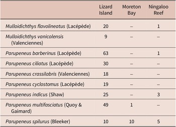
Nine species of mullids were examined for monorchiids (Table 2). Nine species were examined from Lizard Island: Mulloidichthys flavolineatus (Lacépède) (n = 20), Mulloidichthys vanicolensis (Valenciennes) (n = 9), Parupeneus barberinus (Lacépède) (n = 63), Parupeneus ciliatus (Lacépède) (n = 30), Parupeneus crassilabris (Valenciennes) (n = 18), Parupeneus cyclostomus (Lacépède) (n = 19), Parupeneus indicus (Shaw) (n = 25), Parupeneus multifasciatus (Quoy & Gaimard) (n = 49), and Parupeneus spilurus (Bleeker) (n =10). Two species were examined from Moreton Bay: P. multifasciatus (n = 1) and P. spilurus (n = 10). Four species were examined from Ningaloo Reef: M. flavolineatus (n = 1), P. barberinus (n = 1), P. indicus (n = 3), and P. spilurus (n = 5). Adult trematodes consistent with the generic concept of Parachrisomon Madhavi, 2008 were collected from off Lizard Island from M. vanicolensis, P. ciliatus, P. crassilabris, P. indicus, P. multifasciatus, and P. spilurus, and from Moreton Bay and Ningaloo Reef from P. spilurus. Substantial morphological differences were observed among the newly collected specimens, especially in body size, pharynx size, and size and shape of the ovary and testis, between the three locations and also among those from each location.
Numbers of mullid specimens examined at each collection location

Table 2. Long description
The table consists of four columns. The first column lists the species name, followed by three columns for collection locations: Lizard Island, Moreton Bay, and Ningaloo Reef.
* Mulloidichthys flavolineatus (Lacépède): 20 at Lizard Island, 0 at Moreton Bay, 1 at Ningaloo Reef.
* Mulloidichthys vanicolensis (Valenciennes): 9 at Lizard Island, 0 at Moreton Bay, 0 at Ningaloo Reef.
* Parupeneus barberinus (Lacépède): 63 at Lizard Island, 0 at Moreton Bay, 1 at Ningaloo Reef.
* Parupeneus ciliatus (Lacépède): 30 at Lizard Island, 0 at Moreton Bay, 0 at Ningaloo Reef.
* Parupeneus crassilabris (Valenciennes): 18 at Lizard Island, 0 at Moreton Bay, 0 at Ningaloo Reef.
* Parupeneus cyclostomus (Lacépède): 19 at Lizard Island, 0 at Moreton Bay, 0 at Ningaloo Reef.
* Parupeneus indicus (Shaw): 25 at Lizard Island, 0 at Moreton Bay, 3 at Ningaloo Reef.
* Parupeneus multifasciatus (Quoy and Gaimard): 49 at Lizard Island, 1 at Moreton Bay, 0 at Ningaloo Reef.
* Parupeneus spilurus (Bleeker): 10 at Lizard Island, 10 at Moreton Bay, 5 at Ningaloo Reef.
Eighteen new specimens (13 from Lizard Island, two from Moreton Bay, and three from Ningaloo Reef) were sequenced for species delineation. For the cox1 mtDNA region, 18 sequences were generated (13 from off Lizard Island: four from P. crassilabris, two from each of P. ciliatus, P. indicus, P. multifasciatus, and P. spilurus, and one from M. vanicolensis; two from P. spilurus from Moreton Bay; and three from P. spilurus from Ningaloo Reef). These sequences differed at 0–54 base positions (p-distance = 0.0–11.4%), and formed two clades (Figure 1) relating to samples from eastern vs western Australia; sequences from the two clades differ at 46–54 bp (p-distance = 9.7–11.4%). These new sequences differ from the single sequence of P. delicatus at 92–100 bp (p-distance = 19.4–21.1%). For the ITS2 rDNA region, three sequences were generated (two from Lizard Island, one each from P. crassilabris and P. spilurus, and one from P. spilurus from Ningaloo Reef); the two sequences for Lizard Island are identical and differ at 2 bp from the sequence from Ningaloo Reef. For the 28S rDNA region, three sequences were generated for the same individuals sequenced for ITS2. The two sequences from Lizard Island were again identical and differ at 1 bp from the Ningaloo Reef sequence. Sequences of P. delicatus from U. tragula from Moreton Bay differ from the newly generated sequences at 26–28 base positions (p-distance = 5.5–5.9%) and by 33–34 bp (p-distance = 2.7–2.8%) in the ITS2 rDNA and 28S rDNA datasets, respectively.
Relationships between species of Parachrisomon. a, Unrooted neighbour-joining phylogram for the cox1 mtDNA dataset. The scale bar indicates the number of base pair differences. b, Unrooted neighbour-joining phylogram for the cox1 amino acid dataset. The scale bar indicates the number of amino acid substitutions per site.

Figure 1. Long description
Panel a shows a c o x 1 m t D N A phylogram. A scale bar at the bottom left indicates 20 base pair differences. The tree branches from a single root on the left into two main clusters. The bottom branch leads to Parachrisomon delicatus (U. tragula/Moreton Bay/P Z 785831). The upper branch splits into two sub-clusters, both marked with a bootstrap value of 100. The lower sub-cluster contains three specimens of Parachrisomon debonoae n. sp. from Ningaloo Reef. The upper sub-cluster contains twelve specimens of Parachrisomon debonoae n. sp. from Lizard Island and Moreton Bay, showing minor horizontal branch variations.
Panel b shows a c o x 1 amino acid phylogram. A scale bar at the bottom left indicates 0.01 substitutions per site. The tree structure is highly compressed compared to panel a, with almost all specimens of Parachrisomon debonoae appearing on a single vertical line, indicating zero amino acid substitutions between them. Only Parachrisomon delicatus at the bottom is separated by a significant horizontal branch length. The list of 16 specimens on the right matches the taxa in panel a but uses Z-prefixed accession numbers for several entries.
Bayesian inference and maximum likelihood (Figure 2) analyses of the 28S rDNA dataset produced phylograms with similar topologies. In both analyses, the new species resolves as sister to Parachrisomon delicatus, with this clade sister to Ancylocoelium typicum Nicoll, 1912; this larger clade is sister to a clade comprising Madhavia fellaminuta, Ovipusillus geminus Wee, Cutmore & Cribb, 2019, and Ovipusillus mayu Dove & Cribb, 1998. Three minor differences are present between the two analyses, with slight variation in the positions of Alloinfundiburictus haemuli (Overstreet 1969) Wee, Cutmore, Pérez-del-Olmo & Cribb, 2020, Lobucirruatus infloresco, and Monorchis monorchis (Stossich, 1890) Looss, 1902. The nodal support for the placement of these three species is poor in both analyses, and their positions do not affect our interpretation of the new species.
Relationships of the Monorchioidea based on the maximum likelihood analysis of the 28S rDNA dataset. Sequences for the newly characterised species in this study are presented in bold, with members of Parachrisomon marked with a grey box. Posterior probabilities are shown above the nodes, and relationships replicated in the Bayesian inference analysis are shown below or beside them, following a backslash. Nodal support below 0.8/80 is not shown. The scale bar indicates the expected number of substitutions per site. Abbreviations: O, Outgroup taxon.

Figure 2. Long description
The phylogenetic tree branches from a root on the left toward terminal taxa on the right.
At the top, a clade labeled Lissorchiidae includes Palaeorchis incognitus, Asymphylodora species, Asacootrema vietnamiense, and Lissorchis kritskyi.
Below this, a large monophyletic group contains several major clades.
* The first major clade includes Helicometroides species, Hurleytrematoides species, Proctotrema species, and Provitellus species.
* The second major clade includes Lasiotocus species, Diplomonorchis species, and Monorchiidae species.
* The third major clade is the most diverse, containing Pseudohurleytrema, Gerricola, Argenticola, Obscuromonorchis, Retroporomonorchis, Monorchis, Infundiburictus, Paralasiotocus, Allobacciger, Lobucirruatus, Alloinfundiburictus, Sinistroporomonorchis, Postmonorchis, Genolopa, and Ancyclocoelium.
* Near the bottom, a grey box highlights the genus Parachrisomon, containing Parachrisomon debonoae n. sp. in bold text across three entries and Parachrisomon delicatus.
* The final clades include Madhavia fellaminuta and Ovipusillus species.
At the very bottom, Skrjabinopsolus nudidorsalis is marked with an O for Outgroup taxon.
Nodal support is indicated by two numbers separated by a forward slash, such as 100/1 or 97/1, representing maximum likelihood bootstrap and Bayesian posterior probability respectively. A scale bar at the bottom left indicates 0.04 expected substitutions per site.
Although we observed considerable morphological variation, there was no correlation with host or locality. We interpret these molecular and morphological data as consistent with a single species infecting multiple mullids from the Great Barrier Reef, Moreton Bay, and Ningaloo Reef; we formally characterise the species below. Our justification for recognition of a single species is presented in the Discussion.
Parachrisomon debonoae n. sp. (Figures 3 and 4)
Parachrisomon debonoae n. sp. from the eastern and western coasts of Australia. a, Adult worm from P. spilurus, Lizard Island, ventral view, holotype; b, adult worm from P. spilurus, Ningaloo Reef, ventral view; c, terminal genitalia from adult worm from P. spilurus, Lizard Island. Scale bars: a and b, 500 μm; c, 200 μm. Abbreviations: ATO, Anterior part of terminal organ; C, cirrus; CS, cirrus-sac; GA, genital atrium; GP, genital pore; PP, pars prostatica; PTO, posterior part of terminal organ; SV, seminal vesicle; TO, terminal organ; U, uterus; VS, ventral sucker.

Figure 3. Long description
A three-panel anatomical drawing.
Panel a shows a whole adult worm in ventral view with a 500 mu m scale bar. At the anterior end is a circular oral sucker followed by a pharynx. A long esophagus leads to a central V S (ventral sucker). The mid-body contains dark vitelline follicles along the margins and a large posterior region filled with eggs.
Panel b shows a second adult worm specimen with similar morphology, also with a 500 mu m scale bar. The internal organs are arranged identically to panel a, showing the consistency of the species features.
Panel c is a high-magnification view of the terminal genitalia with a 200 mu m scale bar. From top to bottom, the labels indicate:
* G P (genital pore) located just above the large circular V S (ventral sucker).
* G A (genital atrium) positioned to the left of the V S.
* A T O (anterior part of terminal organ) and T O (terminal organ) located below the V S.
* C (cirrus) and C S (cirrus-sac) containing a series of dark, tooth-like structures.
* P T O (posterior part of terminal organ) shown as a large dark sac on the right.
* P P (pars prostatica) and S V (seminal vesicle) at the base of the cirrus-sac.
* U (uterus) containing small oval eggs, winding through the posterior section.
Whole (amended versions of Figure 3a and 3b) and hologenophore adult specimens of Parachrisomon debonoae n. sp. from the eastern and western coasts of Australia demonstrating significant intraspecific morphological variation observed in the species. a, Whole worm from P. spilurus, Lizard Island; b, whole worm from P. spilurus, Ningaloo Reef; c, hologenophore worm from P. crassilabris, Lizard Island; d, hologenophore worm from P. indicus, Lizard Island; e, hologenophore worm from P. multifasciatus, Lizard Island; f, hologenophore worm from P. spilurus, Ningaloo Reef, oral sucker and pharynx not portrayed due to mounting issues. Uterus and eggs from all specimens excluded to show other organs clearly; tegumental spines also not shown. Scale bar: 500 μm.

Figure 4. Long description
A multi-panel line drawing showing six specimens labeled a through f. A vertical scale bar on the right represents 500 micrometers.
* Panel a. A whole worm from P. spilurus at Lizard Island. It has an elongated, spindle-shaped body. At the anterior tip is a circular oral sucker followed by a smaller pharynx. A long esophagus leads to a central cluster of organs including a cirrus sac, ovary, and large posterior testis. Darkly shaded vitelline follicles line the lateral margins.
* Panel b. A whole worm from P. spilurus at Ningaloo Reef. This specimen is significantly longer and more slender than specimen a, particularly in the pre-acetabular region.
* Panel c. A hologenophore worm from P. crassilabris at Lizard Island. It shows a curved body posture with similar internal organ structures as specimen a.
* Panel d. A hologenophore worm from P. indicus at Lizard Island. It has a broader mid-body section compared to specimen c.
* Panel e. A hologenophore worm from P. multifasciatus at Lizard Island. The anterior end is tapered and slightly curved to the left.
* Panel f. A partial hologenophore worm from P. spilurus at Ningaloo Reef. The anterior portion including the oral sucker and pharynx is missing. The drawing focuses on the mid-body organ arrangement and posterior testis.
In all specimens, the uterus, eggs, and tegumental spines are omitted to provide a clear view of the internal anatomy.
Type-host: Parupeneus spilurus (Bleeker), Blackspot goatfish (Perciformes: Mullidae).
Other hosts: Mulloidichthys vanicolensis (Valenciennes), Yellowfin goatfish; Parupeneus ciliatus (Lacépède), Blacksaddle goatfish; Parupeneus crassilabris (Valenciennes), Triplebar goatfish; Parupeneus indicus (Shaw), Indian goatfish; Parupeneus multifasciatus (Quoy & Gaimard), Manybar goatfish (Perciformes: Mullidae).
Type-locality: Off Lizard Island, northern Great Barrier Reef, Australia (14°40′S, 145°27′E).
Other localities: eastern Moreton Bay, Queensland (27°24′S, 153°20′E); Five Fingers, Ningaloo Reef, Western Australia (23°11′S, 113°46′E).
Site of infection: Intestine and rectum.
Prevalence: Lizard Island: M. vanicolensis, 1/9 (11.11%); P. ciliatus, 4/30 (13.33%); P. crassilabris, 4/18 (22.22%); P. indicus, 2/25 (8.0%); P. multifasciatus, 3/48 (6.25%); P. spilurus, 1/10 (10.0%). Moreton Bay: P. spilurus, 1/10 (10.0%). Ningaloo Reef: P. spilurus, 2/5 (40.0%).
Deposition of specimens: Holotype (QM G243017) and 25 paratypes (QM G243018–G243042) in the QM; 8 paratypes (WAM V13834–V13841) in the WAM.
Molecular sequence data: cox1 mtDNA, 15 sequences representative of 10 unique genotypes submitted to (GB PZ785816–PZ785830); ITS2 rDNA (with flanking 5.8S and 28S), three sequences representative of two unique genotypes submitted to GB (PZ785846–PZ785848); partial 28S rDNA, three sequences representative of two unique genotypes submitted to GB (PZ785849–PZ785851).
ZooBank registration: The LSID for P. debonoae is urn:lsid:zoobank.org:act:D3F2E1A3-6FEC-4D20-997C-3C20661D5B78.
Etymology: This species is named in honour of the first author’s wife, Miriam Debono, in recognition of her unwavering support and encouragement over the years.
Description
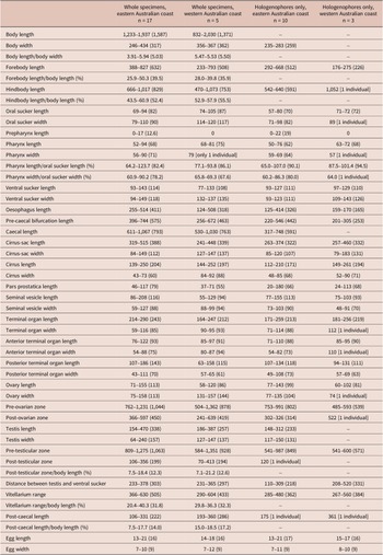
[Based on 34 gravid, unflattened specimens, Table 3; measurements provided in description below relate to specimens from all locations, whereas measurements in Table 3 are separated into eastern and western Australia.] Body elongate, lanceolate to fusiform, slightly tapering both anteriorly and posteriorly, 832–2,030 (1,538) × 235–434 (315), length 3.91–5.94 (5.09) times width, widest at middle of body or middle of hindbody. Forebody 176–827 (558) long, occupying 25.9–50.3 (38.7)% of body length. Hindbody 470–1,073 (804) long, occupying 43.5–60.9 (53.1)% of body length. Tegument thin, covered with small, fine spines. Eye-spot pigment granules observed in forebody in some specimens. Oral sucker terminal, round or oval, with opening distinctly subterminal, 57–105 (79) × 71–120 (90), length 0.95–1.26 (1.13) times width. Ventral sucker in middle third of body, roughly round, 77–143 (112) × 93–149 (118), length 0.87–1.14 (1.04) times width, 0.99–1.76 (1.41) times longer and 1.09–1.63 (1.31) times wider than oral sucker. Prepharynx short, 0–27 (14), not visible in some specimens. Pharynx muscular, elongate oval or subspherical, 50–94 (67) × 51–90 (69); pharynx length 64.2–123.7 (85.5)% of oral sucker length; pharynx width 60.2–90.2 (77.2)% of oral sucker width. Oesophagus typically long, straight or gently sinuous, 124–514 (360) long, occupies 14.9–31.0 (24.9)% of body length. Intestinal bifurcation immediately anterior to ventral sucker; pre-bifurcal zone 201–744 (504) long, occupying 30.8–45.2 (35.6)% of body length. Intestinal caeca blind, long, 317–1,067 (741) long, occupying 37.4–55.4 (49.3)% of body length, terminate near posterior end of testis or in anterior half of post-testicular zone; post-caecal length 106–361 (235), occupying 7.5–20.3 (14.5)% of body length.
Morphometrics of Parachrisomon debonoae measured during this study. Specimens are separated by broad locations, with those from Moreton Bay and Lizard Island grouped under ‘eastern Australian coast’. Measurements are presented in micrometres (μm)

Table 3. Long description
A detailed table presenting measurements in micrometres for forty-eight morphometric categories. The columns are divided into four groups: Whole specimens from the eastern Australian coast (n = 17), Whole specimens from the western Australian coast (n = 5), Hologenophores only from the eastern Australian coast (n = 10), and Hologenophores only from the western Australian coast (n = 3).
Key data points include:
* Body length: Eastern whole specimens range from 1,233 to 1,937 (mean 1,587); Western whole specimens range from 832 to 2,030 (mean 1,371).
* Body width: Eastern whole specimens range from 246 to 434 (mean 317); Western whole specimens range from 356 to 367 (mean 362).
* Oral sucker length: Ranges from 57 to 105 across all groups, with means between 70 and 87.
* Ventral sucker length: Ranges from 77 to 143 across all groups, with means between 108 and 114.
* Egg dimensions: Length ranges from 13 to 21 (mean 16 to 17) and width ranges from 7 to 12 (mean 9) across all categories.
* Other measured features include forebody and hindbody lengths, pharynx dimensions, oesophagus length, cirrus-sac dimensions, ovary and testis dimensions, and various anatomical zones (pre-ovarian, post-ovarian, pre-testicular, and post-testicular).
Cells marked with a dash indicate no data available for that specific category and specimen type.
Note: The three smallest specimens of whole specimens from the western Australian coast were all laterally mounted specimens, and thus, all widths of those specimens were not taken, affecting those metrics.
Testis single, prominent, entire, elongate oval to slightly triangular, mainly in posterior half of hindbody, median, may overlap either or both caeca, separated from ventral sucker by 110–520 (280) or 14.6–29.5 (20.2)% of body length; pre-testicular zone 541–1,351 (957) long, occupying 63.2–74.2 (67.4)% of body length; post-testicular zone 70–413 (194) long, occupying 7.1–21.2 (12.4)% of body length. Cirrus-sac in middle third of body, subcylindrical, mostly median, largely intercaecal, usually slightly overlaps one or both caeca, with posterior end slightly overlapping ovary in some specimens, 241–515 (357) × 79–183 (114). Seminal vesicle ellipsoidal or subspherical, unipartite, 55–208 (110) × 48–127 (87), occupying 22.4–43.3 (30.6)% cirrus-sac length. Pars prostatica simple, straight to strongly curved, with few prostatic cells observed, 20–180 (71) long; prostatic cells only observed to unite with pars prostatica towards anterior end. Cirrus (eversible ejaculatory duct) moderately or very thick, subcylindrical, 112–261 (192) × 43–92 (65), occupying 30.3–66.9 (54.5)% of cirrus-sac length, armed with robust spines. Genital atrium unspined, simple. Common genital pore submedial, immediately anterior to ventral sucker.
Ovary smooth, entire, roughly longitudinally ellipsoidal, in middle third of hindbody, distinctly posterior to ventral sucker, partially overlaps posterior end of cirrus-sac in most specimens, typically anterodextral to and partially overlaps testis, partially overlaps right caecum ventrally in some specimens, 58–155 (103) × 74–158 (112); pre-ovarian zone 485–1,362 (926) long, occupying 60.6–74.8 (65.4)% of body length; post-ovarian zone 241–639 (436) long, occupying 22.9–32.8 (28.8)% of body length. Canalicular seminal receptacle present. Vitellarium composed of two never confluent, branch-like fields of largely tubular follicles, distributed from level of ventral sucker to level of testis, never extends into post-testicular zone, occupying 20.4–40.3 (31.9)% of body length. Uterus restricted to hindbody, thin-walled, extensive, ventral to ovary, testis, caeca and part of cirrus-sac, with coils mostly indiscernible; posterior-most coil reaches to just anterior to posterior body margin; ascending coil enters terminal organ at base of spined anterior section. Metraterm not detected. Terminal organ sinistro-dorsal to cirrus-sac, bipartite, comprising unspined, saccular or slightly tubular posterior chamber, and spined, irregularly shaped anterior section with distinct lobation, 164–290 (229) × 59–116 (87). Posterior chamber ellipsoidal or slightly tubular, contains fibrous mass in most specimens, 63–186 (130) × 43–111 (69). Anterior section irregularly shaped, with distinct irregular lobation, 71–122 (91) × 54–110 (76), armed with fine spines. Eggs small, lightly tanned, operculate, unfilamented, 13–22 (16) × 7–12 (9).
Excretory vesicle small, tubular, reaches just anterior to posterior-most uterine coil. Excretory pore terminal.
Remarks
The specimens that comprise the type series for this species displayed significant morphological variation, particularly in the body size, pharynx size, and form of the ovary and testis; the extent of the variation is explored in the Discussion.
Discussion
Species recognition
In developing the best hypothesis for species recognition among the present samples, we invoked the criteria proposed by Bray et al. (Reference Bray, Cutmore and Cribb2022). These criteria delineate species by an integrative paradigm of reciprocal monophyly at the most discriminating available molecular marker together with consistent differences in morphology or host distribution. Notably, these criteria do not allow specifically for the most problematic of combinations of cryptic species, those with indistinguishable morphology infecting the same host species that demonstrate significant molecular distinction. Here, the molecular distinction in cox1 data between eastern and western Australia was substantial, with specimens from these two locations differing at 46–54 base positions. Such a distinction raises the possibility of recognition of multiple species, as it is greater than the interspecific differences between congeners of some other monorchiid genera (see Wee et al. Reference Wee, Cutmore, Sasal and Cribb2020d) and between congeners of a range of other trematode genera in the tropical Indo-west Pacific (see Cribb et al. Reference Cribb, Bray, Justine, Reimer, Sasal, Shirakashi and Cutmore2022; Cutmore and Cribb, Reference Cutmore and Cribb2021). This distinction, however, is correlated with significant geographic separation; Ningaloo Reef and Lizard Island are separated by over 3,300 km. Further, representative ribosomal data for these two cox1 clades demonstrated only low levels of variation, with specimens from eastern and western Australia differing at just 2 bp in the ITS2 dataset and 1 bp in the 28S dataset.
Host range provides no basis for distinction, with specimens from both cox1 clades found in Parupeneus spilurus.
Initial morphological examination suggested the possibility of multiple species; however, the morphological distinctions did not align with the molecular differences between the east and west coasts of Australia. Of 33 specimens measured, two each from the east and west coasts were significantly larger than all other specimens, measuring 1,925–2,030 μm in length; all other specimens measured only 1,237–1,739 μm. The form of the ovary and testis, size and shape of the pharynx, and extent of the vitellarium and caeca also varied between specimens (see Figure 4). Of these variable characters, the pharynx was most intriguing, almost round in most specimens, but distinctly ellipsoidal and much larger in a few individuals. Ultimately, however, none of these distinctions were reliably associated with the cox1 clades to provide a basis for species distinction between eastern and western samples.
In the light of the overlapping hosts and inconsistent morphological variation, we interpret the specimens from Lizard Island, Moreton Bay, and Ningaloo Reef as representing a single species with significant intraspecific geographical structuring. Comparable interpretations have been made for other monorchiids with wide geographic ranges in the Indo-West Pacific, with intraspecific cox1 variation of 33–36 bp for Helicometroides longicollis Yamaguti, 1934 (see Helicometroides longicollis lineage 1 in Wee et al. Reference Wee, Cutmore, Shirakashi and Cribb2022b), of up to 82 bp for Hurleytrematoides coronatus Manter & Pritchard, 1961 and up to 75 bp for Hurleytrematoides morandi McNamara & Cribb, 2011 (see McNamara et al. Reference McNamara, Miller and Cribb2014). Similar interpretations have also been made for wide-ranging species from other trematode families, such as for Ankistromeces Nolan & Cribb, 2004 and Phthinomita Nolan & Cribb, 2006 [Aporocotylidae] (Cutmore et al. Reference Cutmore, Yong, Reimer, Shirakashi, Nolan and Cribb2021), Emprostiotrema Cianferoni & Ceccolini, 2021 [Emprostiotrematidae] (Huston et al. Reference Huston, Cutmore, Cribb, Sasal and Yong2024), Gorgocephalus Manter, 1966 [Gorgocephalidae] (Huston et al. Reference Huston, Cutmore, Miller, Sasal, Smit and Cribb2021), and Preptetos Pritchard, 1960 [Lepocreadiidae] (Bray et al. Reference Bray, Cutmore and Cribb2022). As mentioned above, some combinations of trematode species from the Indo-West Pacific have been recognised with levels of molecular distinction lower than those reported as intraspecific here. Notably, two combinations of easily morphologically distinguishable combinations of species of Neohexangitrema Machida, 1984 differ by only 27–33 bp and 29–40 bp in cox1 (Cribb et al. Reference Cribb, Martin and Cutmore2025). Despite the inconsistent nature of the available molecular data and the broader problem of interpreting such variation over geographic range, given the absence of distinction in host range and morphology, we conclude that our collection represents a single species.
Parachrisomon debonoae n. sp
Parachrisomon was proposed to accommodate species previously assigned to Chrisomon Manter & Pritchard, 1961 (see Madhavi Reference Madhavi, Bray, Gibson and Jones2008). Chrisomon had been established for species similar to those of the problematic genus Lasiotocus Looss, 1907, with the two genera being differentiated by the form of the vitellarium; species of Chrisomon were reported to possess vitelline follicles arranged in two ‘star-shaped clusters of tubular acini’, rather than the typical and unspecialised arrangement of two lateral fields of follicles. The type-species of Chrisomon was later demonstrated to possess a vitellarium that was consistent with the concept of Lasiotocus, and these genera were thus synonymised (see Bartoli and Bray Reference Bartoli and Bray2004). The remaining eight species of Chrisomon were then transferred to Parachrisomon, with the same vitellarium form of star-shaped tubular acini as the key morphological character separating it from all other monorchiid genera. Notably, our description of P. debonoae vitellarium as ‘branch-like fields of largely tubular follicles’ deviates slightly from that of the generic concept. However, the description of the vitellarium as ‘star-shaped’ is an oversimplification of the shape for some of these species. The vitellarium does branch out from a central point, which is broadly reminiscent of a star, but the generally long branches bear closer resemblance to the description provided here for P. debonoae; regardless, we think the difference in description does not warrant an amended generic diagnosis.
Review of Parachrisomon suggests that its current composition is unlikely to be robust. In addition to P. debonoae and P. delicatus, five species of Parachrisomon, for which no genetic data are yet available, possess the specialised form of the vitellarium characteristic of the genus: Parachrisomon decapteri (Nahhas and Cable, Reference Nahhas and Cable1964) Madhavi, 2008 (type-species), Parachrisomon gaigai (Dove and Cribb, 1998) Madhavi, 2008, Parachrisomon israelensis (Fischthal, Reference Fischthal1980) Madhavi, 2008, Parachrisomon ulua (Yamaguti, Reference Yamaguti1970) Madhavi, 2008, and Parachrisomon weke (Yamaguti, Reference Yamaguti1970) Madhavi, 2008 (see Dove and Cribb Reference Dove and Cribb1998; Fischthal Reference Fischthal1980; Manter and Pritchard Reference Manter and Pritchard1961; Nahhas and Cable Reference Nahhas and Cable1964; Yamaguti Reference Yamaguti1970). However, Parachrisomon albulae (Overstreet, Reference Overstreet1969) Madhavi, 2008 and Parachrisomon brotulidorum (Toman, Reference Toman1973) Madhavi, 2008 apparently lack this specialised vitellarium (see Overstreet Reference Overstreet1969; Toman Reference Toman1973). Parachrisomon albulae reportedly has vitelline follicles arranged in two lateral fields, without any indication of star-shaped tubular acini; rather, they strongly resemble the unspecialised follicles seen in most monorchiid genera. The vitellarium of P. brotulidorum is strikingly different from that of all other species of Parachrisomon, with ‘symmetrical bunches of lobed follicles’ which resemble the vitellarium forms reported for species of Neotimonia Madhavi, 2008, Ovipusillus Dove & Cribb, 1998, and Paramonorcheides Yamaguti, 1938, among other genera. In addition, and perhaps more importantly, P. brotulidorum possesses two testes whereas all other species of the genus possess only one. Parachrisomon brotulidorum was originally described in the genus Paramonorcheides Yamaguti, 1938 (see Toman Reference Toman1973), before being transferred to Parachrisomon by Madhavi (Reference Madhavi, Bray, Gibson and Jones2008). Considering that all species of Paramonorcheides exhibit similar vitellarium form and two testes, we agree with the original designation of the species and recognise it as Paramonorcheides brotulidorum Toman, 1973.
Parachrisomon debonoae is distinguished from P. decapteri, P. delicatus, P. gaigai, P. israelensis, P. ulua and P. weke by the form of the terminal organ. In P. debonoae, the anterior half of the terminal organ is irregularly shaped, with distinct lobes in most specimens; the prominence of these lobes is variable dependent on the orientation in mounted specimens. In all other species of Parachrisomon (including P. albulae), the terminal organ is described and illustrated as bipartite with a spined anterior half and a saccular posterior half, but without any additional specialisation. Parachrisomum debonoae can be further differentiated from P. decapteri, P. israelensis and P. ulua by the distribution of the vitellarium. In P. debonoae, the vitellarium reaches anteriorly to the level of the ventral sucker and posteriorly to the level of the testis, whereas in P. decapteri, P. israelensis and P. ulua it reaches posteriorly into the post-testicular region. Furthermore, P. debonoae can be differentiated from P. israelensis by the form of the prepharynx (short, not always visible vs distinctly long), and from P. ulua in the extent of the uterus (does not extend anteriorly beyond ventral sucker vs extends anteriorly beyond ventral sucker). Parachrisomum debonoae can be further differentiated from P. gaigai by the form of the spines in the terminal organ (narrow vs robust). No consistent feature beyond the specialisation of the terminal organ separates P. debonoae from P. delicatus or P. weke.
Definitive host range of Parachrisomon
Four species of Parachrisomon infect carangids, three infect mullids, and the remaining two infect an albulid and an ophidiid, respectively. Although P. delicatus was reported with Naso sp. or Naso brevirostris (Cuvier) [Acanthuridae] as the type-host, Wee et al. (Reference Wee, Cutmore and Cribb2018) considered the report dubious, suggesting that the infection was spurious or representative of an accidental host. Our team (see Cutmore et al. Reference Cutmore, Bray, Huston, Martin, Miller, Wee, Yong and Cribb2025) has examined many acanthurids in the Indo-West Pacific, including 95 individuals of N. brevirostris, and has found no evidence that species of Naso are infected by monorchiids. The isolated infections of an albulid, Albula vulpes (Linnaeus) for P. albulae, and an ophidiid, Neobythites fasciatus Smith & Radcliffe, 1913 for P. brotulidorum, add to the uncertainty as to whether these species genuinely belong to Parachrisomon. Thus, we consider species of Parachrisomon to principally infect fishes of the Carangidae and Mullidae. No morphological distinction is apparent between the species associated with these two fish families.
Data availability
Sequence data are available on the publicly accessible NCBI GenBank database.
Acknowledgements
For assistance with host collections, we thank Dr Terry Miller, Dr Tim Littlewood, Dr Dan Huston, Dr Delane Kritsky, Dr Dylan Corner, Helen Armstrong, Lenin De Silva, and the various members of the Marine Parasitology Laboratory at the University of Queensland. We greatly appreciate the staff of Lizard Island and Coral Bay Research Stations for their support in the field. This project represents a contribution to Taxonomy Australia (2020), a national initiative organised under the auspices of the Australian Academy of Science that brings together the taxonomic community to develop approaches that will significantly increase the rate at which new species are discovered, resolved and named, with a view to completely documenting the Australian biota within a generation.
Financial support
This project was supported by funding from the Australian Government’s Australian Biological Resources Study National Taxonomy Research Grants Program (NTRGII000024, awarded to NW and THC; 4-H04JDSM, awarded to SCC and THC; and G046WN7, awarded to SBM and THC). Collection in Western Australia was supported by a Pacific Science Foundation Research Grant (APSF21048, awarded to SBM and THC) and funds provided by the Centre for Sustainable Aquatic Ecosystems, Harry Butler Institute, Murdoch University. Species discovery was facilitated by funds from the Nippon Foundation: Nekton Ocean Census programme (https://oceancensus.org).
Competing interests
The authors declare no conflicts of interest.




 Open access

