

Introduction
The affiliation of nematodes with terrestrial molluscs is believed to be ancient and widespread (Morand et al., Reference Morand, Wilson, Glen and GM2004). Seven nematode families have been reported to maintain different types of relationships with terrestrial snails and slugs, ranging from parasitic (obligate or facultative) to phoretic or necromenic relationships (Ross et al., Reference Ross2010b). Slugs are often more parasitized and by a greater diversity of nematode parasites in comparison to snails (Mengert, Reference Mengert1953), mainly because of their increased exposure in soil. A relatively limited amount of research has been carried out on the nematode–mollusc association, with special interest being shown to a rhabditid nematode, P. hermaphrodita, which is used as a biocontrol agent against snails and slugs (Rae et al., Reference Rae2007).
Our knowledge of the diversity of mollusc-associated nematodes around the world comes from various surveys conducted in Australia (Charwat and Davies, Reference Charwat and Davies1999), Belgium (Ross et al., Reference Ross2010b), Bulgaria (Ivanova et al., Reference Ivanova, Panayotova-Pencheva and Spiridonov2013), Chile (Ross et al., Reference Ross2010b), Crimea (Korol and Spiridonov, Reference Korol and Spiridonov1991; Ivanova et al., Reference Ivanova, Panayotova-Pencheva and Spiridonov2013), France (Morand, Reference Morand1986, Reference Morand1992), Germany (Mengert, Reference Mengert1953), Norway (Ross et al., Reference Ross2010b, Reference Ross2016), Slovenia (Laznik et al., Reference Laznik2009; Ross et al., Reference Ross2010b), South Africa (Ross et al., Reference Ross, Ivanova and Malan2011), UK (Ross et al., Reference Ross2010b), USA (Gleich et al., Reference Gleich, Gilbert and Kutscha1977; Ross et al., Reference Ross2010b; Tandingan De Ley et al., Reference Tandingan De Ley2016), and a few other countries from which individual mollusc-associated nematodes were originally described. So far, four species of slug-parasitic nematodes, Agfa flexilis (Dujardin, 1845), Alloionema appendiculatum (Schneider, 1859), Angiostoma dentiferum (Mengert, 1953) and Angiostoma limacis (Dujardin, 1845), have been reported from Belgium (Ross et al., Reference Ross2010b; Ivanova et al., Reference Ivanova, Panayotova-Pencheva and Spiridonov2013). To further add to the information on the diversity of slug parasites in Belgium, an exploratory survey for slug-associated nematodes was conducted in the summer of 2018 at five locations in East and West Flanders.
Six known parasitic species, including the four species already reported, were detected and confirmed based on morphological and molecular analyses. Newly generated sequences for the detected species of slug-associated nematodes were deposited in GenBank. A new species, Angiostoma gandavensis n. sp. (Angiostomatidae), was also found in Ghent (East Flanders) and is described and classified here, bringing the total number of mollusc-associated Angiostoma species to 15. This work thus provides more information on the diversity of slug parasites in Belgium, and in the East and West Flanders provinces in particular.
Materials and methods
Slug collection and nematode extraction
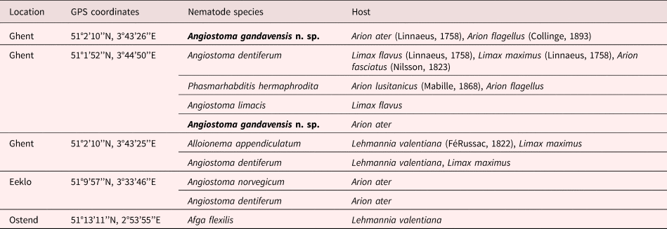
Slugs were collected from household gardens and a forest (see table 1 for locations) and were identified based on morphology (Kerney et al., Reference Kerney, Cameron and Riley1979; Mc Donnell el al., Reference McDonnell, Paine and Gormally2009; Thomas et al., Reference Thomas2010). They were kept in plastic containers lined with filter paper. The filter paper was sprayed with water regularly to retain moisture, and the slugs were fed on carrot. Before dissection, each slug was washed well with water to remove any nematodes adhering to the surface. Each slug was dissected in Ringer's solution and the body's interior was investigated. Nematodes found were extracted and stored in Ringer's solution for a very short period of time before further morphological and molecular analyses.
Slug-parasitic nematodes along with hosts from five locations in East and West Flanders, Belgium, collected in the summer of 2018. A total of six known parasites and a new angiostomatid species (in bold) were detected.

Morphological analysis
Morphological and morphometric characterization was done based on fixed nematodes. For the preparation of permanent slides, a small suspension of nematodes in an embryo dish were killed and fixed by adding a few drops of Trump's fixative (2% paraformaldehyde, 2.5% glutaraldehyde in 0.1m Sorenson buffer (Sodium phosphate buffer at pH 7.5)). The embryo dish was subsequently heated in a microwave (700 W) for 5 s, left to rest for 1 h at 20°C followed by 24 h at 4°C to ensure maximum penetration of the fixative, as described in Singh et al. (Reference Singh2018). Following this, the nematodes were gradually transferred to anhydrous glycerin for permanent slides (Seinhorst, Reference Seinhorst1959) and mounted on glass slides for light microscopic study. Nematodes were examined, photographed, drawn and measured using an Olympus BX51 DIC Microscope (Olympus Optical, Tokyo, Japan) equipped with an Olympus C5060Wz camera and a drawing tube. For scanning electron microscopy, specimens fixed in Trump's fixative were washed in 0.1m phosphate buffer (pH 7.5) and dehydrated in a graded series of ethanol solutions, critical-point-dried with liquid CO2, mounted on stubs with carbon tabs (double conductive tapes), coated with gold of 25 nm, and photographed with a JSM-840 EM (JEOL) at 12 kV (Singh et al., Reference Singh2018).
Molecular analysis
Nematode morphological vouchers were prepared prior to DNA extraction. These vouchers were made with LM of temporary mount using photomicrographs. Vouchered nematodes were subsequently picked from temporary mounts, and each specimen was cut into pieces in distilled water and transferred to a polymerase chain reaction (PCR) tube with 20 μl of worm lysis buffer (50 mm KCl, 10 mm Tris at pH 8.3, 2.5 mm MgCl2, 0.45% NP 40 (Tergitol Sigma), 0.45% Tween 20) followed by incubation at −20°C (10 minutes), adding of 1 μl proteinase K (1.2 mg/ml), incubation at 65°C (1 h) and 95°C (10 minutes) and ending by centrifuging the mixture at 14,000 g for 1 minute (Singh et al., Reference Singh2018). The primer pair D2A: 5′-ACAAGTACCGTGAGGGAAAGTTG-3′ / D3B: 5′-TCCTCGGAAGGAACCAGCTACTA-3′ (Nunn, Reference Nunn1992) was used to amplify D2D3 expansion segment of 28S rDNA following the thermal profile of 95°C for 4 minutes, 5× (95°C for 30 s, 58°C for 30 s, −1°C/cycle, ramp 3°C/s, 72°C for 2 minutes), 45× (95°C for 30 s, 54°C for 30 s, 72°C for 1 minute) and a final hold at 20°C. Amplification of partial sequence of 18S rDNA was done using the primer pair, SSU18A: 5′-AAAGATTAAGCCATGCATG-3′ / SSU26R: 5′-CATTCTTGGCAAATGCTTTCG-3′ (Mayer et al., Reference Mayer, Herrmann and Sommer2007) with the thermal profile as in Singh et al. (Reference Singh2018). For amplifying ITS of rDNA, Vrian2F: 5′-CTTTGTACACACCGCCCGTCGCT-3′ / Vrian2R: 5′-TTTCACTCGCCGTTACTAAGGGAATC-3′ (Vrain et al., Reference Vrain1992; Nguyen et al., Reference Nguyen2017) were used with the following thermal profile: 94°C for 4 minutes, 5× (94°C for 30 s, 64°C for 30 s, −1°C/cycle, ramp 3°C/s, 72°C for 2 minutes), 30× (94°C for 30 s, 60°C for 30 s, 72°C for 1 minute), 72°C for 10 minutes and a final hold at 4°C. Finally, for amplification of COI of mtDNA, the primer pair COI-F1: 5´-CCTACTATGATTGGTGGTTTTGGTAATTG-3´ / COI-R2: 5´-GTAGCAGCAGTAAAATAAGCACG-3´ (Kanzaki and Futai, Reference Kanzaki and Futai2002) was used following the thermal profile of 40× (94°C for 5 minutes, 94°C for 30 s, 51°C for 30 s, 68°C for 2 minutes), 68°C for 10 minutes and final hold at 10°C. The PCR products were purified and sent for sequencing at Macrogen (https://dna.macrogen.com).
Phylogenetic analysis
The phylogenetic relationship of A. gandavensis n. sp. with other related species was analysed based on 18S rDNA and the D2D3 expansion segment of 28S rDNA. Phylogenetic programs implemented in Geneious 10.0.9 (https://www.geneious.com) were used. The consensus sequences of the newly generated sequences were obtained by assembling the forward and backward sequences. BLAST search was used to check for closely related species in GenBank. The newly generated sequences were aligned with closely related species using MUSCLE with default parameters, followed by manual trimming of the poorly aligned ends. Phylogenetic trees were created by using the MrBayes 3.2.6 add-in of Geneious 10.0.9 using a generalized time-reversible (GTR) substitution model with gamma rate variation. The Markov chains were set with 1 × 10⁶ generations, four runs, 20% burn-in, and subsampling frequency of 500 generations (Huelsenbeck and Ronquist, Reference Huelsenbeck and Ronquist2001).
Results
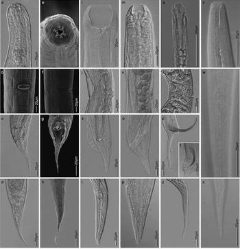
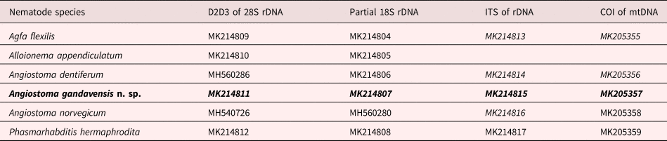
Six known species of parasitic nematodes and a new angiostomatid species were detected from various slug hosts collected from the five locations in Belgium (see table 1). Of 239 slugs that were dissected, 52 (22.8%) were found with parasites in their internal bodies. Agfa flexilis, A. appendiculatum, A. dentiferum and A. limacis were already known in Belgium, whereas A. norvegicum and P. hermaphrodita were recorded for the first time in the country (fig. 1). Only juveniles of P. hermaphrodita were detected in our study. Based on the morphological studies referenced to Hooper et al. (Reference Hooper1999) and a further molecular analysis, the presence of P. hermaphrodita juveniles in some of our slug samples was confirmed. Figure 1 provides photographic documentation of five detected species of slug-parasitic nematodes, focusing mainly on some of the informative characters of the parasites. The photomicrographs show the anterior regions, parts of the reproductive systems and the tail regions. An LM photograph for A. appendiculatum was not included, as no high-quality pictures are available. Two different parasites were sometimes detected from one host. Alloionema appendiculatum was often found together with A. dentiferum on Limax maximus (Linnaeus, 1758) specimens. Juveniles of P. hermaphrodita were also detected alongside A. dentiferum in one host. Angiostoma limacis adults were identified morphologically only, as not enough material was available for molecular work. Besides producing 18S and 28S rDNA sequences for the six slug-parasitic nematodes, this work also generated three new COI and four new ITS sequences from some of the nematode species (see table 2).
Light microscopy (LM) microphotographs and scanning electron microscopy (SEM) images of five slug-parasitic nematode species detected from five locations in Belgium in the summer of 2018. (a–h) Angiostoma dentiferum. (i–l) Angiostoma limacis. (m–p) Angiostoma norvegicum. (q–u) Agfa flexilis. (v–x) Phasmarhabditis hermaphrodita. The figure shows an overview of the head and neck regions (first row), female vulval regions (second row, excluding w), male tail regions, including spicules and bursae (third row, excluding w) and female tail regions (last row excluding w) of the nematodes. The LM microphotographs for P. hermaphrodita are for juveniles only, and the images (b) and (e–h) are SEM images of A. dentiferum.

GenBank accession numbers of submitted sequences of slug-parasitic nematodes found in this study. The new species is in bold. Accession numbers in italics are for sequences generated for the first time.

Angiostoma gandavensis n. sp. was found parasitizing two species of arionid slugs from two locations in Ghent. The nematodes were found in a group of two to three individuals per host in the anterior part of the host's intestine. Only adult stages of both sexes, and no juveniles, were detected and all females were observed with matured eggs.
Angiostoma gandavensis n. sp.
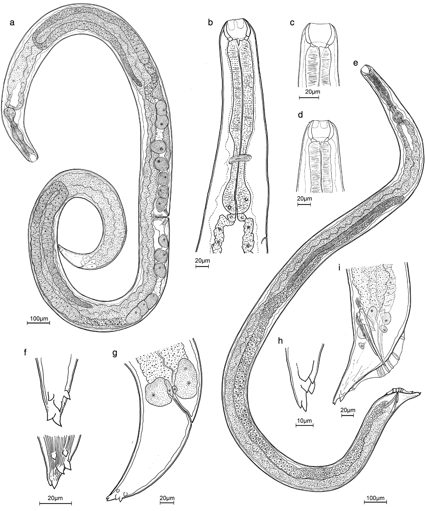
Description ( figs 2–4; table 3)
General. Long cylindrical body with transversely striated body cuticle ornamented with longitudinal ridges. Differentiation of lateral field starting about 2.5 head widths from anterior end as a slightly marked ridge; lateral field showing a narrow mid-ridge (ala), only clearly visible by SEM. Truncated head with differentiated anterior rim bearing six elevations with six outer labial papillae, and adjacent to each lateral papilla the pore-like amphidial aperture. The six inner labial and the four outer cephalic sensilla not observed. Stoma composed of a large barrel-shaped cheilostom with thick sclerotized wall, followed by a slightly narrower short gymnostom with thickened sclerotized wall, and a non-sclerotized funnel-shaped stegostom surrounded by pharyngeal tissues and provided with three tooth-like structures. Muscular and club-shaped pharynx with more or less expanded corpus end, narrow isthmus and rounded to oval bulb without distinct valve. Nerve ring surrounding mid-isthmus region. Prominent cardia. Intestine with strong border of microvilli and rectum with enlarged rectal glands. Secretory–excretory pore posterior to pharynx–intestine junction. Tail short, conical, and tip with five to six unequal mucronate structures. Phasmid indistinct.
Female. (Based on seven female specimens including holotype) Longer body than males. Lateral ala ending just anterior to tail tip. Reproductive system didelphic–amphidelphic, ovaries long and strongly reflexed, with a flexure length of c. 700 μm. Long oviducts containing sperm cells. Uteri of equal length and each branch containing an average of 10–12 eggs. Eggs oval to rounded, with average length and breadth of 61.8 μm and 45.7 μm, respectively. Vulval opening almost at 50% of body length, slit-like vulva with small vulval lips. Straight vagina. Rectum long with thick lining and four rectal glands.
Male. (Based on three male specimens) Lateral ala extending to level of spicule manubrium. Stoma slightly smaller than that of females. Reproductive system monorchic with testis flexure of c. 600 μm (c. 1/4 of body length). Spicules equal, slightly curved ventrally, manubrium weakly differentiated. Pre-anal flap present and a pair of post-cloacal papillae on either side of cloacal opening. Gubernaculum short, c. 1/3 of spicule length. Six rectal glands. Leptoderan bursa, narrow and extending almost to the tail tip. Seven pairs of genital papillae (GP) arranged as 1 + 2/3 + 1, with GP1 and GP7 being the shortest. GP7 close to tail tip.
Line drawings of Angiostoma gandavensis n. sp. in lateral view. (a) Whole body of the female holotype. (b) Anterior region showing head, neck, start of the intestine and secretory–excretory pore. (c, d) Head and neck regions. (e) Whole body of a paratype male. (f, g) Tail regions of female paratypes, showing rectal glands, anus and mucronate structures on tail tips. (h–i) Tail regions of a male paratype, showing spicule and bursa (only right side) and tail tip.

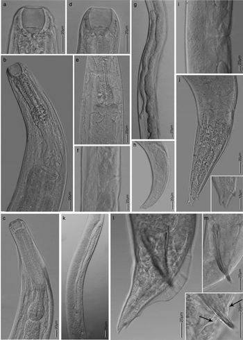
Light microscopy (LM) microphotographs of Angiostoma gandavensis n. sp. in lateral view. (a–d) Head and neck regions. (e) Pharynx–intestine junction. (f) Lateral field with a faintly visible narrow ridge (ala) around vulval region. (g) Reflex of anterior female ovary. (h) Tail region of female, showing rectal gland, rectum and anus. (i) Female vulval region. (j) Tail region of female showing tail tips. (k) Testis of male. (l–n) Tail regions of male, showing spicules, bursa with genital papillae, pre-anal flap and a post-cloacal papilla, indicated by arrows in (n), and tail tip.

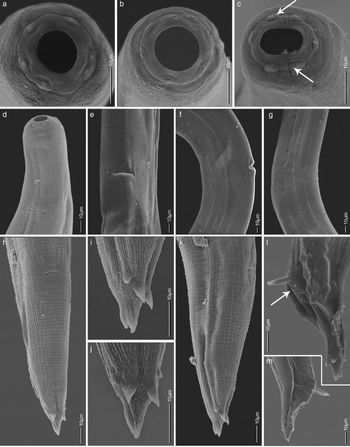
Scanning electron microscopy images of Angiostoma gandavensis n. sp. (a–c) Mouth opening, showing six outer labial papillae and amphidal pores (indicated by arrows). (d) Head and neck region, showing the start of lateral field differentiation. (e) Female vulval region in ventral view. (f) Female vulval region in lateral view, showing a narrow mid-ridge (ala). (g) Lateral field region of male. (h) Tail of female in ventral view, with anal opening. (i, j) Tail tips of female in lateral and ventral views, showing mucronate structures. (k) Tail of female in lateral view. (l, m) Tail regions of male, showing extruded spicules, bursae, pre-anal flap and a post-cloacal papilla (indicated by arrow) and mucronated tail tips.

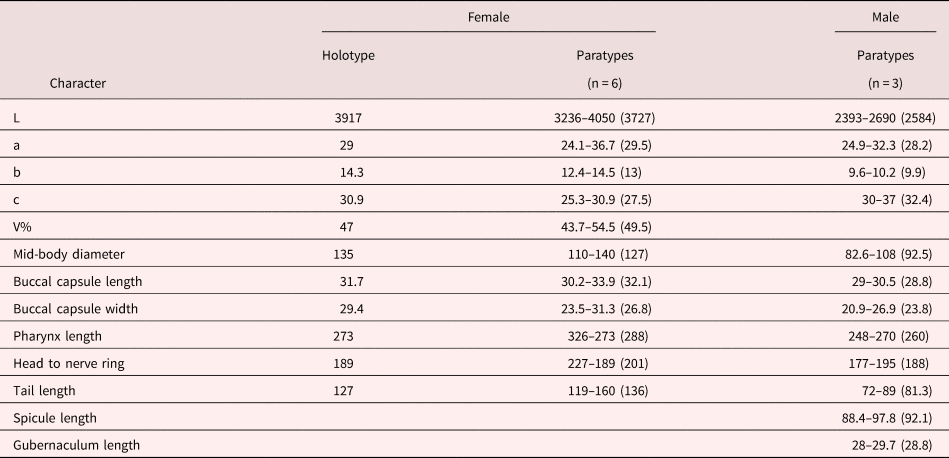
Morphometric data for Angiostoma gandavensis n. sp. Measurements were made on fixed specimens, and are given in μm and in the form range (mean).

Remark: Ten nematode specimens from two different hosts from two locations were measured. No drastic differences in the morphometrics of the individuals from the two hosts and among the populations from the two locations were noted.
Taxonomic summary
Type host. Arion ater.
Other host. Arion flagellus.
Type locality. Ghent (51°1′52″N, 3°44′50″E).
Other localities. See table 1.
Type material. Female holotype and one male paratype collected from Ghent (51°1′52″N, 3°44′50″E) were deposited at Ghent University Museum Zoology Collections, collection numbers UGMD 104396 (holotype) and UGMD 104397 (paratype). Additionally, two female paratypes in two different slides are available in the UGent Nematode Collection (slide UGnem-233-234) of the Nematology Research Unit, Department of Biology, Ghent University, Belgium.
Site in host. Isolated from the anterior part of intestinal lumen of the hosts.
Representative DNA sequences. The sequences obtained for A. gandavensis n. sp. were deposited in NCBI GenBank under accession numbers MK214807 (partial 18S rDNA), MK214811 (D2D3 expansion segment of 28S rDNA), MK214815 (ITS of rDNA) and MK205357 (COI of mtDNA). The sequences for each gene were identical and therefore only one representative sequence per gene was submitted.
Etymology. The species name is derived from the name of the city from which its host was collected.
Diagnosis
Angiostoma gandavensis n. sp. is characterized by average body length of 3727 μm in females and 2584 μm in males, truncated head, presence of a lateral ala, spacious stoma with an anterior large barrel-shaped cheilostom, slightly narrower short gymnostom and a funnel-shaped stegostom surrounded by pharyngeal tissues and provided with three tooth-like structures, club-shaped pharynx with no distinct valve, female reproductive system didelphic–amphidelphic with reflexed ovaries, male monorchic, spicules with an average length of 92 μm and slightly curved ventrally, small gubernaculum of average length 28 μm, presence of pre-anal flap and two post-cloacal papillae on either side of cloacal opening, leptoderan bursa, genital papillae arranged in the pattern 1 + 2/3 + 1, conical tail, tail tip with 5–6 unequal mucronate structures and parasitizing A. ater and A. flagellus.
Morphologically, A. gandavensis n. sp. is similar to A. limacis in the shape of the head, similar shape and structure of stoma, reflexed female ovaries, presence of two post-cloacal papillae on either side of cloacal opening, similar shape of tail in both sexes, and with tail tips supplied by mucronate structures. However, A. gandavensis n. sp. differs from A. limacis by the presence vs absence of lateral alae, the 1 + 2/3 + 1 vs 1 + 2/3 + 3 arrangement of male genital papillae, and the shorter average body length (3727 μm vs 4450 μm for females and 2584 μm vs 4650 μm for males).
Angiostoma gandavensis n. sp. and A. norvegicum have similar head shape and stoma shape and structure; presence of lateral alae, pre-anal flap, a pair of post-cloacal papillae on either side of cloacal opening, conical tails and tail tips with mucronate structures. Angiostoma gandavensis n. sp. and A. norvegicum differ by the 1 + 2/3 + 1 vs 1 + 2/3 + 3 arrangement of male genital papillae, shorter average body length (3727 μm vs 5946 μm for females and 2584 μm vs 4650 μm for males), longer stoma length (33–36 μm vs 14–17 μm), and female ovaries (reflexed vs outstretched).
Angiostoma gandavensis n. sp. is also similar to A. margaretae and A. milacis, by the shape of the head, the presence of lateral alae and similar shape and structure of the spacious stoma. Major differences of the new species from these species include reflexed female ovaries vs outstretched ovaries, presence vs absence of tail mucronate structures, and different arrangements of male genital papillae.
Phylogenetic relationships
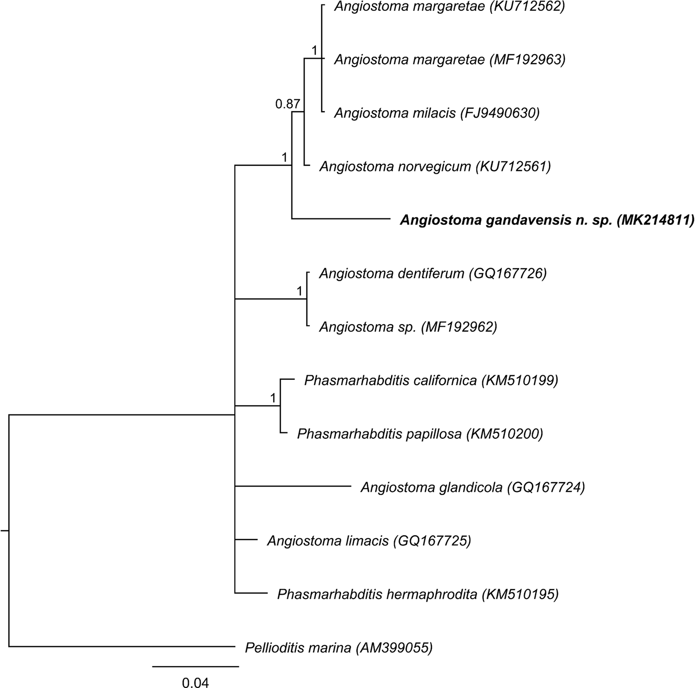
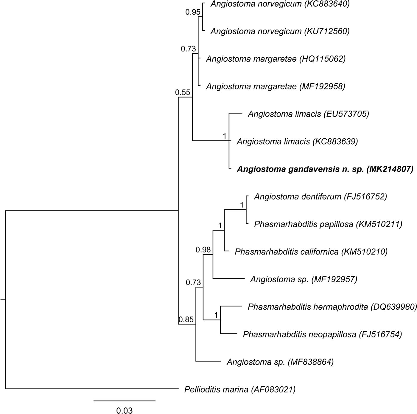
The phylogenetic relationships inferred by D2D3 of 28S rDNA (580 nucleotides) revealed a maximally supported sister relationship of A. gandavensis n. sp. with A. margaretae, A. milacis and A. norvegicum (fig. 5). However, its relationships to A. dentiferum, A. glandicola, A. limacis, Angiostoma sp. and Phasmarhabditis spp. appears to be unresolved. The monophyly of Angiostoma could therefore not be demonstrated. Based on the tree topology based on 18S rDNA (650 nucleotides), A. gandavensis n. sp. and A. limacis form a maximally supported clade; however, their sequences remain unresolved (fig. 6). Angiostoma gandavensis n. sp., A. limacis, A. norvegicum and A. margaretae form a clade, albeit with poor support (PP 0.55). Phasmarhabditis spp. appear to be imbedded within Angiostoma spp., making Angiostoma paraphyletic.
Phylogenetic relationships of Angiostoma gandavensis n. sp. (in bold) with seven Angiostoma spp. and three Phasmarhabditis spp. as inferred from analysis of D2D3 expansion segment of 28S rDNA sequences with the GTR model. The branch support is indicated by posterior probabilities.

Phylogenetic relationships of Angiostoma gandavensis n. sp. (in bold) with six Angiostoma spp. and four Phasmarhabditis spp. as inferred from analysis of partial sequences of 18S rDNA with the GTR model. The branch support is indicated by posterior probabilities.

Discussion
This survey provides an overview of the diversity of slug-associated nematodes in the East and West Flanders provinces of Belgium. Most of the detected parasitic species from this study have also been reported from several parts of Europe and the USA. Besides redetection of Alloionema, three other well-known slug-parasitic nematodes, Agfa, Angiostoma and Phasmarhabditis, which occur within a monophyletic clade (Ross et al., Reference Ross2010b), were also found in this study. Agfa flexilis and A. limacis were reported to be found in Belgium based on unpublished data as mentioned in Ivanova et al. (Reference Ivanova, Panayotova-Pencheva and Spiridonov2013). In addition to photographic documentation of all recovered slug parasites (except A. appendiculatum), this work also generated new molecular data, including COI and ITS sequences, both from previously described parasitic species and the newly found species.
Our results confirm the broad host range of the slug parasites (Ross et al., Reference Ross2010b). For example, A. appendiculatum was reported from Arion lusitanicus (Ross et al., Reference Ross2010b) from Belgium, whereas we found this species in Lehmannia valetiana and Limax maximus. Although not host-specific, all the detected angiostomatid parasites and A. flexilis are confirmed obligate parasites (Ivanova and Wilson, Reference Ivanova and Wilson2009), as fully developed adult stages of these nematodes were detected from the anterior part of the intestinal lumen of the hosts. Phasmarhabditis spp. are facultative parasites (Tan and Grewal, Reference Tan and Grewal2001a; MacMillen et al., Reference MacMillen2009), and only juveniles of P. hermaphrodita were found in our slug samples. Phasmarhabditis hermaphrodita was established as a biological control agent (available commercially as Nemaslug®) (Wilson et al., Reference Wilson, Glen and George1993; Rae et al., Reference Rae2007), and the virulence of its associated bacteria to certain invasive slug hosts (Tan and Grewal, Reference Tan and Grewal2001b), host preference, and chemoattraction (Rae et al., Reference Rae, Robertson and Wilson2009) are relatively well studied. However, Ross et al. (Reference Ross2010a) indicated that many other slug parasites are also present in Europe, with 12 species in the UK alone. They also suggested that the release of parasites might have played a role in the successful invasion of the USA by European slugs. The potential for using A. gandavensis n. sp. and the other recovered species as novel biological control agents for some slug host species remains to be investigated.
With the discovery of Angiostoma gandavensis n. sp., the total number of described species of the genus Angiostoma is now 19, of which 15 are described from molluscan hosts (Dujardin, Reference Dujardin1845; Mengert, Reference Mengert1953; Morand, Reference Morand1986, Reference Morand1988, Reference Morand1992; Morand and Spiridonov, Reference Morand and Spiridonov1989; Korol and Spiridonov, Reference Korol and Spiridonov1991; Morand and Barker, Reference Morand and Barker1995; Luc et al., Reference Luc, Spiridonov and Wilson2005; Ivanova and Wilson, Reference Ivanova and Wilson2009; Ivanova and Spiridonov, Reference Ivanova and Spiridonov2010; Ross et al., Reference Ross, Ivanova and Malan2011, Reference Ross2017). Several traits, such as stoma shape and structure, lateral allae and reproductive systems, have been found to be informative for identification within this genus. Identification of specimens from the Angiostoma species appears to be therefore possible based on morphometrics and morphological characters alone, which is also facilitated by a dichotomous key for the identification of 14 Angiostoma spp. (Ivanova and Wilson, Reference Ivanova and Wilson2009). On the other hand, informative molecular information is limited; only the sequences of seven identified Angiostoma species and two unidentified Angiostoma species were available in GenBank as of December 2018 and, furthermore, some of the assigned sequences appeared to be unreliable. In addition, COI sequences of mtDNA, which is often considered to be a more reliable molecular barcode for animal species identification (Hebert et al., Reference Hebert2003; Deagle et al., Reference Deagle2014), are almost completely lacking.
Most importantly, as some mislabelled sequences in GenBank were detected during our sequence analyses, a correct sequence assignment to slug-parasitic nematodes is of vital importance. For example, partial 18S and D2D3 sequences from the same DNA material were found to be almost 100% similar to sequences from two different species. Nematodes identified morphologically as A. dentiferum and confirmed by D2D3 sequence analyses (571 out of 573 nucleotides similar with GQ167726) were, according to the partial 18S rDNA sequences, 100% similar to an unknown Angiostoma sp. (MF192957) and clearly different from A. dentiferum (FJ516752) (20 out of 673 nucleotides different). Our study also indicated a limitation of 18S rDNA to identify Angiostoma spp., as partial sequences of 18S rDNA (800 bp) from A. gandavensis n. sp. showed 99% similarity to sequences from A. limacis (KC883639). However, the latter is morphologically very different from A. gandavensis n. sp., and their D2D3 sequences also differed by 42 out of 585 nucleotides from the reference (GQ167725).
Thus, although morphological features are useful for species identification within the genus Angiostoma, molecular data should also be generated to facilitate easy identification at the species level and to further facilitate the study of phylogenetic relationships. In a recent work by Ivanova and Spiridonov (Reference Ivanova and Spiridonov2018), a D2D3 of 28S-based phylogenetic relationship analyses revealed unresolved relations of several Angiostoma spp., including A. kimmeriense. Similarly, our study could not establish the exact phylogenetic position of three Angiostoma species based on D2D3 of 28S rDNA. Nonetheless, in the current study, the position of A. gandavensis n. sp. appears to be resolved based on the phylogenetic analyses of both partial 18S and D2D3 of rDNA.
Author ORCIDs
P.R. Singh, 0000-0003-1891-3142
Acknowledgements
We thank Ghent University for all the facilities made available to conduct this work. We are also thankful to Elena S. Ivanova of the A.N. Severtsov Institute of Ecology and Evolution, Russian Academy of Science, Moscow for her valuable input during literature studies, and to all the gardeners for their contributions during slug collections.
Financial support
This research work was funded by a Special Research Fund of Ghent University, Belgium (grant number BOF 01D05918).
Conflict of interest
None.