Introduction
Cystic echinococcosis is a global zoonotic infection that is economically important and is considered as a threat to public health in many countries. Various studies have indicated that hydatid cysts are commonly found in sheep, camels, cattle and goats throughout Iran (Ahmadi, Reference Ahmadi2005; Rokni, Reference Rokni2009; Borji et al., Reference Borji, Azizzadeh and Afsai2011, Reference Borji, Azizzadeh and Afsai2012).
Recent studies have suggested that there are ten distinct genetic types within Echinococcus granulosus, namely E. granulosus sensu stricto (G1, G2, G3), E. equinus (G4), E. ortleppi (G5), E. intermedius (G6, G7, G9) and E. canadensis (G8, G10), which vary in their infectivity in humans and livestock (Thompson, Reference Thompson2008). The life cycle of E. granulosus includes dogs and other canines as the definitive hosts of the adult parasite, while sheep and numerous ungulates (goats, swine and cattle) are intermediate hosts, harbouring the hydatid cyst (Thompson & McManus, Reference Thompson, McManus, Eckert, Gemmell, Meslin and Pawlowski2001). Camels have an important role in the transmission cycle of hydatidosis. Previous studies had indicated that camels harbour the G6 genotype of E. granulosus (Thompson, Reference Thompson2008). Recently, however, several findings have reported the appearance of the E. granulosus G1 and G3 genotypes from camels (Sharbatkhori et al., Reference Sharbatkhori, Mirhendi, Fasihi Harandi, Rezaeian, Mohebali, Eshraghian, Rahimi and Kia2010, Reference Sharbatkhori, Harandi, Mirhendi, Hajialilo and Kia2011; Sharifiyazdi et al., Reference Sharifiyazdi, Oryan, Ahmadnia and Valinezhad2011). According to government officials, the total number of camels in Iran was 153,980 in 2011 and they were distributed in 22 out of the 31 provinces in this country (www.ivo.org.ir). In spite of the fact that 69.7% (107,350) of the total camel population in Iran is distributed in the eastern half of the country including Khorasan-e-Razavi, South Khorasan, Semnan, Sistan-Baluchistan and Yazd (www.ivo.org.ir), there is limited published information on the presence of the different genotypes of E. granulosus infecting the dromedaries (Camelus dromedarius) of these areas.
The aim of this study was to characterize the isolates of the E. granulosus complex infecting dromedaries originating from different parts of the country where most Iranian dromedaries reside, using polymerase chain reaction-restriction fragment length polymorphism (PCR-RFLP) of the internal transcribed spacer 1 (ITS1) gene.
Materials and methods
Collection and examination of hydatid cysts
A total of 50 hydatid cysts from dromedaries were collected from the slaughterhouses of five eastern provinces of Iran, including Khorasan-e-Razavi (15 cysts), South Khorasan (12 cysts), Sistan–Baluchistan (10 cysts), Yazd (8 cysts) and Semnan (5 cysts), where most Iranian dromedaries are bred, during September 2010–October 2011. Cyst contents were examined under light microscopy for the presence of protoscoleces, which were rinsed thoroughly with sterile phosphate-buffered saline and stored in 70% ethanol until DNA isolation.
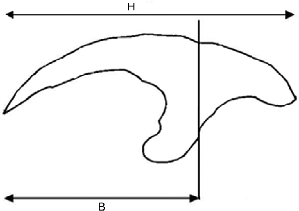
Protoscoleces were mounted in polyvinyl lactophenol with sufficient pressure to flatten the rostellar hooks. The total and blade lengths of two large and two small hooks were measured from protoscoleces of each of 15 E. granulosus cysts (Hobbs et al., Reference Hobbs, Lymbery and Thompson1990). Details of the measured hook components are shown in fig. 1. Morphological differences between sheep and camel strains were analysed graphically by two-dimensional scatter plots.
Dimensions of the large hook, showing hook (H) and blade (B) lengths.

Molecular analysis
Protoscoleces were rinsed several times with sterilized distilled water prior to DNA extraction. According to the manufacturer's instructions, total genomic DNA (gDNA) was extracted from each cyst sample with the aid of a commercial DNA extraction kit (MBST, Tehran, Iran). Briefly, approximately 100 protoscoleces were first lysed in 180 μl lysis buffers, and the proteins were degraded with 20 μl proteinase K for 10 min at 55°C. After adding 360 μl of binding buffer and incubation for 10 min at 70°C, 270 μl of ethanol (100%) were added to the solution and, after vortexing, the complete volume was transferred into the MBST column. The MBST column was first centrifuged and then washed twice with 500 μl of washing buffer. Finally, DNA was eluted from the carrier with 50 μl elution buffer. The concentration of DNA was determined by nanodrop and the samples were stored at − 20°C.
For identification of Echinococcus strain, a PCR-RFLP of the ribosomal DNA ITS1 gene was developed. For this purpose two new primer pairs were created on the basis of published ITS1 sequences of Echinococcus strains (GenBank accession nos AF132701.1, AY389988.2, AF132694.1, AY969-044.1, AF132695.1, AF132696.1, AF132699.1, AJ237773.1, AJ-237774.1, AJ237775.1, AJ237776.1, AJ237835.1, AJ237823.1, AJ237833.1, AJ237834.1, AJ237838.1, AJ237824.1, AJ2378-25.1, AJ237829.1, AJ237822.1, AJ237828.1). The forward and reverse primers employed in this study were as follows: EgF (5′-CAGAGCACTTTTGTATGCA-3′); EgR (5′-ATGGTTGTTA TCG CTG CGA-3′).
The gDNAs (50 ng/sample) were amplified in a final reaction volume of 25 μl as follows: 250 μm of each deoxyribonucleotide triphosphate (dNTP), 2.5 mm of MgCl2, 20 pmol of each primer, 5 μl of 10 × PCR buffer and 1.25 U of Taq DNA polymerase (MBI, Fermentas, Vilnius, Lithuania) in a gradient Thermocycler (Eppendorf, Germany). The following program was used: 5 min incubation at 95°C to denature double-stranded DNA, 35 cycles of 45 s at 94°C (denaturing step), 45 s at 50°C (annealing step) and 45 s at 72°C (extension step). Finally, PCR was completed with an additional extension step for 10 min. The PCR products were analysed by electrophoresis of 5 μl aliquots in 1.5% agarose gels in TBE (Tris/borate/EDTA) buffer with the aid of ethidium bromide staining under ultraviolet conditions. A 100 base pair (bp) ladder was used as a DNA size marker in each gel for estimating the size of the amplicons.
For PCR-RFLP, we digested the DNA directly in the PCR mix. The ITS1 PCR amplification products (16 μl) were digested with 5 units of the restriction endonuclease Bsh1236I (Fermentas) in a final volume of 20 μl for 4 h. Restriction fragments were visualized by gel electrophoresis through a 3% ethidium bromide-stained agarose gel.
Moreover, five cysts for each of the different cutting patterns of ITS1 were analysed with amplification mitochondrial DNA regions, namely the cytochrome c oxidase subunit 1 (cox1) gene. The forward and reverse primers employed with PCRs for cox1 were JB3 (5′-TTTTTTGGGCATC CTGAG GTTTAT-3′) and JB4.5 (5′-TAAAGAAAGAACATAATGAAAATG-3′) (Bowles et al., Reference Bowles, Blair and McManus1992).
Five PCR products from each of the different cutting patterns of ITS1 were purified and submitted to be sequenced in two directions with the use of the same forward and reverse primers employed in the PCR. The sequences were compared with those previously published in the GenBank database using the BLAST system (http://www.ncbi.nlm.nih.gov/blast/Blast.cgi). Moreover, the partial nucleotide sequences of cox1 from two banding patterns (five isolates per pattern) were arranged to confirm the results.
Results
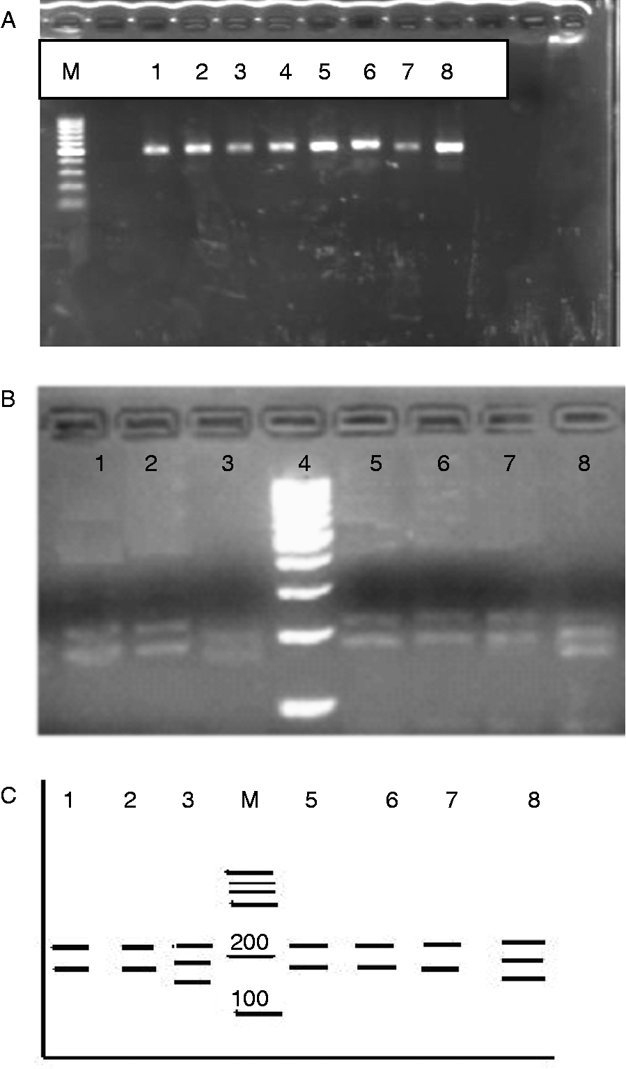
Analysis of all 50 E. granulosus cysts by PCR-RFLP of ITS1, digested with Bsh1236I, showed that two different patterns could be found in the dromedaries. Of the 50 E. granulosus cysts, 27 (54%) were determined as the G1 strain, and the other 23 (46%) were determined as the G6 strain. DNA isolated from selected protoscoleces was amplified by primers derived from a segment of the ITS1 gene, resulting in a PCR product of approximately 462–467 bp in length (fig. 2). As the expected PCR-RFLP analysis showed, there were four Bsh12 361 restriction sites for the G1 genotype and three Bsh12 361 restriction sites for the G6 genotype (fig. 2).
Agarose gel electrophoresis of the internal transcribed spacer 1 (ITS1) polymerase chain reaction (PCR) products of (A) the liver and lung of camels, 462 base pairs; (B) Echinococcus granulosus isolate digested with the restriction enzyme Bsh1236I. G1–G3 genotypes of E. granulosus sensu stricto (lanes 3, 8), G6 Echinococcus genotypes (lanes 1, 2, 5, 6, 7) and 100 bp DNA marker (lane 4), (C) diagrammatic representation of (B).
The sequence of the G6 strain showed 99% homology with the G7 sequence under accession number AJ237829 in GenBank. According to the ITS1 gene, the G6 nucleotide sequence differed from that of the G7 strain by one nucleotide at positions 203, 263 and 275. G6 showed, in comparison with G7, a transition of A to C at position 203, C to T at position 263 and A to G at position 275. Moreover, the nucleotide sequence of the G1 genotype showed 99% homology with the G1 sequence that is accessible under accession number DQ856468 in GenBank. The G1 strain differs from the G1 strain in GenBank by a single nucleotide at positions 212, 383 and 417. G1 showed, in comparison with G1 in GenBank, a transition of G to A at position 212, T to C at position 383 and G to A at position 417. Besides, partial sequences of the mitochondrial genes cox1 (391 bp) of E. granulosus isolates from three- and four-banding patterns revealed complete identity with G6 and G1 sequences under accession numbers DQ856468 and FJ60875901, respectively.
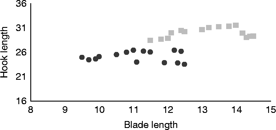
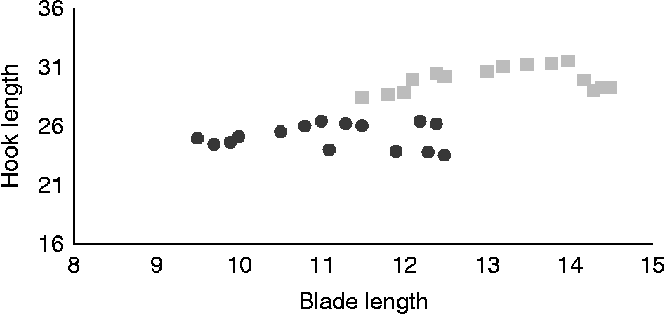
The morphometric analysis showed that 27 cysts belonged to the G1 (sheep) strain and 23 cysts to the G6 (camel) strain. There was a clear distinction between the hook measurements of the sheep and camel strains (fig. 3).
Relationship between mean values of hook and blade lengths from the G1 (circles) and G6 (squares) genotypes of isolates of Echinococcus granulosus from each of the dromedaries; measurements taken from two large hooks of 15 protoscoleces in each strain.
Discussion
An important part of research on hydatidosis is the nature and extent of genotypic variation in Echinococcus spp. (Thompson & McManus, Reference Thompson, McManus, Eckert, Gemmell, Meslin and Pawlowski2001; Thompson, Reference Thompson2008). This is particularly significant in endemic regions such as Iran, where more than one species of intermediate host is present and where there is the possibility of interaction between cycles of transmission (Fasihi Harandi et al., Reference Fasihi Harandi, Hobbs, Adams, Mobedi, Morgan-Ryan and Thompson2002; Thompson, Reference Thompson2008). The design and implementation of control programmes are dependent upon such information and, in particular, the parasite's cycles of transmission are a risk to human health.
A variety of criteria have been used to characterize genotypes of Echinococcus (Thompson & McManus, Reference Thompson, McManus, Eckert, Gemmell, Meslin and Pawlowski2001). The actual benefit of using morphology for differentiating between strains of Echinococcus has been demonstrated on numerous occasions (Constantine et al., Reference Constantine, Thompson, Jenkins, Hobbs and Lymbery1993), but the problems of host-induced morphological variation (Hobbs et al., Reference Hobbs, Lymbery and Thompson1990) and the lack of readily discernible morphological differences between some genotypes of E. granulosus (Eckert et al., Reference Eckert, Thompson, Lymbery, Pawlowski, Gottstein and Morgan1993) have served to emphasize the value of DNA characterization procedures.
In the present study, both morphology and molecular characterization were used to evaluate genotypic variation in a sample of E. granulosus isolates from dromedaries in the eastern half of Iran.
By using this method, we developed a PCR-RFLP to discriminate E. granulosus sensu stricto (G1, G2, G3) and E. intermedius (G6, G7) in DNA samples derived from infected dromedaries. For this aim, a four-cutter restriction endonuclease (Bsh1236I) was needed. The digested fragments were separated by agarose gel electrophoresis and the resulting banding pattern was used to discriminate between the two Echinococcus species. A similar approach was established by Bowles & McManus (Reference Bowles and McManus1993) and an alternative PCR-RFLP approach to discriminate five different Echinococcus species (including, for example, E. granulosus sensu stricto and E. intermedius) using the NAD-1 (NADH dehydrogenase subunit 1) gene was published by Hüttner et al. (Reference Hüttner, Siefert, Mackenstedt and Romig2009). Nevertheless, we used a shorter ITS1 sequence and a new restriction endonuclease in our own approach. One of the problems that may result from the Hüttner method is the time needed for genotyping. The mean time needed for digestion in our study was 4 h, compared with 6 h using the Hüttner method. The results of the new PCR-RFLP were confirmed three times: (1) by ITS1 sequencing; (2) by cox1 sequencing; and (3) by morphometric analysis. Since the PCR-RFLP described here is somewhat similar to others, successful optimization of the protocol would enhance this method. Principally, our system can be optimized. If chromosomal DNA is used for PCR, the cycles can be reduced to 25–30 and a Taq-PCR program for a 462 bp fragment should also work with 94°C for 20 s, 50°C for 20 s, and 72°C for 30 s (saving time). Ten to 20 μl of PCR set-up can be chosen to save material. Interestingly, enzymes are often much faster than expected. Bsh digests 1 μg DNA in 5 min and 1 unit of enzyme is defined as digesting 1 μg λ-DNA in 1 h. Furthermore, it might be possible to use the method to discriminate other species. Specifically, the G4 and G5 strains are shown by the cutting pattern of three pieces with a different banding pattern; however, there are no experimental data or virtual digestions verifying this conclusion.
The results presented here demonstrate that G1 is the dominant genotype of cystic echinococcosis in dromedaries in the eastern half of Iran. This confirms the results of two earlier studies conducted by using a restricted number of isolates collected from Khorasan-e-Razavi province (Sharifiyazdi et al., Reference Sharifiyazdi, Oryan, Ahmadnia and Valinezhad2011) and Isfahan province (Sharbatkhori et al., Reference Sharbatkhori, Mirhendi, Fasihi Harandi, Rezaeian, Mohebali, Eshraghian, Rahimi and Kia2010) in the north-east and the centre of Iran, respectively. In the present study, the G1 strain was reported in dromedaries from Khorasan-e-Razavi, but in a previous study the G3 strain has also been reported (Sharifiyazdi et al., Reference Sharifiyazdi, Oryan, Ahmadnia and Valinezhad2011). We showed the presence of the G1 strain in dromedaries from South Khorasan, Semnan, Sistan-Baluchistan and Yazd provinces for the first time. It should be noted that the G1 strain is closely related to the Tasmanian sheep strain (G2) and the buffalo strain (G3). All three strains do not show any documented fixed intermediate host specificity (Thompson, Reference Thompson2008). The results of this study showed that 23 of 50 camels have the G6 strain. G6 from camels has also been reported by Karimi & Dianatpour (Reference Karimi and Dianatpour2008) in southern Iran, by Ahmadi (Reference Ahmadi2005) in five provinces of Iran, and by Sharbatkhori et al. (Reference Sharbatkhori, Harandi, Mirhendi, Hajialilo and Kia2011) in central Iran. In addition, the camel strain has been reported from sheep, goats, cattle and humans in Iran (Fasihi Harandi et al., Reference Fasihi Harandi, Hobbs, Adams, Mobedi, Morgan-Ryan and Thompson2002; Rajabloo et al., 2012). Based on the morphological characteristics investigated, the total number of protoscoleces and all other morphometric values of the G6 isolate were significantly higher than those of the G1 isolate. All of the camel isolates (G6) identified as the camel strain by molecular methods had mean large hook lengths of 30 μm, fairly similar to the results reported by Eckert et al. (Reference Eckert, Thompson, Michael, Kumaratilake and El-Sawah1989). However, this morphometric parameter was higher than that reported by Fasihi Harandi et al. (Reference Fasihi Harandi, Hobbs, Adams, Mobedi, Morgan-Ryan and Thompson2002) for Iranian camels (28 μm) and was lower than that reported by Derbala & El-Massry (1999) for Egyptian camels (35.2 μm). Furthermore, the mean of total length of small hooks of protoscoleces from camels of the present study (25 μm) was also quite similar to that reported by Eckert et al. (Reference Eckert, Thompson, Michael, Kumaratilake and El-Sawah1989) for camels of Sudan and Chad (24.8 μm) but higher than that reported by Derbala & El-Massry (1999) for Egyptian camels (21.8 μm).
Most of the camel population (69.7%) is distributed in the eastern half of Iran (Khorasan-e-Razavi, South Khorasan, Sistan-Baluchestan, Yazd and Semnan), which is the driest, hottest and least populated part of the country, and climatically a less favourable environment for the survival of Echinococcus eggs. This result could have important epidemiological consequences and a direct impact on Iranian public health, as camel isolates could infect humans. It is known that the majority of human isolates of E. granulosus are the common sheep G1 genotype, although the G6 genotype and others (McManus & Thompson, Reference McManus and Thompson2003) are also known to be infective in humans. Therefore, it is important to define the role of these camel parasite genotypes and their epidemiological implications in E. granulosus zoonotic control.
In conclusion, the molecular protocols used in this study distinguish two genetic groups in the E. granulosus isolates of the camels examined: E. granulosus sensu stricto and E. intermedius. Epidemiological data on the regional occurrence and relative public health importance of different genotypes/species in intermediate hosts are urgently required and the PCR-RFLP system presented here is suitable for this purpose.
Acknowledgements
We thank Mr H. Eshrati for his technical assistance during data collection.
Financial support
This research was supported financially by a grant from the Ferdowsi University of Mashhad (grant number 15794).
Conflict of interest
None.