Transcatheter occlusion has become the treatment of choice for most cases of patent ductus arteriosus in children.Reference Baruteau, Hascoët and Baruteau 1 The results of this intervention are excellent.Reference Backes, Rivera and Bridge 2 However, this technique relies on X-ray imaging that might have some delayed impacts.Reference Ghelani, Glatz and David 3 The risks of excessive exposure to ionising radiation are more and more describedReference Brenner, Doll and Goodhead 4 – Reference Johnson, Hornik and Li 6 and measures are routinely taken to minimise such exposure to both patient and personnel in the catheterisation laboratory. Recently, several teams reported radiation benchmarksReference Ghelani, Glatz and David 3 , Reference Cevallos, Rose and Armsby 7 , Reference Jones, Brennan, Mello-Thoms and Ryan 8 in order to standardise and stratify the procedures. However, data are pooled and extracted from multiple centres, which have different practices, cardiac laboratory, and imaging setting. Moreover, data are given as global data without providing technical aspects, and deep understanding on risks factors of increased radiation exposure are missing at present.
The aims of the present study were to: report our level of radiation exposure in transcatheter closure of patent ductus arteriosus, determine risk factors of increased exposure, and better understand components of radiation exposure in our population.
Methods
Study population and design
All consecutive children (age <18 years old) who came to the cardiac laboratory with the intention to close the patent ductus arteriosus between January 2012 and January 2016 were included in the retrospective study. Data were extracted from our database. The following parameters were collected for each patient: clinical (age; weight; sex; co-morbidities), echocardiographic (left ventricular end-diastolic diameter, mm; ductus arteriosus Vmax with continuous wave Doppler, m/second), per-procedure (anaesthesia type; vascular access; aortic and pulmonary pressures; plane fluoroscopy angle; ductus arteriosus width, mm; ductus arteriosus length, mm; procedure time – from skin to skin – min), and occlusion devices (duct occluder, microvascular plug (Covidien, France), coil (Cook, France), or ventricular septal defect occluder). All complications including embolisation, need for repositioning/changing the device, and failure of the procedure were also collected. This study had been approved by our local ethics committee.
Catheterisation procedure
Cardiac catheterisations were conducted under sedation or general anaesthesia. Patent ductus arteriosus closure was performed according to a standard technique. Briefly, arterial femoral access (4 Fr) was granted. Aortic angiography in the straight lateral view was performed to assess the position, anatomy, shape, length, and size of the patent ductus arteriosus. In most cases, this projection was sufficient. In a minority of patients, a right anterior oblique (30°) angiography was also performed to better delineate the patent ductus arteriosus. After this angiographic assessment, choice of the device was made and venous access was granted if selected device needed insertion from the venous side. Haemodynamic data were collected – i.e. aortic and pulmonary pressures. If pulmonary artery pressures were normal, no additional parameter was collected. Full catheterisation with saturations and pressures to calculate pulmonary vascular resistance was performed in case of pulmonary hypertension. In patients with severe pulmonary arterial hypertension, complete haemodynamic assessment was repeated while occluding the patent ductus arteriosus with a balloon as reported previously.Reference Yan, Zhao and Jiang 9 The choice of device was made by a single operator (Y.B.), based on patient weight, patent ductus arteriosus anatomy and size, anatomy of surrounding vessels – left pulmonary artery and aortic arch – pulmonary artery pressures, and device availability. Asymmetrical occluders were implanted from the venous side as previously described. Advancement of the sheath and device was made using front view as much as possible. A second acquisition – angiography or fluoroscopy, see below – was made after device delivery in a single plane.
During the survey, the equipment used has evolved. Two different SIEMENS catheterisation laboratory suites were used with two different technologies: image intensifier and flat panel. Both catheterisation laboratories – i.e. biplane C-arm Axiom Artis BC system installed in 2003 (Siemens Medical Solution, Forchheim, Germany) and biplane C-arm Artis Zee system installed in March 2013 (Siemens Medical Solution) – were inspected regularly for mechanical integrity and stability of delivered radiation doses in accordance with the Regional Health Center standards in France. Practice changed during the study. Until March 2013 (Axiom Artis), frame rate was set at 15 frames/second (period 1). With the installation of Artis Zee, fluoroscopic images were stored and cineangiography was limited to initial assessment of the patent ductus arteriosus (period 2). From April 2015, frame rate was reduced to 7.5 frames/second (period 3). The reduction of the frame had no impact on the image spatial resolution or on the quality of image.
Radiation doses were analysed for the following variables: total air kerma (mGy), a predictor of deterministic effects, estimates the radiation exposure at the interventional reference point; dose area product (dose area product, Gy.cm2), an indicator of the risk of stochastic effects, represents the products of radiation dose and exposed area (also known as kerma area product); dose area product per body weight (Gy.cm2/kg) a surrogate for energy delivered indexed by body weight; and total fluoroscopic time (min).
Statistical analysis
Analysis was performed using MedCalc software (Mariakerke, Belgium). Descriptive statistics for categorical variables were reported as frequency and percentage, and continuous variables were reported as means, standard deviation, or medians (range, and 1st and 3rd interquartile), and their 95% confidence intervals as appropriate. Level of ionising radiation exposure was evaluated by dose area product. Association between dose area product, as continuous variable, and factors that might influence dose area product was evaluated by univariate and multivariate linear regression analysis. Significance was set at p<0.05, and the Kolmogorov–Smirnov test for normality was performed to determine whether continuous variables were normally distributed, which was the case for each such parameter, to further account for multiple comparisons in univariate analysis and for multivariate regression analysis. Multivariate regression model included variables with significance level p<0.05 in univariate analysis, after a backward selection of relevant variables and excluding collinear variables from the model. For all analyses, a two-tailed p value <0.05 was used as the criterion for statistical significance.
Literature review
We reviewed the scientific literature on radiation dosimetry in patent ductus arteriosus transcatheter occlusion, published between 2000 and March 2016. This review focuses on doses to children. A literature search was conducted using PubMed with broad search terms such as (exposure or radiation) and (children or paediatric) and (dos* or exposure or radiation) and (cardi* or hemodynamic* or cathet* or angiograph* or arteriograph* or angiopla* or intervention*). Only publications speaking on patent ductus arteriosus transcatheter occlusion were considered. In addition, references in each publication were traced back to locate other relevant publications. From each paper, we extracted the number of examinations, the equipment, the kerma area product, also referred to as dose area product, fluoroscopy time, and, where quoted, air kerma. We converted all kerma area product figures to the units of Gy.cm2.
Results
Population characteristics
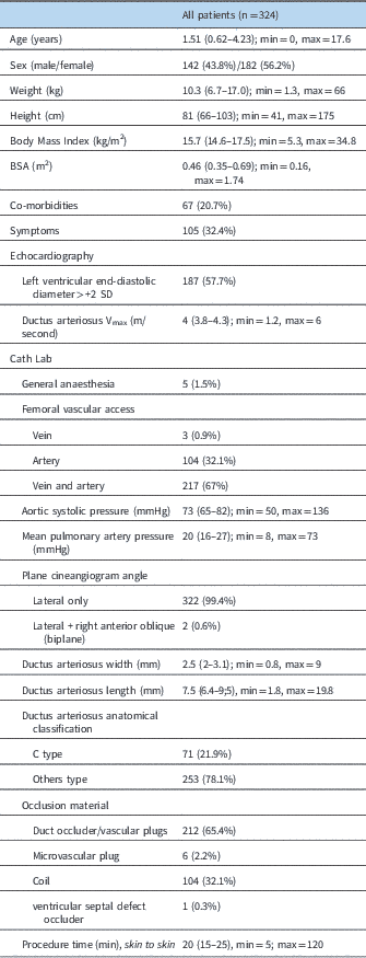
In all, 324 consecutive patients were included. In total, 322/324 (99.4%) procedures were successful. In all, 306 patients (94.4%) had a successful procedure without any complication or failure. A total of 17 complications, in 17 patients, (5.2%) were reported. Seven devices embolised (2.1%): three amplatzer ductus occluders, one vascular plugs II and three coils. In all, six out of seven were immediately snared and patent ductus arteriosus were closed using a different or a larger device – coil: larger coils in two and duct occluder in one; plug: larger plug in one; duct occluder: larger duct occluder in one, plug II in one, failure in one. Eight patients had a significant residual shunt owing to inadequate delivery or inappropriate device. In two, the same device was recaptured and delivered again with success. In two, a second device, i.e. coil, was needed during the same procedure to completely occlude the patent ductus arteriosus. In the remaining, the initial device was judged inadequate before release and exchanged for another device – larger size in two, different device in two. We failed to close two patent ductus arteriosus (0.6%): one needed an immediate surgical after a complication – conversion to retrieve an embolised device and close a very large patent ductus arteriosus) – and the other one had a huge patent ductus arteriosus judged not amenable to transcatheter closure and had a planned surgical closure a few days later. No major bleeding or loss of pulse was observed in the cohort. The patients’ characteristics are summarised in Table 1.
Characteristics of the population.

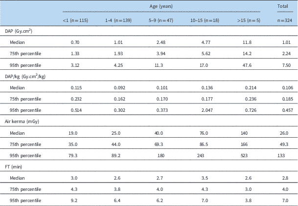
Radiation doses (Table 2)
The median radiation doses were as follows: total air kerma, 26 (Q1–Q3: 14.5–49.3) mGy; dose area product, 1.01 (Q1–Q3: 0.56–2.24) Gy.cm2; dose area product/kg, 0.106 (Q1–Q3: 0.061–0.185) Gy.cm2/kg; fluoroscopic time, 2.8 (Q1–Q3: 2–4) min. The radiation doses stratified by age groups are presented in the Table 2.
Radiation doses stratified by age group.

DAP=dose area product; FT=fluoroscopic time
Frame rate and fluoroscopy/cineangiography impacts (Figure 1)
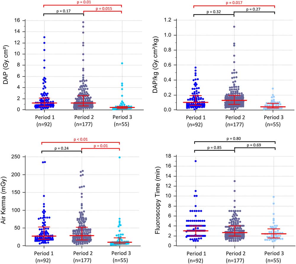
The impact of the three periods on the radiation doses is summarised in Figure 1. Regarding the four radiation parameters – dose area product, dose area product/kg, air kerma, and fluoroscopic time – there was no difference between period 1, two cineangiograms, and period 2, one cineangiogram+one fluoro storage. However, a rate of 7.5 frames/second allowed a significant decrease in comparison with a rate of 15 frames/second for the dose area product (0.44 versus 1.24 Gy.cm2, p=0.015) and the air kerma (9.9 versus 27 mGy, p=0.01), with an equal fluoroscopy use (period 3, one cineangiogram+one fluoro storage).
Radiation exposure in time frame. Period 1 (n=92): before March 2013, 15 frames/second+ cineangiogram only. Period 2 (n=177): From March 2013 to Avril 2015, 15 frames/second+ cineangiogram and fluoro storage. Period 3 (n=55): From April 2015 to January 2016, 7.5 frames/second+cineangiogram and fluoro storage. DAP=dose area product.

Risk factors (Tables 3 and 4)
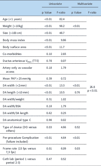
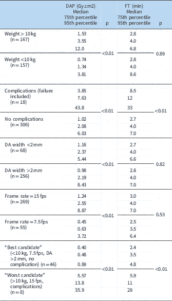
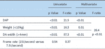
Table 3 summarises the risk factors for elevated dose area product. In the univariate analysis, co-morbidities (p=0.10), vascular access (p=0.18), haemodynamic parameters (ductus arteriosus Vmax, p=0.78; high mean pulmonary artery pressure >25 mmHg, p=0.39), anatomical ductus arteriosus ratio (width/length, p=0.62), and catheterisation laboratory (p=0.47) had no statistical significant impact on the dose area product. The duct occluder devices were the occlusion devices with the higher risk of elevated dose area product (p=0.03). In the multivariate analysis, a weight >10 kg (p<0.01), a ductus arteriosus width <2 mm (p<0.01), complications/failure (p<0.01), and a high frame rate (15 frames/second, p=0.03) exposed the patient to increased risk of prolonged exposure to radiation. Table 4 provides details of radiation exposure, median dose area product, in various situations – i.e. no risk factor, each risk factor, theoretical maximum risks with all the risk factors.
Factors associated with elevated dose area product (DAP); univariate and multivariate analysis.

DA=ductus arteriosus; fps=frames per second; PAP=pulmonary artery pressure
Dose area product (DAP) corresponding to different situations (multivariate risk factors)

DA=ductus arteriosus; fps=frames per second; FT=fluoroscopic time
Table 5 presents the factors associated with a per-procedure complication (n=17). In addition to a relationship between elevated complications and dose area product – due to increased procedural duration, p<0.01 – a weight <10 kg and a ductus arteriosus width >4 mm were the two significant factors in multivariate analysis that increased the complication risks.
Factors associated with a per-procedure complication (n=17); univariate and multivariate analysis.

DA=ductus arteriosus; DAP=dose area product
Literature review
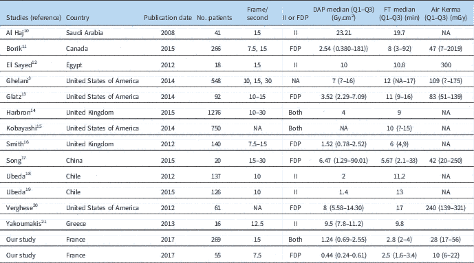
Data were obtained from 14 studies published in the scientific literature, and are summarised in Table 6.
Studies included in the dose review.

DAP=dose area product; FDP=flat panel detector; II=image intensifier; NA=not available
Discussion
Our study presents the results of our centre regarding the level of irradiation during the percutaneous closure of patent ductus arteriosus. We also determined risk factors that expose patients to increased irradiation.
Our irradiation doses are significantly lower than reported studies. The recent US benchmarks published in 2014 by Ghelani et al,Reference Ghelani, Glatz and David 3 the Congenital Cardiac Catheterization Project on Outcomes collaborative multicentre group, presents the radiation doses according to the type of procedures and the age classes. The median dose area product for patent ductus arteriosus closure was 7 (75th percentile: 16 Gy.cm2) or seven times higher than us (and even 16 times current radiation exposure when using 7.5 frames/second), the median fluoroscopic time was 12 (75th percentile: 17) min or 4.3 times higher than us, and the median air kerma was 109 (75th percentile: 175) mGy or 4.2 times higher than us. These differences are therefore really important and deserve to be explained. Four potential explanations are provided in a recent review by Harbron et al on the patient radiation doses in paediatric interventional cardiology procedures.Reference Harbron, Dreuil and Bernier 22 First, the authors advocated that variations of dose area product may result from a mix of procedure types, each having different complexities and degrees of irradiation. This is why we decided to focus on one specific type of procedure – i.e. patent ductus arteriosus closure. We anticipated that patent ductus arteriosus complexity may require more skills, time to close, and as a result radiation exposure. No patient selection was made. All patients undergoing this procedure as an intention to treat were included in the present study. Results are divided in device type, patent ductus arteriosus anatomy, weight, and age, all variables that have been shown to affect rate of success and complexity. The rate of large patent ductus arteriosus and use of devices over than coils was quite high, demonstrating the level of complexity in our cohort. Papers reporting radiation do not report the level of complexity, nor related stratification, but it is highly improbable that this would explain the differences seen here. Second, the age distribution within the population may vary between studies. We and others have demonstrated that it is an important criterion affecting the dose area product. This distribution is relatively similar between the two studies, so it could not explain the observed differences in radiation exposure. A third potential explanation advances by Harbron et al is incorrect recording, or reporting of dose indicators. Here again, it is unlikely to explain the dose area product variation reported in our study, as all data were prospectively and automatically recorded. The fourth explanation for dose area product variation is related to the fluoroscopic equipment. There are major differences between manufacturers but also with the setting of the machine. Various parameters such as frame rate, dose rate, use of filtration, or antiscatter grid use may vary. In the Congenital Cardiac Catheterization Project on Outcomes collaborative multicentre group, no data are provided except for frame rates that varied between 10 and 30 frames/second depending on the centres. In our study, we were able to compare two different technologies, as well as different frame rates with the same equipment. We were able to see that flat panel technology is more “irradiant” compared with intensifier image technology. By simply setting the frame rate to 7.5 frames/second, we were able to significantly reduce dose area product by 2.8 (1.24–0.44 Gy.cm2). This is not the only reason for reduction of radiation exposure as dose area product was even lower with 15 frames/second. Harbron et al falsely assumed that practices are similar between groups. We, in contrary, think that practice variation explains most of the reduction of dose area product. Even if there is no strict relationship between fluoroscopic time and dose area product, our level of fluoroscopic time was far lower than previously reported. Moreover, cineangiogram is known to be a big contributor to dose area product. Our policy is to limit the number of cineangiograms. Unfortunately, most of current equipment, including ours, do not allow measurement on stored fluoroscopy. We only performed one initial cineangiogram to delineate and measure the patent ductus arteriosus. No cineangiogram in the right anterior oblique projection is done in the vast majority of patients (99.4%).
Despite variation of practice, the rate of successful closure is high (99.4%). Indeed, we have only two failed procedures (0.6%) and seventeen “complications” (5.2%). This rate of complications might appear high but we included patients in whom a second or a different device was necessary to successfully close the patent ductus arteriosus. In all, 7 devices embolised, with six successfully recaptured and patent ductus arteriosus closed using another device. Backes et al recently stated in a meta-analysisReference Backes, Rivera and Bridge 2 that the technical success of percutaneous patent ductus arteriosus closure was 92.2%, which was very similar to what we report. It supports the fact that decreasing dose area product and fluoroscopic time do not expose the patient to an increased risk of complications.
It is clear that even before establishing benchmarks of irradiation dose during these procedures, it is essential to present to the centres carrying out these procedures simple and applicable methods making it possible to reduce these exposures. For example, the As Low as Reasonably Achievable concept in paediatric cardiac catheterisation, published by Henri Justino in 2006, expose 20 tactics for radiation dose reduction and image quality improvement.Reference Justino 23 His fourth tactic was to use the lowest acceptable frame rate during pulsed fluoroscopy and cineangiography. We believe that this comment is fundamental, especially in the management of patent ductus arteriosus closure procedure. The fluoroscopic time and the frame rate should be drastically controlled, because an efficient procedure can be achieved well below the benchmarks proposed by the Congenital Cardiac Catheterization Project on Outcomes collaborative multicentre group, as shown in our study.
Perspectives
Transcatheter occlusion of patent ductus arteriosus being currently the first line treatment, the question now is to reduce radiation exposure. To this end, we believe it is essential to increase our requirement for the risk of irradiation. Our study shows that benchmarks need to be challenged, and that different strategies are possible for the team in order to reduce this irradiation. For us, three simple factors seem to decrease drastically the radiation dose while maintaining the efficiency of the procedure: reduction of frame rate, avoidance of cineangiography in biplane, and limitation of fluoroscopy time. Besides these factors controlled by the interventionists, manufactures can help us to improve these data. With the current cath lab, the precise assessment of patent ductus arteriosus size can only be done using cineangiogram that encounters for a large part of the total radiation exposure. Manufacturers should make possible measurement on stored fluoroscopic images in order to be able to further reduce radiation. Additionally, post-device angiographies routinely applied for the assessment of device position and residual shunt should be performed using fluoroscopic mode rather than cineangiogram. Although this procedure can be performed with limited fluoroscopic exposure nowadays, potential injury still cannot be ignored. Currently, transthoracic echocardiography plays a small role in non-premature babies. However, it can be used to guide in patent ductus arteriosus closureReference Ewert, Berger and Daehnert 24 , Reference Pan, Ouyang and Wang 25 and reduce or even avoid radiation exposure. The use of echo was very limited in our study, but this is clearly the way to go if we want to improve our level of radiation exposure.
We also believe that this comment about echo-guiding is particularly important concerning percutaneous patent ductus arteriosus closure in premature babies. Although our study and our results are not focused on this specific population, we know that radiation will be an important issue for these procedures. Further specific studies on the radiation exposure and/or the echo-guiding for percutaneous patent ductus arteriosus closure in premature babies are needed, and we will try to realise them from our centre.
Conclusion
The benchmarks for radiation doses concerning patent ductus arteriosus closure have to be daily improved. Lower doses of radiation can be achieved with subsequent recommendations: reduction of frame rate, avoidance of biplane cineangiogram, and limitation of fluoroscopy time. Technical improvements are important to lower radiation exposure. We have identified main parameters that have an impact on radiation exposure during the patent ductus arteriosus closure in our population. The next level of guidance optimisation should be the increased use of echocardiography, but additional and specific studies will be required to see how much impact it has on success rate, complication rate, and radiation exposure.
Acknowledgements
None.
Financial Support
This research received no specific grant from any funding agency, commercial or not-for-profit sectors.
Conflicts of Interest
None.
Ethical Standards
The authors assert that all procedures contributing to this work comply with the ethical standards the Helsinki Declaration of 1975, as revised in 2008, and has been approved by the institutional committees (Paris VI).