Introduction
The inflammatory myofibroblastic tumour is a rare neoplasm which has been found in virtually every site in the body. The first cases were described in the lung. Later, extrapulmonary inflammatory myofibroblastic tumours were reported, most of which were located in the abdominal region. Rarer still are inflammatory myofibroblastic tumours involving the pharynx or larynx.
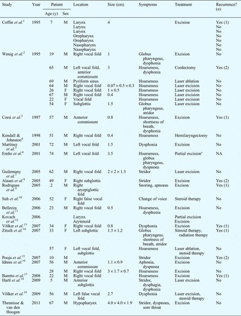
We found 35 previously reported cases of pharyngeal or laryngeal inflammatory myofibroblastic tumour (Table I), mainly tumours restricted to the vocal folds or subglottis. Reports included one tumour located in the pyriform sinus and two located on the aryepiglottic fold. Furthermore, Coffin et al. reported four cases, within a larger case series, with tumour localisation in the oro- or nasopharynx.Reference Coffin, Watterson, Priest and Dehner1
Reports of pharyngeal and laryngeal inflammatory myofibroblastic tumour

*Radical surgery could not be performed due to the patient's medical condition. y = years; M = male; F = female; NA = not applicable
Here, we present a case of inflammatory myofibroblastic tumour of the posterior wall of the hypopharynx. To our knowledge, this is the first reported case of an inflammatory myofibroblastic tumour in this location.
Case report
A 67-year-old man was referred to our university hospital by another otolaryngologist because of inspiratory stridor, dyspnoea and sore throat due to a spherical mass in the hypopharynx. The patient's general practitioner had referred him to the otolaryngologist the same day because of stridor.
The patient's symptoms had begun three weeks earlier, with a sore throat, hoarse voice and periods of fever, for which he had received antibiotics from his general practitioner. However, his symptoms had deteriorated: he had developed progressive shortness of breath, and he had not been able to swallow solid food for at least a week and a half prior to the current presentation.
On fibre-optic laryngoscopy, we found a spherical mass located at the level of the epiglottis, which almost occluded the entire airway. The mass seemed to be pedunculated from the posterior pharyngeal wall. Below the mass, we observed a normal larynx with intact vocal folds.
The patient was admitted to our hospital for observation.
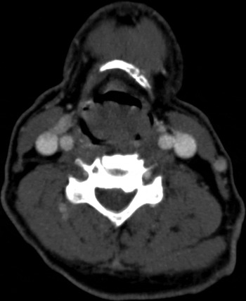
A computed tomography (CT) scan (Figure 1) showed a supraglottic, solid, polypoidal process with a maximum diameter of 4 cm, presumably originating from the posterior laryngeal wall. No pathologically enlarged lymph nodes were seen.
Axial computed tomography scan showing a large hypopharyngeal mass obstructing the airway.

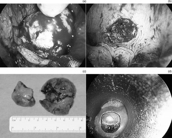
The next day, direct laryngoscopy was performed in order to resect the tumour. After induction of general anaesthesia, inspection during apnoea revealed that the tumour did indeed originate from the posterior pharyngeal wall (Figure 2a). As the histology of the tumour was unknown and maximal debulking was necessary to ensure the airway, we decided to use a tonsillectomy snare for the resection, leaving the remainder of the tumour stalk for further treatment depending on the histology report (Figure 2b).
Intra-operative views showing (a) the tumour mass in the hypopharynx, (b) the posterior pharyngeal wall after initial resection, (c) the tumour specimen after resection and (d) the re-resection procedure.

After the procedure, the patient experienced no shortness of breath and had no complaints whatsoever. He was fed via a nasogastric tube until a lateral neck X-ray showed no signs of perforation. The patient was discharged a few days later after tolerating a normal diet.
Macroscopic examination of the tumour specimen revealed two irregular, yellow-grey tissue fragments measuring 4.0 × 4.0 × 1.9 cm and 2.5 × 2.0 × 1.5 cm in size (Figure 2c). Histologically, the tumour was covered with a normal mucous membrane of squamous epithelium. The tumour itself consisted of medium-sized, spindle-shaped cells with eosinophilic cytoplasm and polymorphic nuclei, admixed with an inflammatory infiltrate of lymphocytes and some neutrophils and foamy histiocytes. Sporadic mitoses were seen, and the tumour extended into the muscle layer. Upon immunohistochemical analysis, the tumour cells were strongly positive for vimentin and smooth muscle 1, focally positive for desmin, and incidentally positive for anaplastic lymphoma kinase.
In consultation with the patient, a re-resection of the pharyngeal wall was planned. Using a CO2 laser, the remaining lesion was excised with a wide margin, in an uncomplicated procedure (Figure 2d). The same post-operative protocol was followed, and the patient was discharged three days post-operatively.
Histological examination of the re-resection specimen showed residual tumour in the centre of the resected tissue. The resection margin was free of tumour.
At the time of writing, two years after the second procedure, the patient showed no sign of recurrence.
Discussion
The nomenclature of inflammatory pseudotumours and related sarcomas has been very confusing over the years. Terms such as ‘plasma cell granuloma’, ‘inflammatory pseudotumour’, ‘xanthogranuloma’, ‘histiocytoma’ and ‘inflammatory myofibroblastic tumour’ have been used interchangeably, making literature review very challenging. The term ‘inflammatory myofibroblastic tumour’ was introduced in 1990, when Pettinato et al. presented 20 cases of plasma cell granuloma in which they found myofibroblasts to be the main cell type.Reference Pettinato, Manivel, De Rosa and Dehner20 The changing terminology of this tumour is related to a long dispute about its nature. Until the late 1990s, inflammatory myofibroblastic tumours were thought to represent a postinflammatory process rather than a neoplasm. Nowadays, inflammatory myofibroblastic tumour is considered to be a distinct entity within the broad range of inflammatory pseudotumours, which are classified in a continuous spectrum with inflammatory fibrosarcoma.Reference Corsi, Ciofalo, Leonardi, Zambetti and Bosman3
The inflammatory myofibroblastic tumour is regarded as a neoplastic tumour of intermediate biological potential. The incidence of recurrence of extrapulmonary tumours is reported to be as high as 25 per cent, probably due to incomplete resection because of the multinodular aspect of many intra-abdominal lesions, and/or the proximity to vital structures such as the larynx and heart.Reference Coffin, Watterson, Priest and Dehner1
Metastasis of inflammatory myofibroblastic tumours is extremely rare (<5 per cent), and has never been reported in cases of inflammatory myofibroblastic tumour of the larynx or pharynx.Reference Coffin, Fletcher, Fletcher, Unni and Mertens21
Histologically, inflammatory myofibroblastic tumour consists of spindle cells, mainly myofibroblastic in appearance, which are embedded in a myxoid stroma with an inflammatory infiltrate predominantly consisting of plasma cells. About half of all inflammatory myofibroblastic tumours show expression of anaplastic lymphoma kinase, due to a rearrangement of the anaplastic lymphoma kinase gene, which was first described in anaplastic large cell lymphoma. This further contributes to the conception of inflammatory myofibroblastic tumour as a neoplasm; however, there does not seem to be a relationship between anaplastic lymphoma kinase expression and the clinical course or chance of recurrence.Reference Gleason and Hornick22
• Pharyngeal inflammatory myofibroblastic tumours are extremely rare, with only five previously reported cases
• This is the first report of an inflammatory myofibroblastic tumour originating from the posterior wall of the hypopharynx
• Inflammatory myofibroblastic tumour of the larynx and pharynx is regarded as a neoplasm of intermediate biological potential, with a local recurrence incidence of up to 25 per cent; metastasis has not been reported
• Surgery is the treatment of first choice; no adjuvant therapy is necessary
We performed a literature search using the PubMed database and the terms ‘inflammatory myofibroblastic tumour/tumor’, ‘larynx’ and ‘pharynx’. All relevant English language citations were selected, full text articles were obtained and references were checked for additional material, resulting in identification of 19 papers. It should be noted that we did not include cases of ‘inflammatory pseudotumour’ or ‘plasma cell granuloma’ in our review. While some of these cases are probably histopathologically identical to inflammatory myofibroblastic tumour, this diagnosis cannot be made retrospectively.
We found one published series that included four cases of pharyngeal inflammatory myofibroblastic tumour (in the naso- or oropharynx); however, no further information on these cases was available.Reference Coffin, Watterson, Priest and Dehner1 Wenig et al. reported the only other case of an inflammatory myofibroblastic tumour of the hypopharynx, in a 69-year-old man who presented with hoarseness due to a tumour located in the pyriform sinus, which was treated with laser ablation.Reference Wenig, Devaney and Bisceglia2
We found a total of 30 cases of inflammatory myofibroblastic tumour of the larynx, most of which originated from the vocal folds.Reference Coffin, Watterson, Priest and Dehner1–Reference Völker, Scheich, Zettl, Hagen, Müller-Hermelink and Gattenlöhner19 The most common symptom was hoarseness or dysphonia. Nine patients presented with stridor and/or shortness of breath, and one patient complained of snoring and apnoeic episodes. Seven patients required tracheotomy because of severe dyspnoea. In most cases, the tumour was identified by physical examination. Some patients underwent CT or magnetic resonance imaging. Several patients underwent biopsy before definitive excision of the tumour. Treatment consisted of ‘cold steel’ or laser excision of the tumour. One patient underwent hemilaryngectomy, one patient received radiation therapy, three patients received adjuvant corticosteroid therapy and one patient was treated with corticosteroid therapy alone. Tumour size varied from 0.4 to 4.0 cm. Nine patients (28 per cent) had a recurrence after initial treatment; of these nine, three had a second recurrence.
Initially, our patient presented with a sore throat and periods of fever. In 15–30 per cent of inflammatory myofibroblastic tumour patients, a syndrome of fever, weight loss and malaise is reported, accompanied by laboratory findings including microcytic anaemia, elevated erythrocyte sedimentation rate and thrombocytosis.Reference Gleason and Hornick22 In our patient, we found only an elevated C-reactive protein (of 34 mg/l) pre-operatively.
Because of our patient's threatened airway, we decided to perform a debulking procedure. This was supported by the appearance of the tumour (smooth-surfaced and pedunculated), which suggested a benign or low-grade malignant tumour. This approach enabled us to avoid tracheotomy, but we did need to perform a re-resection after the histological diagnosis had been established. If an inflammatory myofibroblastic tumour is suspected and the airway is not under threat, an initial biopsy is required in order to establish a pre-operative diagnosis so that a second procedure can be avoided. For inflammatory myofibroblastic tumour, radical surgery is the treatment of first choice, and no adjuvant therapy is necessary.