


INTRODUCTION
Individuals who experience the same age-related changes or damage to the brain due to neurological conditions can vary greatly in their cognitive response (e.g., Stern, Reference Stern2002, Reference Stern2009; Lindenberger, Burzynska, & Nagel, Reference Lindenberger, Burzynska, Nagel, Stuss and Knight2013; Jokinen et al., Reference Jokinen, Melkas, Madureira, Verdelho, Ferro, Fazekas and Erkinjuntti2016). The cognitive reserve (CR) hypothesis attempts to explain some of this variability. It suggests that premorbid efficacy, aptitude, and flexibility of cognitive processing can aid the brain’s ability to cope with change or damage (e.g., Stern, Reference Stern2002; Jones et al., Reference Jones, Manly, Glymour, Rentz, Jefferson and Stern2011; Barulli & Stern, Reference Barulli and Stern2013; Levi, Rassovsky, Agranov, Sela-Kaufman, & Vakil, Reference Levi, Rassovsky, Agranov, Sela-Kaufman and Vakil2013). Early environmental influences such as education and childhood socio-economic status (SES) have been found to be predictors of cognition in later life, suggesting those with higher education or SES might be less susceptible to cognitive decline because of their initially higher levels of cognition (Deary & Brett, Reference Deary and Brett2015; Greenfield & Moorman, Reference Greenfield and Moorman2019). For example, education has been found to be related to overall cognition, episodic, and semantic memory as well as perceptual abilities in older adults and adults with possible dementia (Jefferson et al., Reference Jefferson, Gibbons, Rentz, Carvalho, Manly, Bennett and Jones2011). Further life experiences such as occupational achievement, literacy attainment, and engagement in cognitively and socially stimulating activities are also known to play an important role in increasing the effectiveness of cognitive processing (Suchy, Kraybill, & Franchow, Reference Suchy, Kraybill and Franchow2011; Stern, Reference Stern2012; Levi et al., Reference Levi, Rassovsky, Agranov, Sela-Kaufman and Vakil2013; Liu, Cai, Xue, Zhou, & Wu, Reference Liu, Cai, Xue, Zhou and Wu2013; Okonkwo et al., Reference Okonkwo, Schultz, Oh, Larson, Edwards, Cook and Sager2014; for a review, see Arenaza-Urquijo, Wirth, & Chételat, Reference Arenaza-Urquijo, Wirth and Chételat2015). Literacy attainment, a CR proxy often assessed using single-word reading tasks, such as the National Adult Reading Test (NART; Nelson & Willison, Reference Nelson and Willison1991), has been related to overall cognition, working memory, and episodic memory (Siedlecki et al., Reference Siedlecki, Stern, Reuben, Sacco, Elkind and Wright2009).
CR may explain some of the individual differences among the vulnerability to brain damage and may increase resistance to age- and disease-related brain changes (Jokinen et al., Reference Jokinen, Melkas, Madureira, Verdelho, Ferro, Fazekas and Erkinjuntti2016). The heterogeneity of brain pathology presents a challenge for clinicians to be able to predict patients’ cognitive outcomes, and following focal brain injury, better understanding of the mechanisms underlying recovery of cognitive function is important (Green et al., Reference Green, Colella, Christensen, Johns, Frasca, Bayley and Monette2008). CR continues to develop across the lifetime and so even late-stage interventions can potentially enhance CR to mitigate the effects of brain damage (Tucker & Stern, Reference Tucker and Stern2011). Research has shown that individuals with comparable levels of brain pathology demonstrate differences in their cognitive impairment, depending on whether they have high or low educational attainment and/or NART IQ (e.g., Grafman, Salazar, Weingartner, Vance, & Amin, Reference Grafman, Salazar, Weingartner, Vance and Amin1986; Bennett et al., Reference Bennett, Wilson, Schneider, Evans, Mendes de Leon, Arnold and Bienias2003; Stern, Reference Stern2006; Singh-Manoux et al., Reference Singh-Manoux, Marmot, Glymour, Sabia, Kivimäki and Dugravot2011; Serra et al., Reference Serra, Musicco, Cercignani, Torso, Spanò, Mastropasqua and Bozzali2014; Bozzali et al., Reference Bozzali, Dowling, Serra, Spano, Torso, Marra and Cercignani2015). Darby et al. (Reference Darby, Brickhouse, Wolk and Dickerson2017) found that higher years of education were related to performance on executive tasks in patients with mild cognitive impairment (MCI), but not Alzheimer’s disease (AD), whereas higher years of education were associated with performance on semantic tasks in MCI and AD. Individuals with low levels of education are at a higher risk of dementia compared to individuals with higher levels of education, especially AD (Schmand, Smit, Geerlings, & Lindeboom, Reference Schmand, Smit, Geerlings and Lindeboom1997; Meng & D’Arcy, Reference Meng and D’Arcy2012; Lo & Jagust, Reference Lo and Jagust2013; see for a review Xu, Yu, Tan, & Tan, Reference Xu, Yu, Tan and Tan2015).
Compared to diffuse lesions associated with degenerative conditions, few studies have examined the influence of CR on cognitive performance in neurological conditions that result in focal lesions such as stroke (see Nunnari, Bramanti, & Marino, Reference Nunnari, Bramanti and Marino2014) or brain tumour. CR may not have the same neuroprotective benefit in the context of focal brain damage due to brain tumour or stroke. In healthy and pathological ageing, there may be more plasticity and functional reorganisation due to their slow progressive nature (Morris, Reference Morris2005; Ryan & Rossor, Reference Ryan and Rossor2011). As stroke and tumour are associated with a more rapid disease process, they may have limited effects of CR proxies. Yet, in stroke, patients who received a higher number of years of formal education had less cognitive decline than stroke patients with fewer years of formal education (e.g., Sachdev, Brodaty, Valenzuela, Lorentz, & Koschera, Reference Sachdev, Brodaty, Valenzuela, Lorentz and Koschera2004; Elkins et al., Reference Elkins, Longstreth, Manolio, Newman, Bhadelia and Johnston2006; Zieren et al., Reference Zieren, Duering, Peters, Reyes, Jouvent, Hervé and Dichgans2013; see Kessels et al., Reference Kessels, Eikelboom, Schaapsmeerders, Maaijwee, Arntz, van Dijk and de Leeuw2017 for a meta-analysis). Moreover, stroke patients with a higher number of years of formal education were found to have a lower risk of developing clinically diagnosed cognitive impairment (Kessels et al., Reference Kessels, Eikelboom, Schaapsmeerders, Maaijwee, Arntz, van Dijk and de Leeuw2017) and less severe aphasia (González-Fernández et al., Reference González-Fernández, Davis, Molitoris, Newhart, Leigh and Hillis2011). Recently, Makin et al. (Reference Makin, Doubal, Shuler, Chappell, Staals, Dennis and Wardlaw2018) reported that NART IQ and years of education were better predictors of cognition post-stroke compared to vascular risk factors or stroke severity.
Yet, to our knowledge, CR studies have not examined whether particular brain areas are responsible for the ability to compensate for brain damage. CR has been associated with the scaffolding theory of ageing and cognition (STAC; Park & Reuter-Lorenz, Reference Park and Reuter-Lorenz2009). Scaffolding is a process that takes place throughout the lifespan and involves the formation and enhancement of existing and new neural connections to achieve specific cognitive goals (Alexander et al., Reference Alexander, Furey, Grady, Pietrini, Brady, Mentis and Schapiro1997; Perneczky et al., Reference Perneczky, Drzezga, Diehl-Schmid, Schmid, Wohlschläger, Kars and Kurz2006). In healthy ageing and neurodegenerative diseases, higher levels of CR are thought to result in more effective scaffolding as compensation for cognitive decline (Reuter-Lorenz & Park, Reference Reuter-Lorenz and Park2014). Research suggests that both CR and scaffolding are thought to rely on the integrity of the prefrontal cortex (Park & Reuter-Lorenz, Reference Park and Reuter-Lorenz2009; Robertson, Reference Robertson2014; see Anthony & Lin, Reference Anthony and Lin2017 for a review of the neuroimaging literature). Therefore, the prefrontal cortex may be a potential brain area for sustaining the ability to protect or compensate for cognitive decline.
Neuroimaging studies have also provided evidence of potential neural substrates for CR, including the frontal lobes. For example, a review of PET studies by Morbelli and Nobili (Reference Morbelli and Nobili2014) found that AD patients with high CR tend to show hypermetabolism in the dorsolateral prefrontal cortex but hypometabolism in the temporo-parietal cortex. Studies examining CR based on education have reported greater frontal lobe thickness associated with higher education (Vaqué-Alcázar et al., Reference Vaqué-Alcázar, Sala-Llonch, Valls-Pedret, Vidal-Piñeiro, Fernández-Cabello, Bargalló and Bartrés-Faz2017), with greater loss in the left anterior cingulate cortex and left dorsomedial prefrontal cortex in individuals with exceptionally low years of education (Rzezak et al., Reference Rzezak, Squarzoni, Duran, Alves, Tamashiro-Duran, Bottino and Busatto2015). However, other studies examining CR have shown higher occupation, SES, and leisure activities are associated with less hippocampal atrophy (Staff et al., Reference Staff, Murray, Ahearn, Mustafa, Fox and Whalley2012; Suo et al., Reference Suo, Lèon, Brodaty, Trollor, Wen, Sachdev and Valenzuela2012). In large cohort studies, education but not occupation or leisure activities significantly correlates with frontal and parieto-temporal regions (Foubert-Samier et al., Reference Foubert-Samier, Catheline, Amieva, Dilharreguy, Helmer, Allard and Dartigues2012).
If the prefrontal cortex plays a role in CR, lesions in the prefrontal cortex should reduce the ability to compensate for brain damage. Therefore, patients with prefrontal lesions are less likely to demonstrate differences in their cognitive impairment depending on whether they have higher or lower levels of education and/or NART IQ. Yet, few studies have examined CR in patients with lesions restricted to specific cortical areas. While higher educational attainment has not been shown to attenuate cognitive impairment in brain tumour patients, younger age and having a frontal tumour were associated with better performance on speed, executive, and working memory measures (Kaleita et al., Reference Kaleita, Wellisch, Cloughesy, Ford, Freeman, Belin and Goldman2004). In a recent study, we retrospectively examined the effects of years of education and literacy attainment measured by the NART IQ on the cognitive performance of patients with unilateral prefrontal lesions due to stroke or brain tumour (MacPherson et al., Reference MacPherson, Healy, Allerhand, Spano, Chan, Tudor-Sfetea and Cipolotti2017). NART IQ predicted executive and naming performance but not fluid intelligence, processing speed, verbal short-term memory, or perceptual abilities. Importantly, however, our study showed that the effects of education and/or NART IQ on our cognitive measures did not interact with lesion severity, arguing against a frontal theory of CR effect, that is, the effect of lesion severity on cognitive impairment was not altered by either CR proxy in our frontal patients. One limitation of our previous study was that data from patients with non-frontal lesions were not available. This would have allowed us to directly compare whether the degree of variance accounted for by CR is reduced in frontal patients when compared to non-frontal patients.
In the current study, we examined the effect of CR, as measured using NART IQ, on the cognitive performance of a large sample of patients with focal, unilateral frontal, or non-frontal brain regions due to stroke or tumour. Our aim was to compare the influence of lesion location (frontal vs. non-frontal) on cognitive performance in order to determine whether CR differentially safeguards against focal neuropathology according to lesion location. If the frontal theory of CR is to be supported, NART IQ will account for less variance on the cognitive tests in frontal patients compared to non-frontal patients.
MATERIALS AND METHODS
Participants
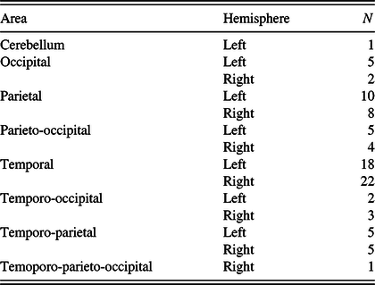
The patient database within the Neuropsychology Department at the National Hospital for Neurology and Neurosurgery was retrospectively examined for patients with frontal or non-frontal lesions who could be included in the study. Patients were identified as having a unilateral lesion confined to either the frontal or non-frontal brain regions due to a stroke or a brain tumour by a neurologist on the basis of clinical magnetic resonance imaging (MRI) scans (or CT scans where MRI was unavailable). Lesions were localised by operation site in the case of surgical patients or by gross lesion characterisation in the nonsurgical patients. Tumour grade was confirmed by histopathological studies following resection or biopsy and patients had undergone tumour resection prior to neuropsychological assessment. Exclusion criteria were (i) age ≥80 years at the time of testing; (ii) current or previous psychiatric disorders; (iii) previous neurological disorders including previous stroke or tumours; (iv) presence of metastatic tumours; (v) previous chemotherapy; (vi) gross visual, perceptual, language, or motor impairment; (vii) previous head trauma; (viii) history of excessive alcohol or drug use; (ix) no MRI or CT scan results available; (x) no or limited neuropsychological data available; and (xi) a score below the 5th percentile on a test of general intelligence (Wechsler Adult Intelligence Scale-III, WAIS-III; Wechsler, Reference Wechsler1997, Wechsler Adult Intelligence Scale-R, WAIS-R; Wechsler, Reference Wechsler1981 or Raven’s Matrices; Raven, Reference Raven1976). Non-native English speakers were only included in the study if they obtained a score ≥25th percentile on the NART (Nelson, Reference Nelson1982) to ensure their English abilities were able to cope with task demands. One hundred and sixty-six frontal patients were included in the study: stroke, N = 53; high-grade tumour, N = 27; low-grade tumour, N = 37; and meningioma, N = 49. Some clinical and cognitive aspects of these patients have been previously reported (MacPherson et al., Reference MacPherson, Turner, Bozzali, Cipolotti and Shallice2010, Reference MacPherson, Turner, Bozzali, Cipolotti and Shallice2016, Reference MacPherson, Healy, Allerhand, Spano, Chan, Tudor-Sfetea and Cipolotti2017; Robinson, Shallice, Bozzali, & Cipolotti, Reference Robinson, Shallice, Bozzali and Cipolotti2012, Reference Robinson, Cipolotti, Walker, Biggs, Bozzali and Shallice2015; Murphy et al., Reference Murphy, Shallice, Robinson, MacPherson, Turner, Woollett, Bozzali and Cipolotti2013; Cipolotti et al., Reference Cipolotti, Healy, Chan, Bolsover, Lecce, White and Bozzali2015a). Ninety-one non-frontal patients were included in the study: stroke, N = 30; high-grade tumour, N = 19; low-grade tumour, N = 22; and meningioma, N = 20. See Table 1 for the lesion localisation of the non-frontal patients. Data from 136 healthy controls (HC) were also included (see below). The study was approved by the National Hospital for Neurology and Neurosurgery and the Institute of Neurology Joint Research Ethics Committee (UK), all procedures were in accordance with the Declaration of Helsinki, and all participants provided informed written consent.
Distribution of the non-frontal patients according to lesion area and hemisphere
Cognitive Investigation
All patients had previously undertaken a single neuropsychological assessment in the Neuropsychology Department of the National Hospital for Neurology and Neurosurgery which involved the administration of established neuropsychological tests assessing executive abilities (phonemic fluency S – number of words produced; Tombaugh, Kozak, Rees, Reference Tombaugh, Kozak and Rees1999), intelligence (WAIS-III – full Scale IQ; Wechsler, Reference Wechsler1997), speed of information processing (Trail Making Test Part-A, Trail-A – number of seconds to complete; Reitan, Reference Reitan1992), and naming (Graded Naming Test, GNT – number of pictures correctly named; McKenna & Warrington, Reference McKenna and Warrington1983). Test administration was conducted in accordance with the procedures outlined in test manuals. The neuropsychological tests selected and administered during the assessment were at the discretion of the different clinical neuropsychologists; hence, data for the various tests were not available for all participants. A pairwise deletion method was used with no substitutions made to the dependent variables. Fewer neuropsychological tests were considered compared to MacPherson et al. (Reference MacPherson, Healy, Allerhand, Spano, Chan, Tudor-Sfetea and Cipolotti2017) to allow the inclusion of more patients. Of the 166 frontal patients, the individuals who had data for each cognitive measure were as follows: executive function: N = 147; intellectual abilities: N = 82; speed of information processing: N = 77; and naming: N = 156. For the 91 non-frontal patients, the individuals who had data for each cognitive measure were as follows: executive function: N = 56; intellectual abilities: N = 71; speed of information processing: N = 20; and naming: N = 57. For the 136 HC, the individuals who had data for each cognitive measure were as follows: executive function: N = 43; intellectual abilities: N = 0; speed of information processing: N = 81; and naming: N = 131.
CR Proxy
Literacy attainment was included as our proxy of CR. A test of single-word reading was adopted (e.g., Scarmeas, Albert, Manly, & Stern, Reference Scarmeas, Albert, Manly and Stern2006; Stern et al., Reference Stern, Zarahn, Habeck, Holtzer, Rakitin, Kumar and Brown2008). This was based on NART IQ, which has a split-half reliability coefficient of .93, inter-rater reliability of .96–.98, and test–retest reliability of .98 (O’Carroll, Reference O’Carroll1987; Crawford, Parker, Stewart, Besson, & De Lacey, Reference Crawford, Parker, Stewart, Besson and De Lacey1989a; Schlosser & Ivison, Reference Schlosser and Ivison1989). In terms of validity, the NART loads highly (.85) on g, the general factor of intelligence from the WAIS (Crawford et al., Reference Crawford, Stewart, Cochrane, Parker and Besson1989b).
Statistical Analysis
The statistical analyses were carried out using R version 3.6.0. The effect of our CR proxy on performance on the cognitive measures was examined by fitting separate multiple linear regression models for each measure using R function “lm”. In the first step of the analysis, age (step 1) was entered as a continuous predictor variable. In step 2, lesion group was entered as a categorical predictor variable with three levels (frontal, non-frontal, and HC). Here, two dichotomous dummy-coded variables were created and directly entered into the model: frontal versus HC and non-frontal versus HC. In the case of WAIS-III where HC data were not available, there was only one dichotomous variable comparing frontal versus non-frontal. The third step involved NART IQ (step 3) being entered as a predictor variable to examine the contributions of our CR proxy to cognitive performance, in addition to any effect of age and lesion group. In the final step, the interaction term between lesion group (dichotomous variables: frontal vs. HC and non-frontal vs. HC) and NART IQ (step 4) was added to the model to determine whether any association between the CR proxy and cognitive performance differed across groups.
As the assumption of normality of the residuals was violated, log10 transformations of the dependent variables were carried out prior to conducting the regression analyses. For all models, the contribution and significance of each predictor was estimated at each step and exponentiated betas values are reported. As multiple regression models were fitted for each neuropsychological test (i.e., fluency, WAIS Full-Scale IQ, Trail Making Test Part-A, and GNT), the p-value was Bonferroni corrected (.05/4 = .0125). For each linear regression model, the variance inflation factor (VIF) was used to examine multi-collinearity. In all instances, the VIF was below 2, indicating that there were not high intercorrelations among predictor variables. Missing values for our dependent variables were not imputed as the imputation process is not thought to provide additional information and may introduce additional error (von Hippel, Reference Von Hippel2007).
RESULTS
Table 2 demonstrates the means and standard deviations for the demographic and neuropsychological performance of the frontal, non-frontal, and HC groups.
Means and standard deviations (SDs) for the demographic and neuropsychological performance of the frontal, non-frontal, and HC groups
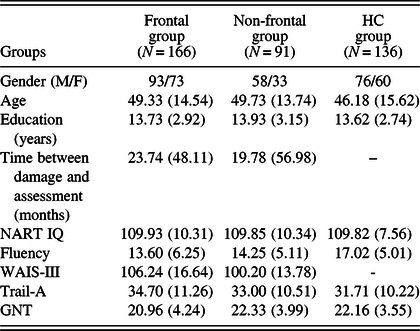
HC = healthy controls; WAIS-III = Wechsler Adult Intelligence Scale-III; Trail-A = Trail Making Test Part-A; GNT = Graded Naming Test.
Prior to running the regression models, we demonstrated that the different aetiology subgroups (i.e., stroke, high-grade glioma, low-grade glioma, and meningioma) did not significantly differ in their performance on the neuropsychological tests (except our low-grade glioma frontal patients who were significantly faster on Trail Making Test Part-A than the other frontal aetiology groups; see Tables 1 and 2 in the online Supplementary Materials). Of note, this group of patients was also significantly younger. On the whole, it appears methodologically justifiable to group together patients with different aetiologies for the purpose of cognitive analyses (for similar conclusions in frontal patients see Cipolotti et al., Reference Cipolotti, Healy, Chan, Bolsover, Lecce, White and Bozzali2015a). Table 3 shows the results of the multiple linear regression models testing for the effect of NART IQ on each cognitive test.
Regression models for the four cognitive tests

F = frontal patients; NF = non-frontal patients; group factor baseline level = healthy controls; WAIS = Wechsler Adult Intelligence Scale; Trail-A = Trail Making Test Part-A; GNT = Graded Naming Test; agroup factor baseline level = frontal patients. Exponentiated betas are reported. Bonferroni adjusted p-value < .0125. Bold typeface denotes significant p-values.
Letter Fluency “S” Test
In the case of letter fluency, NART IQ significantly predicted performance where the higher the NART IQ, the more words were produced. Lesion group also significantly contributed to the model fit with frontal patients producing significantly fewer words than HC. Non-frontal patients did not significantly differ from HC. Age also contributed to the fit of the model, where younger individuals produced more words. The final model explained 17% of the variance (F(4,241) = 11.93, p < .0001). The interaction between lesion group and NART IQ did not significantly contribute to participants’ fluency performance.
WAIS-III Full-Scale IQ
NART IQ significantly contributed to performance on WAIS IQ, where the higher the NART IQ, the higher the WAIS-III full-scale IQ. Lesion group also independently predicted performance with the frontal patients having significantly higher full-scale IQ scores than the non-frontal group. Age did not contribute to the model. NART IQ and lesion group accounted for 39% of the variance on WAIS IQ (F(3,149) = 32.15, p < .0001). The interaction term between lesion group and NART IQ did not significantly contribute to the models.
Trail Making Part-A (Trail-A)
Age significantly predicted Trail-A performance, accounting for 17% of the variance (F(1,176) = 35.53, p < .0001), where the younger the individual, the faster the performance. Entering lesion group or NART IQ did not significantly improve the fit of the model.
Graded Naming Test
Again, NART IQ was a significant predictor of performance, where the higher the NART IQ, the higher the GNT performance. Lesion group also significantly contributed to performance on the GNT where the frontal patients performed significantly more poorly than HC. Non-frontal patients did not significantly differ from HC. Age did not contribute to the model at any stage. NART IQ accounted for 36% of the variance on the GNT (F(4,339) = 47.21, p < .0001). Again, lesion group did not contribute to the model as an interaction term with NART IQ.
DISCUSSION
In this retrospective study, we examined the influence of literacy attainment based on NART IQ on neuropsychological test performance in a large sample of patients with unilateral frontal or non-frontal lesions and HCs. Our analyses revealed that our frontal group performed significantly poorer than HCs on the executive (i.e., fluency) and naming tests (i.e., GNT). In contrast, our frontal patients were not significantly slower than HCs on the test of processing speed and had significantly higher full-scale IQs compared to our non-frontal group. The reduced fluency performance in our frontal patients supports previous patient studies (e.g., Milner, Reference Milner, Warren and Akert1964; Perret, Reference Perret1974; Robinson et al., Reference Robinson, Shallice, Bozzali and Cipolotti2012; Stuss et al., Reference Stuss, Alexander, Hamer, Palumbo, Dempster, Binns and Izukawa1998; Troyer, Moscovitch, Winocur, Alexander, & Stuss, Reference Troyer, Moscovitch, Winocur, Alexander and Stuss1998; see Henry & Crawford, Reference Henry and Crawford2004; Cipolotti et al., Reference Cipolotti, Molenberghs, Dominguez, Smith, Smirni, Xu and Chan2020).
In terms of the contribution of NART IQ on neuropsychological test performance, after adjusting for age and lesion group (frontal and non-frontal vs. HC), NART IQ predicted performance on fluency, intelligence, and naming. Our previous work involving only frontal patients has demonstrated that NART IQ predicts executive and naming performance (MacPherson et al., Reference MacPherson, Healy, Allerhand, Spano, Chan, Tudor-Sfetea and Cipolotti2017). Here, we provide further support for the predictive nature of the NART in terms of cognition following frontal and non-frontal lesions. Importantly, however, the influence of NART IQ on neuropsychological test performance does not differ across lesion groups suggesting that the frontal lobes do not play a role in mediating the CR effect.
Of course, it remains possible that there may be specific frontal subregions associated with CR and cognitive performance and only damage to these specific subregions may hinder any benefits of CR. For example, regions such as the superior, middle, and inferior frontal gyri, as well as frontal lobe-associated networks (e.g., left anterior intraparietal sulcus; Bastin et al., Reference Bastin, Yakushev, Bahri, Fellgiebel, Eustache, Landeau and Salmon2012), have been associated with CR (for a review see Anthony & Lin, Reference Anthony and Lin2017), and damage to these specific regions may prevent compensation from CR after brain injury. In addition, we did not consider parameters such as white matter intensities (WMH) and cortical atrophy in our patients. Patients with high CR estimates have been found to have greater quantities of WMH than patients with low CR estimates and yet may perform equally well or better on cognitive tasks (e.g., Brickman et al., Reference Brickman, Siedlecki, Muraskin, Manly, Luchsinger, Yeung and Stern2011; Jokinen et al., Reference Jokinen, Melkas, Madureira, Verdelho, Ferro, Fazekas and Erkinjuntti2016; Murray et al., Reference Murray, Staff, McNeil, Salarirad, Ahearn, Mustafa and Whalley2011). However, given the heterogeneous neuroimaging data that were available for our retrospective study through clinical scans, as well as our sample size, it was not possible to investigate focal damage to specific frontal or non-frontal subregions. Yet, the major strength of our retrospective study is that it follows on and supports our previous findings examining the effects of CR proxies in frontal patients due to stroke or tumour (MacPherson et al., Reference MacPherson, Healy, Allerhand, Spano, Chan, Tudor-Sfetea and Cipolotti2017). To our knowledge, the current study is the first to examine the influence of a CR proxy on the performance of a large group of patients with unilateral frontal and non-frontal lesions across different cognitive measures.
It should also be pointed out that patients with more severe brain lesions were not included in our study due to their inability to cope with the demands of our cognitive tests. Therefore, we cannot rule out the possibility that more severe frontal lesions may not safeguard against focal neuropathology and moderate cognitive impairment across these various cognitive measures. In our previous work (MacPherson et al., Reference MacPherson, Healy, Allerhand, Spano, Chan, Tudor-Sfetea and Cipolotti2017), we observed that the patients who were not included in our retrospective study tended to have extensive frontal lobe lesions. Moreover, those frontal patients with high lesion severity performed significantly more poorly on fluency and speed of processing tasks than frontal patients with low lesion severity, despite being matched on education and NART IQ. Future prospective studies examining the effects of CR in patients with focal frontal and non-frontal lesions are needed to examine the role of lesion severity on cognition.
Age independently predicted performance on fluency and Trail-Making Part-A but not WAIS-III and GNT. This is in line with our previous work demonstrating that age and NART IQ influence performance on a range of cognitive measures in a smaller group of frontal patients, some of whom have also participated in the current study (Cipolotti et al., Reference Cipolotti, Healy, Chan, MacPherson, White, Woollett and Shallice2015b; MacPherson et al., Reference MacPherson, Healy, Allerhand, Spano, Chan, Tudor-Sfetea and Cipolotti2017). However, age did not predict our frontal and non-frontal patients’ intellectual abilities. As our data are age-scaled, these findings suggest that there is not a further effect of age on our patient population over and above the adjustments made using normative data.
Our analyses indicate that performance on Trail-Making Test Part-A is predicted only by age. Yet, we acknowledge that our sample size for the Trail-Making Test Part-A is small, particularly for the non-frontal patients (i.e., non-frontal = 20, frontal = 77 and HC = 81), so caution should be taken when concluding that NART is selectively unrelated to processing speed. Nonetheless, in our MacPherson et al. (Reference MacPherson, Healy, Allerhand, Spano, Chan, Tudor-Sfetea and Cipolotti2017) study involving frontal patients only, we similarly reported that age was the only significant contributor to the fit of the Trail Making Part-A model and education and NART IQ made no significant contributions to the model at any stage. Future prospective work is needed to examine further the relationship between CR proxies and speed of processing, as well as other aspects of cognition.
Following a common practice in neuropsychology, we mixed different aetiologies in our patients’ samples to obtain a large enough group. Previously, we have reported that there was not a significant difference between 100 frontal patients with four different types of aetiology (i.e., stroke, high-grade glioma, low-grade glioma, and meningioma) on four frontal-executive tasks (Cipolotti et al., Reference Cipolotti, Healy, Chan, Bolsover, Lecce, White and Bozzali2015a). Critically, it remained unknown if the effects of strokes and tumours were roughly equivalent when affecting the non-frontal cortex. In our Supplementary Materials, we document for the first time that the cognitive performance in our non-frontal patients was not affected by variability in lesion aetiology. Our subgroups of non-frontal patients with stroke, high- or low-grade tumour, or meningioma did not differ in their performance on tests of frontal executive (fluency), intelligence (WAIS-III), processing speed (Trail-A), or naming (GNT). Similarly, our patients with frontal lesions due to stroke, high- or low-grade tumour, or meningioma did not differ in their performance on the neuropsychological tests except our test of processing speed (Trail Making Test Part-A) where the low-grade tumour patients were significantly faster than the other frontal aetiology groups. This is perhaps not surprising given our low-grade glioma frontal group was significantly younger than the other aetiology subgroups and individuals start to show age-related decline in processing speed as early as their 30s (Baxendale, Reference Baxendale2011). Previous research examining cognition in glioma patients has also reported that processing speed is less impaired in low-grade compared to high-grade glioma, although this impairment was not specific to frontal lesions (Dehcordi, Mariano, Mazza, & Galzio, Reference Dehcordi, Mariano, Mazza and Galzio2013; Miotto et al., Reference Miotto, Junior, Silva, Cabrera, Machado, Benute and Teixeira2011; van Kessel et al., Reference van Kessel, Emons, Wajer, van Baarsen, Broekman, Robe and Van Zandvoort2019). While grouping patients with different aetiologies is likely to suffer from potential confounds, it remains necessary to obtain large groups of patients to investigate cognitive impairments (for similar approaches see Aridan, Pelletier, Fellows, & Schonberg, Reference Aridan, Pelletier, Fellows and Schonberg2019; Aron, Monsell, Sahakian, & Robbins, Reference Aron, Monsell, Sahakian and Robbins2004; Gläscher et al., Reference Gläscher, Adolphs, Damasio, Bechara, Rudrauf, Calamia and Tranel2012; Roca et al., Reference Roca, Parr, Thompson, Woolgar, Torralva, Antoun and Duncan2010; Stamenova et al., Reference Stamenova, Gao, Black, Schwartz, Kovacevic, Alexander and Levine2017; Stuss et al., Reference Stuss, Alexander, Shallice, Picton, Binns, Macdonald and Katz2005; Thompson–Schill et al., Reference Thompson-Schill, Swick, Farah, D’Esposito, Kan and Knight1998; Urbanski et al., Reference Urbanski, Brechemier, Garcin, Bendetowicz, Thiebaut de Schotten, Foulon and Volle2016). Some other studies favour the use of a single aetiology (e.g., Baldo, Schwartz, Wilkins, & Dronkers, Reference Baldo, Schwartz, Wilkins and Dronkers2006; Campanella et al, Reference Campanella, Skrap and Vallesi2016; Sperber & Karnath, Reference Sperber and Karnath2017; Varjačić et al., Reference Varjačić, Mantini, Levenstein, Slavkova, Demeyere and Gillebert2018). However, there is no consensus in the field of neuropsychology regarding what is the best approach to adopt. As a minimum, we have attempted to demonstrate that certain aetiologies do not result in more severe impairments than others (see also Cipolotti et al., Reference Cipolotti, Healy, Chan, Bolsover, Lecce, White and Bozzali2015a).
In summary, our CR analyses suggest that age and NART IQ provide protective effects of focal brain pathology in patients with lesions due to stroke or brain tumour. However, importantly, the relationship between NART IQ and cognitive performance following focal brain damage does not differ between frontal and non-frontal lesions. Therefore, environmental factors shape resilience to cognitive decline in both patients who have experienced focal frontal or non-frontal lesions. Future work involving prospective studies should be conducted to examine further the complex relationship between CR, age, and frontal/non-frontal regions when attempting to understand impairments and recovery on cognitive tasks. CR may influence the degree of recovery post-stroke or brain tumour, which is critical for our understanding of the recovery process. CR may also be a predictive factor of the efficacy of neuropsychological rehabilitation training in individuals who have experienced focal brain damage, regardless of the brain area damaged.
ACKNOWLEDGEMENTS
Financial support: This project was funded by Wellcome Trust (grant numbers 066763, 089231/A/09/Z) and supported by the National Institute for Health Research University College London Hospitals Biomedical Research Centre.
CONFLICT OF INTEREST
The authors have nothing to disclose.
SUPPLEMENTARY MATERIAL
To view supplementary material for this article, please visit https://doi.org/10.1017/S1355617720000326



