Introduction
The idea that Mars could be the first extraterrestrial body to yield evidence of life beyond Earth is nowadays widespread among planetary scientists. If life was once present on Mars, the so-called biosignatures, which can be preserved for billions of years under favourable conditions, may still exist. Biosignatures may be linked both with organic and inorganic compounds. In particular, minerals of fossils may display distinctive morphologies, isotope signatures, chemicals composition or defect microstructures that can reveal their biological origin (Banfield et al. Reference Banfield, Moreau, Chan, Welch and Little2001).
Among the minerals, calcium carbonates (CaCO3) are particularly interesting because they can be produced either by abiotic processes or by biologically induced or controlled mineralization (Mann Reference Mann2001). Many living organisms on Earth, prokaryotes and eukaryotes, are able to biomineralize calcite or aragonite and such biomineralized carbonates are among the oldest terrestrial evidence of life (Wacey Reference Wacey2009; Westall Reference Westall, Gargaud, López-Garcìa and Martin2011). On the other hand, it is well known that carbonates are also produced by chemical precipitation following different processes not related to the presence of any life form (Wilkinson & Given Reference Wilkinson and Given1986).
The increasing evidence for the presence of carbonates on Mars (Pollack et al. Reference Pollack, Roush, Witteborn, Wooden, Stoker and Toon1990; Bandfield et al. Reference Bandfield, Glotch and Christensen2003; Ehlmann et al. Reference Ehlmann2008; Boynton et al. Reference Boynton2009; Palomba et al. Reference Palomba, Zinzi, Cloutis, D'Amore, Grassi and Maturilli2009; Michalski & Niles Reference Michalski and Niles2010; Morris et al. Reference Morris2010; Carter & Poulet Reference Carter and Poulet2012; Michalski et al. Reference Michalski, Cuadros, Niles, Parnell, Rogers and Wright2013), suggests that a number of locations may have existed where surface conditions would have been favourable for microbial habitability. Some of these sites may be good candidates for the exploration in search for signs of extinct or extant life both on the surface and in the near-subsurface. Anyway, if life existed on Mars, then the extreme conditions may have favoured the development of chemotrophic organisms (Summons et al. Reference Summons, Amend, Bish, Buick, Cody, Des Marais, Dromart, Eigenbrode, Knoll and Sumner2011; Westall et al. Reference Westall, Loizeau, Foucher, Bost, Bertrand, Vago and Kminek2013, Reference Westall2015).
The problem of discriminating between biominerals and their abiotic counterparts is far from trivial. Nevertheless by means of thermal processing it is possible to distinguish, using differential thermal analysis (Cabane et al. Reference Cabane2004; Stalport et al. Reference Stalport2005, Reference Stalport, Coll, Szopa, Person, Navarro-Gonzalez, Cabane, Ausset and Vaulay2007) or infrared (IR) spectroscopy (Orofino et al. Reference Orofino, Blanco, D'Elia, Licchelli and Fonti2007), abiotic CaCO3 minerals (i.e. aragonite or calcite) from the corresponding biominerals. Furthermore, Berg et al. (Reference Berg, Ronholm, Applin, Mann, Izawa, Cloutis and Whyte2014) and Ronholm et al. (Reference Ronholm2014) have recently described diverse approaches aimed to distinguish different biogenic carbonates from abiotic materials.
In a series of papers, we have developed and applied our method to different carbonate samples, in the form of fresh shells and fossils of different ages found in different places and easily recognizable as of biotic origin (D'Elia et al. Reference D'Elia, Blanco, Licchelli, Orofino, Fonti, Pomati and Burns2006; Orofino et al. Reference Orofino, Blanco, D'Elia, Licchelli and Fonti2007, Reference Orofino, Blanco, D'Elia, Fonti and Licchelli2009, Reference Orofino, Blanco, D'Elia, Licchelli, Fonti and Marzo2010). The method has been then successfully applied to bio-induced carbonates deposit, i.e. microbialites (Blanco et al. Reference Blanco, Orofino, D'Elia, Fonti, Mastandrea, Guido and Russo2011, Reference Blanco, Orofino, D'Elia, Fonti, Mastandrea, Guido and Russo2013), and very recently, by studying different parts of the same rock sample, we have been able to discriminate the different origin of the fine carbonate fractions, namely the micrite types (Blanco et al. Reference Blanco, D'Elia, Orofino, Mancarella, Fonti, Mastandrea, Guido, Tosti and Russo2014). The term micrite, introduced by Folk (Reference Folk1959), is the abbreviation of ‘microcrystalline calcite’ and is commonly used to describe the particles <4 µm in size (Tucker & Wright Reference Tucker and Wright1990). On the basis of the degree of biogenicity we distinguished the micrites deriving from physical processes (i.e. erosion and transport of pre-existing carbonate), named detrital or allochthonous micrite, from those deriving by biotic processes (i.e. in situ precipitation via bacterial mediation), named autochthonous micrites or microbialites. Stromatolites, the laminated fabric of microbialites, are well known to be typical examples of very primitive forms of life on Earth, hence samples of biocarbonates that can be considered as analogues of fossils of putative Martian life forms.
An alternative and ancillary approach to distinguish biotic from abiotic carbonates could be the study of their morphological aspect at different scales (Cady et al. Reference Cady, Farmer, Grotzinger, Schopf and Steele2003; Stolarski & Mazur Reference Stolarski and Mazur2005; Bianciardi et al. Reference Bianciardi, Rizzo and Cantasano2014; Ronholm et al. Reference Ronholm2014). The complexity of biomineralized structures gives an indication of the potential of organic constituents for controlling energetic factors during crystal synthesis. Many organisms mediate crystallization through the selective application of organic compounds that exert a detailed control over the structure (Belcher et al. Reference Belcher, Wu, Christensen, Hansma, Stucky and Morse1996), orientation (Berman et al. Reference Berman, Addadi and Weiner1988), growth kinetics (Mann et al. Reference Mann, Archibald, Didymus, Douglas, Heywood, Meldrum and Reeves1993) and nucleation sites of crystals (Winter & Seisser Reference Winter and Seisser1994). Researchers studied biominerals for decades and found that crystal growth in vitro, in the presence of a biomineral matrix, have a distinct morphology in comparison with those grown without matrix. These findings generally support the hypothesis, suggested also by morphological observations, that matrix influences crystal growth and may well influence the structure of biominerals.
D'Elia et al. (Reference D'Elia, Blanco, Licchelli, Orofino, Fonti, Pomati and Burns2006) examining at micrometer scale the morphology of two fossil shells composed of aragonite and calcite, with a scanning electron microscope (SEM), showed that they exhibit a well-defined crystal pattern quite different from the massive micromorphologies of the mineral crystals imaged at the same scale. In this work, we extend the morphological analysis to biotic carbonates, mostly microbialites, linked to primitive living microrganisms which could be considered as good analogues of rock minerals remains linked to present or past life on Mars.
The intent of this paper is to highlight and suggest a line of research aimed to find a key to distinguish biogenic from abiogenic carbonates. The study has been prompted by the need to provide images and data concerning mineral structures and textures. Such data may be useful for the analysis of Martian samples coming from sample return missions or investigated by future explorations in situ, with the aim to characterize the near-subsurface of Mars in search for past or present life.
In ‘Sample description and preparation’ the main characteristics of the analysed samples are described. In section ‘SEM analysis and conclusions’, the experimental results are reported together with discussion and conclusions.
Sample description and preparation
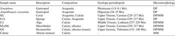
The samples analysed in this work are listed in Table 1. The first two are fossil shells composed of aragonite, a metastable phase of CaCO3, while the others are skeletal fossils of organisms (coral, sponge and algae) and microbialites. All samples are made of CaCO3, calcite or aragonite, with some silicates in the case of the microbialites. A calcite mineral rock of abiotic origin is also included for comparison. The estimated geological ages of the samples are also reported in Table 1 where the beginning and the end of each period/epoch are those established by the International Commission on Stratigraphy (ICS) deputed to the terrestrial stratigraphy on a global scale (Ogg et al. Reference Ogg, Ogg and Gradstein2008).
Studied samples listed in order of confidence of biological origin

In the last column, we report the types of micromorphology of each sample (DP, defined pattern; MM, massive micromorphology).
The fossil shells of two marine gastropod molluscs, already studied by Orofino et al. (Reference Orofino, Blanco, D'Elia, Licchelli, Fonti and Marzo2010), have been collected in clay deposits located at two different sites, about 30 km apart, in the Salento Peninsula, Italy. The Xenophora gastropod comes from a Pleistocene deposit and in particular, in relation to stratigrapy, from a whitish calcarenite with an upper greenish clayey–sandy interval, having an elevated paleontological interest for the abundance of the fossil associations with excellently preserved taxa.
The Ampullinopsis crassatina, that biostratigraphical indications allowed us to assign to the late Oligocene, was collected from a lignite clayey deposit, overlapped on a blanket of mineralized residual deposits (bauxitic residual deposits) resting on the local carbonatic basement.
The samples S/L, S1A and U2 are skeletal organisms (coral, sponge and algae, respectively) embedded in microbial carbonates such as sample S/L(M). They have been selected within three rock specimens that developed, in time, in two distinct palaeoecological conditions, characteristic of Alpe di Specie and Punta Grohmann carbonate outcrops, in the Dolomites, Italy.
The S/L and S1A samples were extracted from two different Alpe di Specie rock samples in which skeletal organisms (Tubiphytes, skeletal cyanobacteria, sphinctozoan and inozoan sponges, etc.) represent a minor component of the rock (usually <40%). The composition is dominated by the micritic fraction (about 60%), mainly represented by autochthonous micrite (microbialite), with subordinate amounts of micrite interpreted as detrital (allochthonous micrite) (Russo et al. Reference Russo, Neri, Mastandrea and Laghi1991; Tosti et al. Reference Tosti, Guido, Demasi, Mastandrea and Russo2011, Reference Tosti, Guido, Mastandrea, Demasi and Russo2012, Reference Tosti, Mastandrea, Guido, Demasi, Russo and Riding2014). The microbialites or autochthonous micrites, which may exhibit both dense microcrystalline (aphanitic) or peloidal microfabric, are sometimes organized in stromatolitic laminae or thrombolitic fabric.
The U2 was selected from a rock sample collected in the basinal section cropping out at the base of Punta Grohmann mountain, belonging to the Sasso Piatto Massif, in the province of Bolzano, Italy. The analysed sample belongs to the Punta Grohmann buildups. These buildups represent the last records of the bioconstructions that first appeared in the Late Pennsylvanian. They are characterized by subcentimetre skeletons (mainly calcified microbes, Tubiphytes and other problematica, small sponges) intimately associated with microbialites and cements. The organic-induced nature of microbialite was supposed on the base of micromorphological and biogeochemical evidence (Russo et al. Reference Russo, Neri, Mastandrea and Baracca1997; Tosti et al. Reference Tosti, Guido, Demasi, Mastandrea and Russo2011, Reference Tosti, Guido, Mastandrea, Demasi and Russo2012, Reference Tosti, Mastandrea, Guido, Demasi, Russo and Riding2014). Their biotic origin has been confirmed by Blanco et al. (Reference Blanco, Orofino, D'Elia, Fonti, Mastandrea, Guido and Russo2013, Reference Blanco, D'Elia, Orofino, Mancarella, Fonti, Mastandrea, Guido, Tosti and Russo2014) with independent methods.
The stromatolitic sample GE, Upper Jurassic in age (Tithonian, 151–146 Ma) was collected from Thüste Quarries, south of Hanover, Germany. In this area, stromatolites developed in stressed environments, probably represented by a lagoonal setting, with alternate deposition of oolitic limestone and evaporites (Jahnke & Ritzkowski Reference Jahnke and Ritzkowski1980).
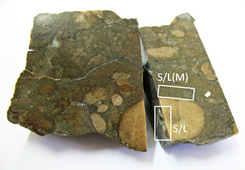
In order to obtain fine particulate materials for SEM morphological analyses, the fossil shells (Xenophora and A. crassatina) and the mineral calcite were ground with a mechanical mortar grinder and then the size fraction between 20 and 50 µm was sieve selected for our investigation. The other specimens, instead, were collected from specific areas of the rock samples using a thin drill bit. For instance, in Fig. 1 the original rock sample is shown along with the areas where the powders, pertaining to S/L skeletal organism and S/L(M) microbialite, were picked up.
The rock sample where the skeletal grains of the coral (S/L) and microbialite S/L(M) particles have been picked up. The specific areas are shown by white rectangles.

The grains thus obtained were comparable in size to those derived from sieve selection as derived by SEM observation. Double-stick and electrically conductive adhesive carbon tabs were pressed to the surface of pin stub mounts of aluminium allowing the adhesion of diced silicon wafers (5 × 5 mm2). Afterwards the particulate materials were placed in small glass vessel containing ethyl alcohol and introduced in an ultrasonic unit (Elma Transsonic 890/H) for about 30 s to separate any agglomerates of grains. Using a pipette, a drop of this suspension was added to the silicon matrix. After the evaporation of the alcohol, the grains of the different samples remained attached to the matrix and then, since specimens viewed using a conventional SEM need to be coated with electrically conductive material, we sputtered the stubs with a nanometric gold layer using an Emitech K550X Sputter Coater. Finally, the various stubs were placed inside the SEM chamber in high vacuum mode and morphologically analysed with the secondary electron detector.
The mineral composition, reported in Table 1, has been determined using both the IR spectroscopy and the energy dispersive X-ray elemental analysis performed in our laboratory on all samples (for details see Blanco et al. Reference Blanco, D'Elia, Orofino, Mancarella, Fonti, Mastandrea, Guido, Tosti and Russo2014).
SEM analysis and conclusions
The morphological analysis has been done using a SEM JEOL JSM 6480LV, equipped with an iXRF Systems EDS Sirius SD spectrometer for the elemental composition.
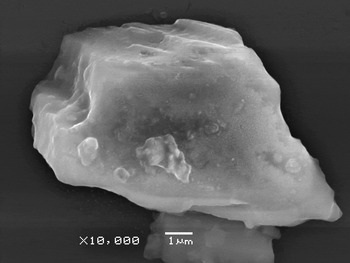
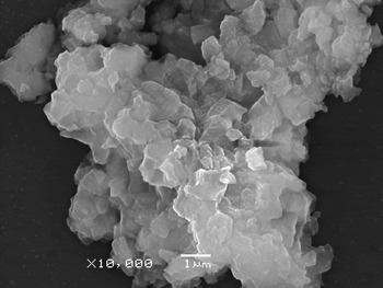
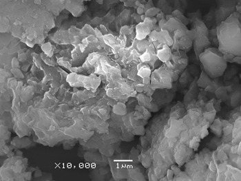
In Figs 2 and 3, typical SEM images of particles of the two fossil shells are reported, while in Fig. 4 the micrograph of mineral calcite grains is shown. It is evident the well-defined crystal pattern of the shells particles compared with the massive micromorphology of the mineral crystals imaged at the same scale.
SEM image of the Xenophora particles.

SEM image of the Ampullinopsis crassatina particles.

SEM image of abiotic calcite mineral particles.

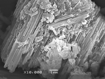
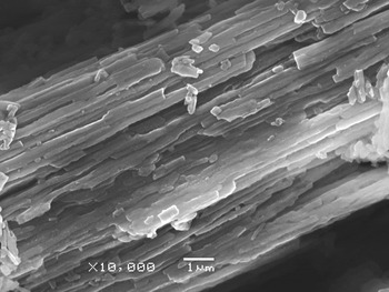
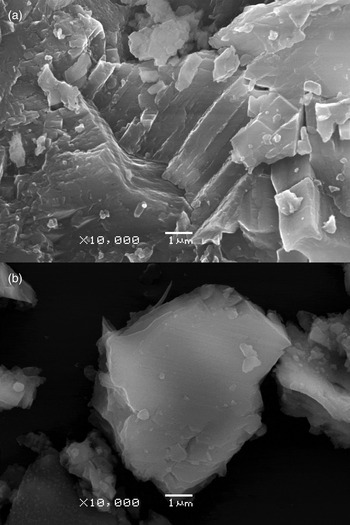
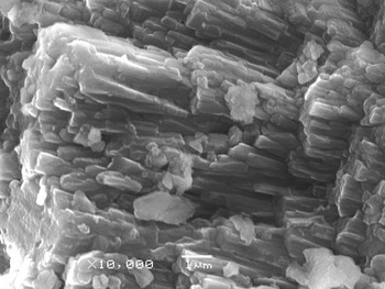
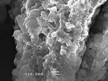
Similar morphologies at micrometric scale are observable in Fig. 5(a) and from Figs 6 to 9 for the skeletal organisms (samples S/L, S1A and U2), microbialites and stromatolites (samples S/L(M) and GE, respectively). Except for the case of S1A (sponge), in which all the particles have the same crystalline structure (see Fig. 6 and last column of Table 1), in the other samples some grains exhibit a well-defined pattern (labelled DP in Table 1), while others show a massive micromorphology (labelled MM) as the abiogenic calcite crystals. As an example, we can refer to the difference between panels (a) and (b) in Fig. 5 (S/L sample) and the comparison between Figs 5(b) and 4 (calcite) that indicates an analogous arrangement.
SEM images of the coral particles (samples S/L). Some particles show a well-defined pattern (a) while others a massive micromorphology (b).

SEM image of the sponge particles (sample S1A).

Furthermore, we note that within those biogenic samples showing a well-defined pattern, as evident, the biogenic samples can be divided in two categories: those having a ‘laminar’ pattern (coral and sponge, Figs 5(a) and 6) similar to those of the fossil shells and those having a granular pattern of small crystals (algae, microbialites and stromatolites, Figs 7–9). Even if such differences could be ascribed to a diverse degree of complexity of the involved forms of life, we tentatively speculate that they can be linked to the diverse type of biomineralization. In fact, microorganisms are remarkably adept at forming mineral phase and this process can occur in two different ways. The first involves mineral precipitation in the open environment, without any apparent control by the cell over the mineral product. This process was defined by Lowenstam (Reference Lowenstam1981) as ‘biologically induced biomineralization’, with mineral forming only as a by-product of the cells metabolic activity or through its interaction with the surrounding aqueous environment (Konhauser Reference Konhauser2007). By contrast, the second way, ‘biologically controlled biomineralization’, is completely regulated, allowing the organisms to precipitate mineral that serve some physiological purpose. This process is specifically designed to form minerals through the development of intracellular (within the cytoplasm) or epicellular (on the cell wall) organic matrices, into which specific ions are actively introduced and their concentrations controlled in order to achieve the right mineral saturation states. Since the mineralization site is isolated from outside the cell by a barrier through which ions cannot freely diffuse, minerals can form anyway, in spite of thermodynamically unfavourable external conditions (Konhauser Reference Konhauser2007).
SEM image of the algae particles (sample U2).

SEM image of the microbialite particles (sample S/L(M)).

SEM image of the stromatolites particles (sample GE).

To investigate the distribution of these different crystal patterns of particles, we have morphologically examined more than 50 grains of each sample and we have summarized the results of this analysis in the last column of Table 1.
It is worthwhile to note that these results cannot be conclusive although they give precise indications on the main differences between the crystal micromorphologies of biogenic and abiogenic carbonates. This means that, in order to reach meaningful conclusions, we need to analyse the morphology of other samples as well as a statistically significant number of particles. Furthermore, special attention should be given to diagenesis (Tucker & Wright Reference Tucker and Wright1990), namely the chemical and physical post-depositional transformation processes that alter the original chemistry and microstructure of the carbonate minerals, producing a uniform final product difficult to discriminate between biotic versus abiotic origin. However, we think that the effort towards this line of research may possibly provide a method useful for discriminating the origin of Martian carbonates with the aim to characterize the surface and near-subsurface of the red planet in search for extraterrestrial life.
Acknowledgements
This research has been partially supported by the Italian Space Agency (ASI) and the Italian Ministry of University and Research (MIUR).