Introduction
Fasciola jacksoni (Cobbold, Reference Cobbold1869) is one of four recognized species within the genus Fasciola Linnaeus, 1758 (Cobbold, Reference Cobbold1882; Mas-Coma et al., Reference Mas-Coma, Valero and Bargues2009) and a major health problem for elephants in Sri Lanka, causing fatal fascioliasis in wild elephant populations. The size of the natural populations of Sri Lankan Asian elephants (Elephas maximus maximus) is estimated to be 2500–3000 individuals; approximately 15% of the total number of elephants found across Asia. Sri Lankan elephant herds vary in size with some made up of a few adults, juveniles and calves through to those composed of several hundred individuals in the Forest Reserves, Sanctuaries, National Parks and Orphanages distributed across the country. However, elephants are one of the most endangered large animal species in the world, in part associated with their continued long-term conflict with people for land. In Sri Lanka there is intense human-wild elephant conflict (HEC), resulting in the death of an elephant every 2 days and a human death every week (Fernando et al., Reference Fernando, Wikramanayake, Weerakoon, Jayasinghe, Gunawardene and Janaka2005). Each year elephants destroy human properties and crops valued at millions of rupees leaving thousands of people living in financial uncertainty and insecurity (Perera, Rajapakse, Reference Perera, Rajapakse, Wibbelt, Kretschmar, Hofer and Seet2009; Lorimer, Reference Lorimer2010).
Owing to continued HEC elephant numbers have dramatically decreased due to an ever increasing lack of space, as well as infectious diseases (Fernando, Reference Fernando2000; Lorimer, Reference Lorimer2010; Perera et al., Reference Perera, Rajapakse, Silva-fletcher, Thewarage and Jinadasa2015). Consequently, emerging parasitic diseases, including F. jacksoni are now major factors contributed to the dramatic decline of elephant populations (Bhalerao, Reference Bhalerao1933; Alahakoon, Reference Alahakoon1994; Perera et al., Reference Perera, Rajapakse, Silva-fletcher, Thewarage and Jinadasa2015). Such flukes contribute to the high burden of gastro-intestinal parasitism in elephants, which has led to high mortality and morbidity of calves and adults alike across Sri Lanka (Alahakoon, Reference Alahakoon1994). Despite F. jacksoni being one of the major risk factors contributing to the decrease of wild Asian elephants, the parasite is completely neglected, regardless of continued reports of infection in India, Sri Lanka and Malaysia causing severe pathology (Windsor and Scott, Reference Windsor and Scott1976; Caple et al., Reference Caple, Jainudeen, Buick and Song1978; Alahakoon, Reference Alahakoon1994; Islam, Reference Islam1997; Perera and Rajapakse, Reference Perera, Rajapakse, Wibbelt, Kretschmar, Hofer and Seet2009; Hing et al., Reference Hing, Othman, Nathan, Fox, Fisher and Goossens2013).
The systematics of Fasciola and the Fasciolidae has been fiercely debated over the past decade with the majority of studies depending on tenuous and plastic morphological characteristics of both adult flukes and eggs, which has ultimately affected the accuracy of identification and diagnosis. Molecular phylogenetic approaches have been employed to resolve the relationships between members of the Fasciolidae illustrating the complex taxonomic associations between members of the genus Fasciola and their association with the genus Fascioloides (Nolan and Cribb, Reference Nolan and Cribb2005; Wey-Fabrizius et al., Reference Wey-Fabrizius, Podsiadlowski, Herlyn and Hankeln2013; Tkach et al., Reference Tkach, Kudlai and Kostadinova2016; Le et al., Reference Le, Nguyen, Nguyen, Doan, Dung and Blair2017). Recently, such approaches have suggested that F. jacksoni should be moved to the genus Fascioloides and thus renamed as Fascioloides jacksoni (Lotfy et al., Reference Lotfy, Brant, DeJong, Le, Demiaszkiewicz, Rajapakse, Perera, Laursen and Loker2008; Heneberg, Reference Heneberg2013). Phylogenetic analyses of the Fasciolidae family have been dependant on the mitochondrial markers cytochrome oxidase subunit 1 gene (cox1) and the NADH dehydrogenase subunit 1 gene (nad1) as well as the nuclear ribosomal transcription units particularly ITS1, ITS2, 18S rDNA and partial 28S rDNA. These genetic markers have greatly contributed to molecular identification, diagnostic, epidemiological, phylogenetic and evolutionary studies of the Fasciolidae, especially the highly pathogenic Fasciola hepatica and Fasciola gigantica, the invasive Fascioloides magna, and the neglected but re-emerging Fasciolopsis buski (Králová-Hromadová et al., Reference Králová-Hromadová, Špakulová, Horáčková, Turčeková, Novobilský, Beck, Koudela, Marinculić, Rajský and Pybus2008; Lotfy et al., Reference Lotfy, Brant, DeJong, Le, Demiaszkiewicz, Rajapakse, Perera, Laursen and Loker2008; Heneberg, Reference Heneberg2013; Mucheka et al., Reference Mucheka, Lamb, Pfukenyi and Mukaratirwa2015). Yet, despite their economic importance and the impact of fascioliasis on the health of wild elephant populations to date, molecular data associated with F. jacksoni is limited (Lotfy et al., Reference Lotfy, Brant, DeJong, Le, Demiaszkiewicz, Rajapakse, Perera, Laursen and Loker2008; Singh et al., Reference Singh, Srivastava, Prasad and Pandey1994; Perera and Rajapakse, Reference Perera, Rajapakse, Wibbelt, Kretschmar, Hofer and Seet2009; Heneberg, Reference Heneberg2013).
Thus, we present one of the first detailed applications of molecular approaches for the identification and phylogenetic analysis of F. jacksoni from wild elephants in Sri Lanka. This study was part of a larger epidemiological survey of F. jacksoni infection in National Parks in Sri Lanka and aimed to address the taxonomic position of F. jacksoni species in the Fasciolidae and ultimately the Trematoda.
Materials and methods
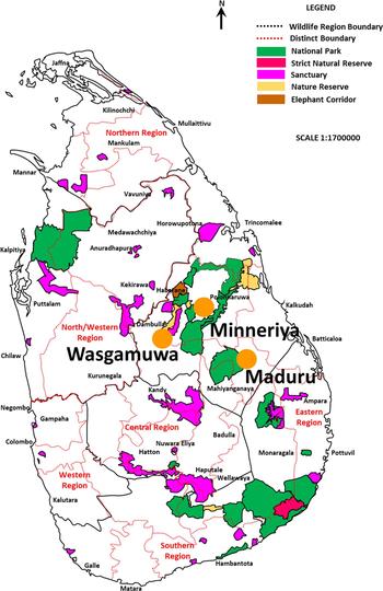
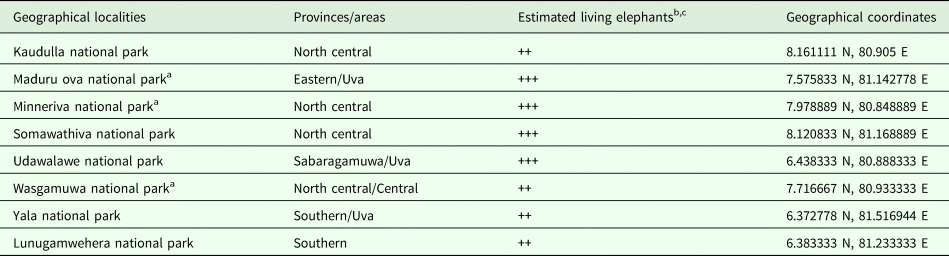
Post-mortem pathological examination was performed on a total of 47 elephants during a large-scale epidemiological study carried out from January 2000 to April 2003 (39 months) across six districts of Sri Lanka (Fig. 1; Table 1) (Perera and Rajapakse, Reference Perera, Rajapakse, Wibbelt, Kretschmar, Hofer and Seet2009). The liver was opened and separated with scissors, and the bile ducts were examined for the presence of adult Fasciola. Flukes were collected from the infected animals, washed with saline several times and fixed with 70% ethanol. Morphological species identification was performed by microscopy after adult flukes were stained with carmine (Fig. 2). Total worm burden within the liver was used to assess the intensity of the parasitic infestation in the affected animals (Perera and Rajapakse, Reference Perera, Rajapakse, Wibbelt, Kretschmar, Hofer and Seet2009). In addition, fecal samples from 48 living elephants were also collected randomly from the Maduruoya, Minneriya and Wasgamuwa National Parks (see map in Fig. 1), and Fasciola eggs were harvested for identification by filtration and examined using standard microscopy and fecal egg count techniques. Eggs were washed and centrifuged several times in normal saline (0.9% NaCl), then a further three times in phosphate buffered saline before storage at −20 °C until use. A total of three to four adult worms from each post-mortem elephant from each locality were individually fixed in 70% molecular grade ethanol. One ethanol-fixed worm and eggs samples from the selected localities were separately subjected to genomic DNA extraction and molecular analysis (Table 1).
Schematic map of living elephants in Sri Lanka and the study localities of National Parks where Fasciola jacksoni samples were collected (indicated by solid circles).

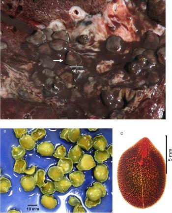
Adult Fasciola jacksoni collected from elephants. Where (A) are adult Fasciola jacksoni flukes (indicated by arrow) in the bile duct of the infected liver in a dead elephant. Bar (10 mm) indicates the size of the flukes; (B) are adult flukes collected from bile duct of the liver from a post-mortem elephant in Sri Lanka and illustrate gross morphology; (C) an adult F. jacksoni fluke stained (5X) with carmine. Bars indicate the size of the flukes. Bars (100 and 5 mm) indicate the size of the flukes.

Information of geographical localities with estimated living elephants for Fasciola jacksoni infection survey

a Geolocalities where Fasciola jacksoni samples (adult and eggs) were collected.
b Scientific name: Elephas maximus maximus.
c Estimated numbers of living elephants in the area: +: <50; ++: 50–250; +++: >250 elephants based on the information in public media and statistics (see the map of living areas for elephants in Fig. 1), geographical coordinates provided in latitude and longitude.
DNA extraction, PCR amplification and sequencing
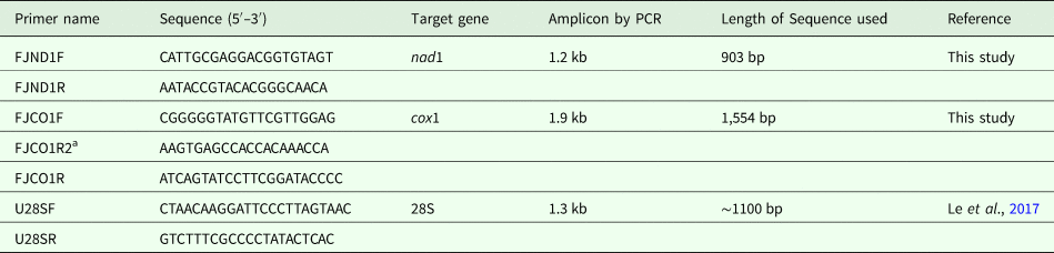
Total genomic DNA was extracted from approximately 10 mg lateral tissue from individual adult flukes or pooled eggs (about 1000 eggs) using the GeneJET™ Genomic DNA Purification Kit (Thermo Fisher Scientific Inc., MA, USA), according to the manufacturer's instructions. Genomic DNA was eluted into 50 µL of the elution buffer provided in the kit and stored at −20 °C. Complete gene fragments of both the mitochondrial markers cox1, 1.9 kb and nad1, 1.2 kb, where amplified using novel primers designed during this current study (Table 2). These were coupled with the amplification of a 1.4 kb portion of 28S ribosomal DNA and additional internal primers were designed and used for sequencing (see Le et al., Reference Le, Nguyen, Nguyen, Doan, Dung and Blair2017) (Table 2). The PCR reactions were performed in a 50 µL volume containing 25 µL of DreamTaq PCR Master Mix (2×) (Thermo Fisher Scientific Inc.) and 2 µL DNA template (50 ng µL−1), 2 µL of each primer (10 pmol µL−1), 2 µL DMSO (dimethyl sulfoxide) and 17 µL H2O. All PCRs were performed in an MJ PTC-100 thermal cycler with the following cycling conditions: initiation at 94 °C for 5 min, followed by 35 cycles consisting of denaturation for 30 s at 94 °C, annealing for 30 s (at 52 °C for nad1 and cox1; at 56 °C for 28S rDNA), extension at 72 °C for 3 min; and a final extension at 72 °C for 7 min. The PCR products (10 µL of each) were examined on a 1% agarose gel, stained with ethidium bromide and visualized under UV light (Wealtec, Sparks, NV, USA). All the purified or gel-extracted PCR amplicons were sent for sequencing (Macrogen Inc., South Korea) using amplifying/flanking and internal primers (Table 2) in both directions. All sequences obtained from adult or eggs of F. jacksoni samples were identical, regardless of the adult or eggs stage or locality.
Primers for amplification and sequencing of the mitochondrial protein-coding and nuclear ribosomal genes used in this study

F, forward; R, reverse.
a Internal primer used for sequencing.
Sequence analyses and phylogenetic reconstruction
An alignment of 21 concatenated nad1 and cox1 DNA sequences, approximately 2439–2457 nucleotides in length, containing representations of trematode isolates from the Fasciolidae including F. jacksoni from three localities, the Echinostomatidae and Schistosoma haematobium as the outgroup (Supplementary Table S1) was constructed using GENEDOC 2.7 (available at: http://iubio.bio.indiana.edu/soft/molbio/ibmpc/genedoc-readme.html). The final alignment was 2470 nucleotide (nt) long of which 21 nt positions were trimmed from either end, leaving 2449 characters for analyses. Edited alignments were used to perform phylogenetic reconstructions using the maximum likelihood (ML) method in MEGA 7 (Kumar et al., Reference Kumar, Stecher and Tamura2016). Pairwise distance analysis, was also performed on the concatenated nad1 and cox1 alignment as a measure of genetic distances (p-distance) between 12 representative species across three families of the Echinostomatoidea (Fasciolidae, Echinochamidae, Echinostomatidae) (Supplementary Table S1). DNA sequences of 28S rRNA genes (listed in Supplementary Table S2) were also aligned using GENEDOC 2.7. The resultant alignment of 1349 nucleotide long was edited by the removing 282 nt positions from both ends leaving to a total of 1067 characters for analyses. The final 28S rDNA alignment was composed of 43 species/isolates and MEGA 7 was again used to perform ML phylogenetic reconstruction construction (Kumar et al., Reference Kumar, Stecher and Tamura2016). For both alignments, MEGA 7 identified the general time reversible GTR + G + I model (γ rate heterogeneity and a proportion of invariant sites) as the most appropriate model for phylogenetic reconstruction based on the lowest Bayesian information criterion score. Thus phylogenetic analyses for both alignments were performed under the conditions of the above mentioned model with nodal support values calculated using 1000 bootstrap replicates (Kumar et al., Reference Kumar, Stecher and Tamura2016).
Results
Cause of mortality of elephants, description and incidence of Fasciola jacksoni
The survey was performed on a total of 47 post-mortems of elephants across all age groups over the duration of the 4-year study period. There were a variety of causes of fatalities of these elephants which included septicaemia associated with gunshot wounds (n = 23), gunshot leading to severe damage of the brain and lungs (n = 7), electrocution (n = 9), drowning (n = 1), suffocation through airway obstruction (n = 1), severe pneumonia (n = 2), old age (n = 1), unknown causes (n = 1) and fatal infection with F. jacksoni (n = 2) (Perera and Rajapakse, Reference Perera, Rajapakse, Wibbelt, Kretschmar, Hofer and Seet2009).
The parasitological investigation revealed that 27 out of the 47 elephant post-mortems were infected with F. jacksoni, with worm burdens varying from seven to 325 flukes (Fig. 2A). Livers of the affected animals showed cholangitis and fibrous tissue proliferation on the bile duct wall. The liver flukes present in the elephants were identified as typical for F. jacksoni species, according to the morphological characters described by Bhalerao (Reference Bhalerao1933). The average size of each F. jacksoni fluke was 12–14 mm × 9–12.5 mm (Fig. 2A–C) and the severity of infection varied greatly between individual elephants. There were two elephants with heavy worm burdens (>100 flukes), four with moderate (51–100 flukes), seven with mild (10–50 flukes) and 14 with low burden (<10 flukes). It was clear that the severity of the Fasciola infection was higher in the weaker animals compared to those considered to be healthy. Fecal examination of 48 live animals revealed that 60% of the dung samples harboured eggs of F. jacksoni and fecal egg counts in the infected animals were varied from six to 30 EPG (eggs per gram).
Phylogenetic analyses of F. jacksoni isolates
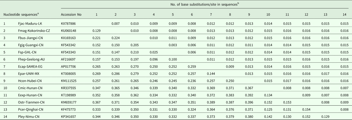
Four of each representative F. jacksoni adult flukes from three localities (Maduruoya, Minneriya and Wasgamuwa National Parks) and fecal eggs from live elephants were subjected to molecular characterization. The p-distance calculations for the concatenated nad1 + cox1 nucleotide sequences showed the lowest level of divergence to be between F. jacksoni and Fa. magna at 12.9%. This was contrasted to the 15.0–15.6% divergence seen when F. jackosoni was compared to other members of the Fasciola genus, including F. gigantica, F. hepatica and an intermediate Fasciola sp. Within the Fasciolidae F. jacksoni had the highest rate of divergence with F. buski at 21.9%, however, this rate of divergence increased substantial to 26.2–27.9% among echinostomids; and with the highest rate at 28.2% with Echinochasmus japonicus, within the superfamily Echinostomoidea (Table 3).
Estimation of pairwise genetic distances (%) between Fasciola jacksoni and the published or GenBank deposited representative species of the superfamily Echinostomatoidea (Fasciolidae, Echinochasmidae, Echinostomatidae) inferred from the concatenated nucleotide sequence of mitochondrial nad1 and cox1

a Sequence abbreviation is listed in Supplementary Table S1.
b The percentage representing the number of base substitutions per site from all sequence pairs between species is shown. Genetic distances were inferred by the analysis of 2,439–2,457 nucleotides of the nad1 and cox1 genes, conducted using the maximum likelihood method in MEGA 7 (Kumar et al., Reference Kumar, Stecher and Tamura2016). Nodal values were obtained by a bootstrap procedure (1000 replicates).
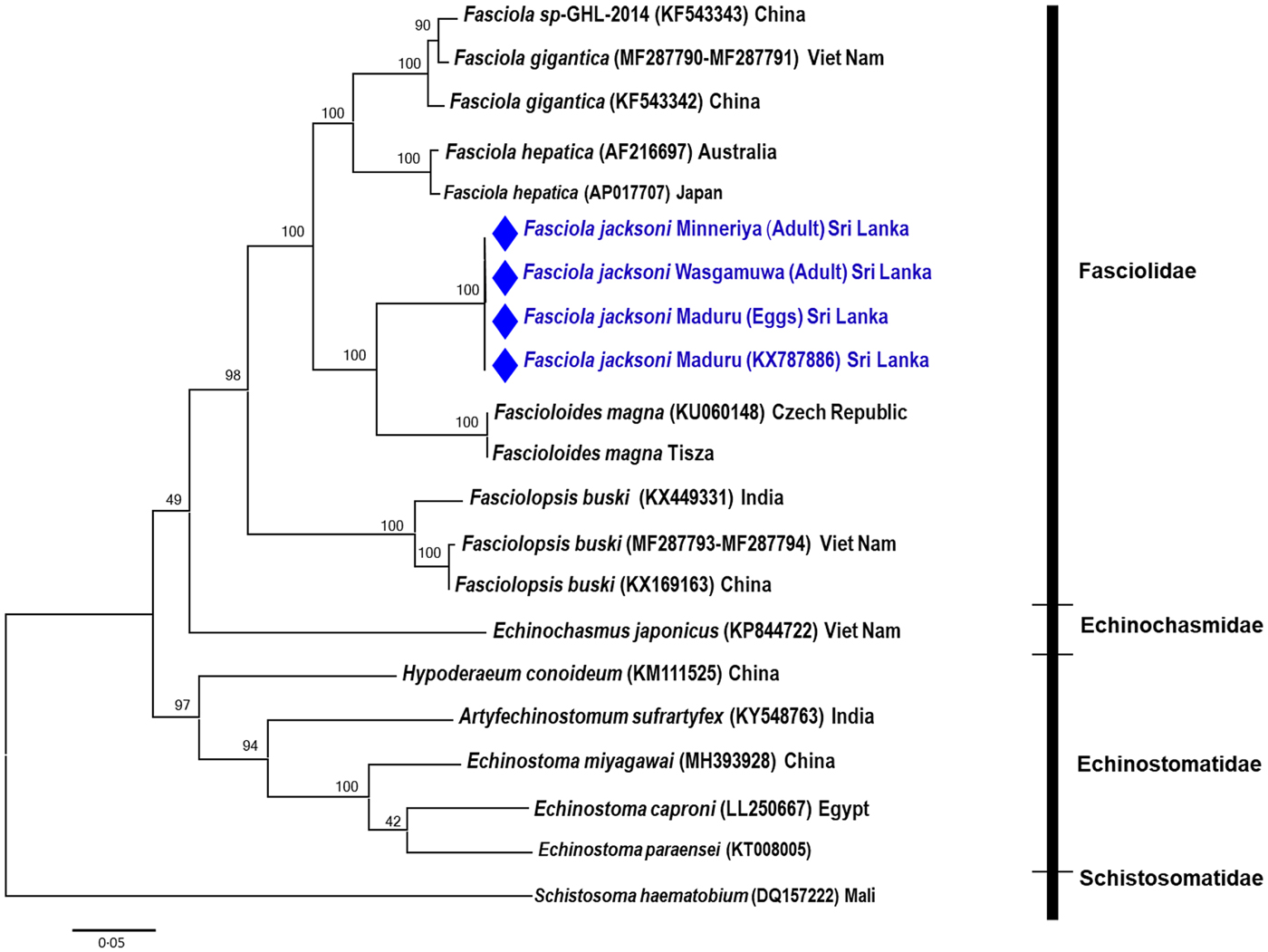
To examine the phylogenetic position of F. jacksoni in the family Fasciolidae and in the superfamily Echinostomatoidea, an ML tree was constructed from 21 complete nad1 and cox1 nucleotide sequences from 13 trematode species belonging to four families including Fasciolidae, Echinochasmidae, Echinostomatidae and Schistosoma haematobium of the Schistosomatidae as the outgroup (Fig. 3; Supplementary Table S1). Sequences of nad1 and cox1 from all F. jacksoni samples were identical regardless of being generated from adults or eggs and no geographical differentiation was shown. The ML analyses clustered F. jacksoni firmly within the Fasciolidae and closely associated with trematodes of ruminants. It was placed in a monophyletic clade as a sister taxon to the European isolate of Fa. magna from the Czech Republic and Hungary; while Fasciola species and F. buski clustered into separate groups, respectively, with strong nodal support of 98–100% (Fig. 3). This rendered the genus Fasciola polyphyletic as all other species within the genus formed a Fasciola specific clade, which contained F. gigantica, F. hepatica and the intermediate form of Fasciola (Fig. 3).
Maximum likelihood phylogenetic reconstruction showing the position of Fasciola jacksoni within the Fasciolidae based on concatenated cox1 and nad1 DNA sequences. Three trematode families are represented Fasciolidae, Echinostomatidae, Echinochasmidae, and the position of F. jacksoni indicated by a diamond symbol. Nodal support values are shown based on 1000 bootstrap replicates.

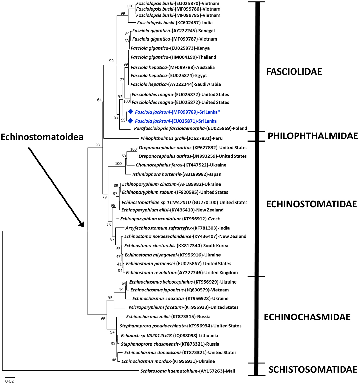
The 28S rDNA alignment produced a well-supported phylogeny of four families in Echinostomatoidea produced topology for 43 sequences of 33 species, with S. haematobium as an outgroup (Fig. 4; Supplementary Table S2). Sequences of 16 fasciolid species/isolates were consistently grouped together, forming a discrete Fascioloidae clade, distinct from Philophthalmidae, Echinostomatidae and Echinochasmidae (Fig. 4). Fasciola jacksoni sequences generated in this study clustered into a single group with a published reference sequence of the same species (Acc: EU025871) (Lotfy et al., Reference Lotfy, Brant, DeJong, Le, Demiaszkiewicz, Rajapakse, Perera, Laursen and Loker2008). However, the topology of the 28S rDNA placed F. jacksoni basal to the Fasciola genus, which also included Fa. magna as the paraphyletic sister taxon to a distinct F. gigantica/F. hepatica clade, again illustrating a lack of monophyly in the genus Fasciola (Fig. 4). In Echinostomatoidea, broadly, the family Echinochasmidae clearly is distinct from the cluster formed by Echinostomatidae; Fasciolidae and Philophthalmidae. The sensu lato Philophthalmus gralli is closer to Fasciolidae than other echinostomids and echinochasmids with a high nodal support (Fig. 4).
Maximum likelihood phylogenetic reconstruction showing the position of Fasciola jacksoni within the Fasciolidae based on partial 28S rDNA sequences. Four families of the superfamily Echinostomatoidea (Fasciolidae, Philophthalmidae, Echinostomatidae, Echinochasmidae) are represented and the position of Fasciola jacksoni is indicated by a diamond symbol. Nodal support values are shown based on 1000 bootstrap replicates.

Discussion
To date, F. jacksoni has been found to be highly host specific residing in the bile ducts of the liver and causing severe fascioliasis only in Asian elephants (E. maximus maximus and E. maximus indicus) (Cobbold, Reference Cobbold1882; Islam, Reference Islam1997; Heneberg, Reference Heneberg2013; Perera et al., Reference Perera, Rajapakse, Silva-fletcher, Thewarage and Jinadasa2015). In this current study, among the 27 infected dead elephants examined, there were 22.2% of individuals with more than 50 flukes (6/27) and only 50% with low worm burdens with less than 10 flukes (14/27). There was a high rate of infection of F. jacksoni in living elephants with 60% of the 48 elephants examined, although egg shedding seemed to be moderate, with 6–30 EPG. Despite the high burden of infection, generally F. jacksoni has low egg counts and has also been shown to have reduced numbers of rediae and cercariae relative to those of Fasciola spp infecting ruminants, indicating a low rate of transmission (Islam, Reference Islam1997; Perera and Rajapakse, Reference Perera, Rajapakse, Wibbelt, Kretschmar, Hofer and Seet2009). Such low levels of transmission could possibly be related to the infrequent consumption of food contaminated with metacercariae owing to the wide-ranging habit of the elephant, with herds taking advantage of large territories and transmission sites being visited infrequently (Lorimer, Reference Lorimer2010; Hing et al., Reference Hing, Narayan, Thompson and Godfrey2016). However, it could be argued that even with low infection and transmission rates, F. jacksoni has evolved to by highly virulent with infections of elephants being highly pathogenic causing lesions not only in the liver parenchyma and bile ductules, but also in other organs such as lungs and kidneys leading to fatalities, particularly in calves (Singh et al., Reference Singh, Srivastava, Prasad and Pandey1994; Islam, Reference Islam1997; Perera and Rajapakse, Reference Perera, Rajapakse, Wibbelt, Kretschmar, Hofer and Seet2009; Heneberg, Reference Heneberg2013; Perera et al., Reference Perera, Rajapakse, Silva-fletcher, Thewarage and Jinadasa2015).
The family Fasciolidae Railliet, 1895 comprises of nine recognized species of which only three are found in the genus Fasciola including F. jacksoni, F. hepatica and F. gigantica which are considered to be taxonomically valid (Mas-Coma et al., Reference Mas-Coma, Valero and Bargues2009). Although listed in the genus Fasciola, taxonomic consideration of F. jacksoni is limited and its phylogenetic position within the Fasciolidae continues to be debated (Lotfy et al., Reference Lotfy, Brant, DeJong, Le, Demiaszkiewicz, Rajapakse, Perera, Laursen and Loker2008; Heneberg, Reference Heneberg2013). In both molecular phylogenetic reconstructions in this study the members of the genus Fasciola collected from ruminants and humans always clustered together in one monophyletic clade that did not include F. jacksoni, placing it with other fasciolid species (Lotfy et al., Reference Lotfy, Brant, DeJong, Le, Demiaszkiewicz, Rajapakse, Perera, Laursen and Loker2008; Le et al., Reference Le, De, Agatsuma, Nguyen, Nguyen, Mcmanus and Blair2008; Nguyen et al., Reference Nguyen, De, Vercruysse, Dorny and Le2009; Mas-Coma et al., Reference Mas-Coma, Valero and Bargues2009; Heneberg, Reference Heneberg2013; Tkach et al., Reference Tkach, Kudlai and Kostadinova2016). Using three ribosomal markers, 28S rDNA, ITS1, ITS2 and a single mitochondrial nad1 marker Heneberg (Reference Heneberg2013) indicated the high similarity of F. jacksoni to Fa. magna rather than to the other ruminant Fasciola spp. group and suggested re-classification of F. jacksoni as Fascioloides jacksoni comb. nov. In this current study mitochondrial and ribosomal 28S DNA analyses based on increased numbers and longer sequences of the nad1 and 28S, as well as representations of cox1, once again illustrated the close evolutionary relationships of F. jacksoni to Fa. magna (Figs 3 and 4). Similarly, evolutionary distance analyses (shown in Table 3) in this study found the lowest rate of divergence (12.9%) between F. jacksoni and Fa. magna, rather than 15.0–15.6% among the ruminant Fasciola spp. and other echinostomatids (around 27%) in Echinostomatoidea. Interestingly, the close phylogenetic relationship between F. jacksoni and Fa. magna is also supported by several shared morphological characteristics such as a thick body, lack of distinct cephalic cone and have long extensive median intestinal branches as highlighted by Lotfy et al. (Reference Lotfy, Brant, DeJong, Le, Demiaszkiewicz, Rajapakse, Perera, Laursen and Loker2008).
The close evolutionary relationship between F. jacksoni and Fa. magna makes it therefore challenging to disentangle the origins of these two species. Fascioloides magna is a parasite of North American cervids and it has been argued that proboscideans brought liver fluke with them into the Neoarctic as they moved from Asia into North America (Lotfy et al., Reference Lotfy, Brant, DeJong, Le, Demiaszkiewicz, Rajapakse, Perera, Laursen and Loker2008). Eventually, the parasite would have possibly undergone a host shift into North American cervids and persisted in deer after the North American elephants became extinct (Lotfy et al., Reference Lotfy, Brant, DeJong, Le, Demiaszkiewicz, Rajapakse, Perera, Laursen and Loker2008). However, only by sampling liver flukes from elephants, antelope and deer across Asia and cervids in the Americas will provide a deeper understanding of the evolutionary relationships of F. jacksoni and Fa. magna and ultimately resolve the taxonomic relationship between F. jacksoni, the ruminant Fasciola spp. and monotypic Fa. magna of the genus Fascioloides.
The present study revealed the utility of molecular taxonomic approaches for the accurate identification of highly pathogenic liver flukes in Asian elephants from different localities in Sri Lanka. However, despite the accuracy of the species identification it was challenging to resolve the taxonomic relationships of F. jacksoni within the Fasciolidae. Molecular data analysed in this study suggested the reappraisal of the family Fasciolidae and recognition of the right taxonomic position of F. jacksoni in the superfamily Echinostomatoidea. Such molecular approaches not only provide vital DNA sequence reference data to develop tools but also provide a foundation for a detailed reconciliation of the taxonomy of these parasites, which is crucial to aid in accurate diagnostics and monitoring of the spread of such an important, but a neglected disease of wild and domestic elephants in Asia.
Supplementary material
The supplementary material for this article can be found at https://doi.org/10.1017/S0031182019000519.
Author ORCIDs
S. P. Lawton, 0000-0003-4055-6524.
Acknowledgements
We express our thanks to colleagues and technicians for contribution to our laboratory work. We would also like to thank Dr David Blair for advising on data analysis and reviewing the manuscript before submission. We would also like to express our thanks to the external reviewers for their extremely informative and constructive comments.
Author contributions
Rajapakse, R.P.V.J, Thanh Hoa Le and Scott P. Lawton conceived the study, analyses of final data and wrote the manuscript. K. J. K. Karunathilake, Nga Thi Bich Nguyen conducted laboratory work and preliminary sequence analyses. B.V.P. Perera provided specimens and collected field data. All authors read and approved the final manuscript.
Financial support
This work was funded by the National Research Council of Sri Lankan (Rajapakse, R.P.V.J). This work was supported by the National Foundation of Science and Technology of Vietnam (NAFOSTED) to Nguyen TBN (grant No 106-YS.02-2013.06).
Conflict of interest
None.
Ethical standards
The study had ethical approval from the Sri Lankan Government. Appropriate permission was obtained from the commune authorities before the collection of parasite specimens from elephants.