

Introduction
The world's egg industry is primarily based on eggs from chickens (Gallus domesticus) (Stadelman, Reference STADELMAN1995). Chicken eggs are valuable foodstuffs for humans since they represent a source of complete protein, supplying all essential amino acids. They also provide several nutrients, including vitamins A (retinol), B2 (riboflavin), B6 (pyridoxine), B9 (folic acid), and B12 (cobalamin), coenzyme Q10 (ubiquinone, abbreviated CoQ10), choline and several minerals such as iron, phosphorus, calcium, and potassium (Cherian et al., Reference CHERIAN, HOLSONBAKE and GOEGER2002; Adeyeye, Reference ADEYEYE2012).
Chicken eggs are composed of about 9.5% protective shell, 63% albumen (egg white), and 27.5% vitellus (egg yolk). The calcium carbonate in shell makes it tough and, as a consequence, protects the integrity of the inner egg. Egg white consists primarily of 88% water into which are dissolved 11% proteins, including albumins, mucoproteins, and globulins. It contains almost no fat and the carbohydrate content is less than 1%. Egg yolk is surrounded by vitelline membrane and contains significant amounts of protein and choline. It is also rich in lipids. Egg yolk coagulates when it reaches temperatures between 65 and 70°C, while egg white coagulates at temperatures between 63 and 73°C (Li-Chan et al., Reference LI-CHAN, PAWRIE and NAKAI1995).
In 2009 an estimated quantity of 62.1 million metric tons of eggs was produced worldwide from a total laying flock of approximately 6.3 billion hens (WATTAgNet, 2010). However, there are regional variations and continuing debates concerning the methods of mass production, such as the European Union's ban on battery farming of chickens (Elson, Reference ELSON2008; European Directive 99/74/EC, 1999) that can influence yearly production.
Contamination of eggs
Whereas bacteria and viruses are naturally present on any raw food, there are concerns related to the contamination of chicken eggs. Eggs have been found to be 90% sterile when laid. However, eggs have the potential to become contaminated with bacteria from the hen's intestinal tract, faeces, and from the surrounding environment (Egg Safety Center, 2010). The egg content is an ideal growth medium for pathogenic bacteria which are hazardous to humans (e.g. Salmonella, Escherichia or Enterobacter). It has been found that the microbiota of the egg shell is dominated by Gram-positive bacteria, which are spoilage microorganisms, while Gram-negative bacteria are best equipped to strike the antimicrobial defence system of the egg (De Reu et al., Reference DE REU, MESSENS, HEYNDRICKX, RODENBURG, UYTTENDAELE and HERMAN2008; Chousalkar et al., Reference CHOUSALKAR, FLYNN, SUTHERLAND, ROBERTS and CHEETHAM2010).
There are two possible routes of bacterial contamination of egg shells: either vertically or horizontally (De Reu, Reference DE REU2006; De Reu et al., Reference DE REU, GRIJSPEERDT, MESSENS, HEYNDRICKX, UYTTENDAELE, DEBEVERE and HERMAN2006b). Vertical transmission takes place in the transovarian route where the yolk, albumen and membranes are directly contaminated as a result of bacterial shedding infection from the hen's reproductive organs, which takes place before the shell covers the eggs (Messens et al., Reference MESSENS, GRIJSPEERDT and HERMAN2005). Horizontal contamination begins with the passage of the eggs through the highly contaminated cloaca area at the moment of lay and leads to the shells penetration by microorganisms (De Reu, Reference DE REU2006). Egg shells may additionally become contaminated from any surface with which it comes into contact. When laid, the egg shells are wet and at the same temperature as the hen's body temperature (Patterson et al., Reference PATTERSON, KOELKEBECK, ANDERSON, DARRE, CAREY, AHN, ERNST, KUNEY and JONES2008). They are then released in an environment with a temperature of approximately 20°C, where they cool immediately and their content contracts creating a negative pressure inside which can allow for the contaminant to move through the shell (De Reu et al., Reference DE REU, GRIJSPEERDT, MESSENS, HEYNDRICKX, UYTTENDAELE, DEBEVERE and HERMAN2006b).
Much of the current research on egg shell and content contamination focuses on salmonellosis. Numerous outbreaks of this disease in humans have been caused by Salmonella enteritidis, from the consumption of food containing contaminated eggs or their products, and still represents a major food safety problems (De Reu et al., Reference DE REU, MESSENS, HEYNDRICKX, RODENBURG, UYTTENDAELE and HERMAN2008). As a result, Salmonella spp. studies have used inoculated egg shells to study the influence of eggs characteristics on microbial decontamination and the behaviour of bacteria to chemical sanitizers.
Berrang et al. (Reference BERRANG, FRANK, BUHR, BAILEY, COX and MAULDIN1998) reported on the influence of egg weight, specific gravity, conductance and hen's age on the ability of Salmonella spp. to penetrate the shell and its membranes and found no relationship. Due to the fact that shell characteristics did not change over time and in relation to hens’ age, they concluded that factors other than shell's quality are involved in egg shells penetration by Salmonella spp.
De Reu et al. (Reference DE REU, GRIJSPEERDT, MESSENS, HEYNDRICKX, UYTTENDAELE, DEBEVERE and HERMAN2006b) studied the movement of the bacteria through egg shells for seven selected bacterial strains recovered from egg contents; Staphylococcus warneri, Acinetobacter baumannii, Alcaligenes sp., Serratia marcescens, Carnobacterium sp., Pseudomonas sp. and Salmonella enteritidis. They determined that the area and the porosity of the egg shells did not influence the contamination of the egg and that Gram-negative, motile and non-clustering bacteria most frequently penetrate egg shells. All selected strains were able to penetrate most frequently after approximately five days. The first organism to penetrate the egg shells were Pseudomonas sp. (60%) and Alcaligenes sp. (58%) followed by S. enteritidis (43%). S. enteritidis can be classed as a primary invader of whole eggs, contaminating up to 32% of the membranes and/or the content of the whole eggs. De Reu et al. (Reference DE REU, GRIJSPEERDT, MESSENS, HEYNDRICKX, UYTTENDAELE, DEBEVERE and HERMAN2006b) observed that penetrated egg shells and contaminated whole eggs showed a significantly higher bacterial contamination on the shell compared to non-penetrated egg shells and non-contaminated whole eggs.
Decontamination of egg shells
Sanitisation of egg shells is important both for consumable and hatching eggs due to the higher incidence of contamination with pathogens. It has been shown that Salmonella and other bacteria can rapidly penetrate the shell and associated membranes of the chicken egg and their elimination is very difficult (Cox et al., Reference COX, BERRANG and BAILEY1998). When an egg at hatch is contaminated with Salmonella spp. they can cross-contaminate chicks in the incubator via circulating air (Cevoli, Reference CEVOLI2010). Sanitisation has traditionally been done in two ways, either washing with water (Hutchison et al., Reference HUTCHISON, GITTINS, WALKER, SPARKS, HUMPHREY, BURTON and MOORE2004) or immersing in disinfectant solutions.
Many studies have been conducted to examine different chemicals such as egg sanitizers, including quaternary ammonium disinfectants (Kaudia, Reference KAUDIA1999), sodium hydroxide 1-2% (Berrang et al., Reference BERRANG, COX, FRANK, BUHR and BAILEY2000), phenols (Cox et al., Reference COX, BERRANG and BAILEY1998; Reference COX, BERRANG, BAILEY and STERN2002a), antibiotic flumisol (Kaudia, Reference KAUDIA1999), hydrogen peroxide (H2O2) 1.4-1.5% (Cox et al., Reference COX, BERRANG, BUHR and BAILEY2000a; Reference COX, BERRANG, BAILEY and STERN2002a; Reference COX, MAULDIN, KUMARAJ and MUSGROVE2002b; Bailey et al., Reference BAILEY, COX and BERRANG2001), timsen (Cox et al., Reference COX, MAULDIN, KUMARAJ and MUSGROVE2002b), polyhexamethylenebiguanide hydrochloride (PHMB) 0.035% (Bailey et al., Reference BAILEY, COX and BERRANG2001), formalin as a 0.5% solution, formaldehyde applied by fumigation (Kaudia, Reference KAUDIA1999), and electrolysed oxidizing (EO) water (Bialka et al., Reference BIALKA, DEMIRCI, KNABEL, PATTERSON and PURI2004; Fasenko et al., Reference FASENKO, O'DEA CHRISTOPHER and MCMULLEN2009).
Most disinfectants were found to be helpful in the control of Salmonella spp. and other microorganisms without reducing egg hatchability. However, chemical solutions are not able to eliminate contamination. As long as disinfectants were applied to eggs before or at the same time as artificial contamination, penetration of egg shells was prevented. However, when Salmonella spp. were present on the eggs prior to the application of the disinfectant, bacteria penetrated the shell deeply enough to avoid direct contact with the chemical disinfectant solution. Available data demonstrates that chemical treatments do not consistently kill all Salmonella spp. on or in eggs and that their effectiveness appears to diminish as the amount of time between contamination and treatment increases (Berrang et al., Reference BERRANG, FRANK, BUHR, BAILEY, COX and MAULDIN1998; Reference BERRANG, COX, FRANK, BUHR and BAILEY2000; Cox et al., Reference COX, BERRANG and CASON2000b). Moreover, many chemicals have noxious characteristics and their use in current practice is not recommended (e.g. formaldehyde, PHMB).
Another aspect is the concern regarding deterioration of the cuticle when intensive washing of egg shells and chemical sanitizers are used, which creates conditions for bacterial penetration through the shell (Berrang et al., Reference BERRANG, FRANK, BUHR, BAILEY, COX and MAULDIN1998; Cox et al., Reference COX, BERRANG and CASON2000b; Messens et al., Reference MESSENS, GRIJSPEERDT and HERMAN2005). However, Leleu et al. (Reference LELEU, MESSENS, DE REU, DE PRETER, HERMAN, HEYNDRICKX, DE BAERDEMAEKER, MICHIELIS and BAIN2011) showed that washing performed on fresh eggs from young hens did not lead to cuticle damage.
To overcome the drawbacks of chemical sanitisation, other methods of decontamination have been tested, including immersion in boiling water for three seconds (Himathongkham et al., Reference HIMATHONGKHAM, RIEMANN and ERNST1999), pulsed light (Dunn, Reference DUNN1996; Lasagabaster et al., Reference LASAGABASTER, ARBOLEYA and DE MARAÑÓN2011), UV light treatment (Kuo et al., Reference KUO, RICKE and CAREY1997b; Chavez et al., Reference CHAVEZ, KNAPE and CAREY1999; Reference CHAVEZ, KNAPE, COUFAL and CAREY2002; Favier et al., Reference FAVIER, ESCUDERO and DEGUZMAN2001; Coufal et al., Reference COUFAL, CHAVEZ, KNAPE and CAREY2003; Rodrigues-Romo and Yousef, Reference RODRIGUES-ROMO and YOUSEF2005; De Reu et al., Reference DE REU, GRIJSPEERDT, HERMAN, HEYNDRICKX, UYTTENDAELE, DEBEVERE, PUTIRULAN and BOLDER2006a; Wells et al., Reference WELLS, COUFAL, PARKER, KIESS, YOUNG and MCDANIEL2011), hot air (180°C for eight seconds), hot water (95°C for 10 seconds), infra-red (210°C for 30 seconds) and steam (100°C for two seconds) (James et al., Reference JAMES, LECHEVALIER and KETTERINGHAM2002), pulsed UV light treatment (Keklik et al., Reference KEKLIK, DEMIRCI, PATTERSON and PURI2009), warm air (Cevoli, Reference CEVOLI2010) and ozone (Rodrigues-Romo and Yousef, Reference RODRIGUES-ROMO and YOUSEF2005). Combined methods have included UV light and H2O2 (Wells et al., Reference WELLS, COUFAL, PARKER and MCDANIEL2010; Reference WELLS, COUFAL, PARKER, KIESS, YOUNG and MCDANIEL2011), UV light and ozone (Rodrigues-Romo and Yousef, Reference RODRIGUES-ROMO and YOUSEF2005).
In the USA there are specific regulations requiring decontamination of egg shells. The Food and Drug Administration (FDA) issued a ruling in 2009 that requires shell egg producers to implement measures to prevent Salmonella enteritidis from contaminating eggs on the farm and during storage and transportation (U.S. FDA, 2009). In the same year, the European Food Safety Authority (EFSA) delivered a scientific opinion on special measures to reduce the risk for consumers through Salmonella spp. transmission in table eggs (EFSA, 2009). While egg washing decontamination is approved in the United States, Canada, Australia and Japan, it is not usually allowed in the European Union (EC 589/2008, 2008).
Principles of UV light treatment
UV light is found in sun's electromagnetic radiation being situated between visible light and X-rays. The range of UV wavelength is between 10 nm and 400 nm. UV light waves have frequencies higher than those the human eye identifies as the violet colour; therefore, they are called ‘ultraviolet’.
UV light is emitted by electric arcs and specialized lights such as mercury lamps and black light systems. These systems are designed to produce UV light with a specific wavelength range, in accordance with the purposed use. The range of the UV spectrum is situated between 200 and 400 nm and is divided in three segments: UV-A, UV-B and UV-C (Table 1).
Division of UV light (from ISO 21348-2007).

UV-A (315-400 nm) contains long length UV waves which are normally responsible for changes in melanin human skin called tanning. UV-B (280-315 nm) contains medium length UV waves. A smaller fraction of this region (295-297 nm) is responsible for the formation of vitamin D in all organisms that synthesise this vitamin, including humans. UV-B light is absorbed by the ozone layer in proportion of around 97%. A small part of UV-B light reaches the surface of the Earth and may cause damage to living organisms. Finally, UV-C (200-280 nm) contains short UV waves and is called the germicidal domain because it effectively inactivates microorganisms. It is completely absorbed in the upper and middle parts of atmosphere by ozone and molecular oxygen (Koutchma et al., Reference KOUTCHMA, FORNEY and MORARU2009).
Microbial inactivation by UV light
UV light is lethal to most microorganisms found in air, water or on hard surfaces. Cell inactivation is based on the damage of nucleic acids (DNA and RNA) under UV light. Nucleic acids absorb UV light from 200 to 310 nm. Absorbed UV light causes the breaking of some bonds and the formation of pyrimidine dimers, which are bonded to adjacent pairs of thimine or cytosine pyrimidines on the same DNA or RNA strand. These dimers prevent cells from replicating, so microorganisms become inactive and unable to proliferate (Koutchma et al., Reference KOUTCHMA, FORNEY and MORARU2009). However, damage of a nucleic acid does not totally kill the cells. This means that cells are unable to replicate but they are still able to metabolise and perform other cell functions. Some of the damage to nucleic acid can be repaired by enzyme mechanisms within the cell. Therefore, microorganisms can repair themselves using either a light repair mechanism called photoreactivation, or dark repair mechanism. After reactivation, microorganisms become via pathogens once more. Consequently, UV treatment has to provide a high enough level of disruption to ensure that the nucleic acid is damaged beyond the stage where it can be repaired (Kim et al., Reference KIM, ANDERSON, MUELLER, GAINES and KENDALL2002; Koutchma et al., Reference KOUTCHMA, FORNEY and MORARU2009).
Koutchma et al. (Reference KOUTCHMA, FORNEY and MORARU2009) showed this behaviour is different from destroying microorganisms which happens when chemical disinfectants are used. Although these chemical disinfectants destroy bacterial cells, there are concerns about their retention on shells or penetration whereby they reach the egg contents, and hence cause potential toxicity in the case of human consumption and/or hatching. Therefore, UV light treatment for microbial inactivation of egg shells is increasingly preferred and applied in research and processing. It is expected to behave as an effective sanitation method that maintains the cuticle on the egg (Kuo et al., Reference KUO, CAREY and RICKE1997a) prevents bacteria from causing internal contamination and eliminate the use of chemical disinfectants with known toxic effects.
UV light inactivation of microorganisms on egg shells
UV light of different intensities has been shown to be effective in reducing various microbial populations on the surface of egg shells (Goerzen and Scott, Reference GOERZEN and SCOTT1995; Gao et al., Reference GAO, STEWART, JOSEPH and CARR1997). A significant reduction of bacteria on egg shells can be achieved with the increase of UV exposure time as reported by Kuo et al. (Reference KUO, RICKE and CAREY1997b). Exposing egg shells to a UV-C light intensity of 1.72 mW/cm2 whilst rotating the eggs at one revolution per min resulted in aerobic bacteria, yeast, and mould populations being significantly reduced. Rotated eggs exposed to 15 min of 4.35 mW/cm2 UV light had a 2.80 log CFU/egg reduction of bacterial load. These results were compared to those obtained for eggs dipped in 200 ppm chlorine treated water solution for 1 min, exposed to 3× formaldehyde fumigation for 20 min, sprayed with Bioguard®, a commercial sanitizer, for 3 min, using untreated eggs as control sample. The mould and yeast population of UV-treated eggs were significantly lower than those on the untreated eggs. Aerobic bacteria were most effectively reduced by chemical disinfectants, however, UV light treatment produced a significantly greater reduction in the total number of the bacterial populations than the other methods (Kuo et al., Reference KUO, RICKE and CAREY1997b).
Kuo et al. (Reference KUO, CAREY and RICKE1997a) investigated the effects of UV-C light (λ = 254 nm) on populations of Salmonella typhimurium, aerobic bacteria and moulds on the egg shells. First, egg shells inoculated with S. typhimurium were exposed to 1, 3, 5, and 7 min of 0.62 m μW/cm2 UV-C light and to one minute alternating light and dark cycles for 5 min (three light and two dark cycles). S. typhimurium CFU were significantly reduced by the UV light treatments. No significant difference in microbial population reduction was observed among UV treatments and light and dark cycles.
The same UV treatments were used in a subsequent experiment aiming to evaluate photoreactivation. Eggs treated with UV light were exposed to 1 h of fluorescent light or 1 h of darkness. No significant differences between microbial populations exposed to UV light and UV light plus photoreactivation were detected. Further studies were performed for aerobic bacteria and moulds evaluation. Egg shells were exposed to 0.62 mW/cm2 for 15 and 30 min and to different intensities (0.62, 1.35, and 1.72 mW/cm2) for 15 min. A 99% CFU/egg reduction of the aerobic bacteria was observed for all the UV treatments. Another important conclusion of Kuo et al. (Reference KUO, CAREY and RICKE1997a) was that S. typhimurium treated by UV light on egg shells did not recover after subsequent incubation under either dark or light conditions.
The results of these studies indicate that UV-C light can significantly reduce populations of S. typhimurium, aerobic bacteria, yeast and moulds on egg shell surfaces (Kuo et al., Reference KUO, CAREY and RICKE1997a; Reference KUO, RICKE and CAREY1997b). However, this treatment may be of little practical use, as the most important pathogen for hen's eggs, namely S. enteritidis, would be inside the egg and protected by the shell (Bintsis et al., Reference BINTSIS, LITOPOULOU-TZANETAKI and ROBINSON2000).
At a meeting of the International Ultraviolet Association, Koutchma (Reference KOUTCHMA2008) has asserted that UV light has been documented to be effective in reducing various bacterial populations on egg shells including Yersinia enterocolitica (Favier et al., Reference FAVIER, ESCUDERO and DEGUZMAN2001), total aerobic plate count (APC) (Chavez et al., Reference CHAVEZ, KNAPE, COUFAL and CAREY2002), S. typhimurium and E. coli (Coufal et al., Reference COUFAL, CHAVEZ, KNAPE and CAREY2003).
The study conducted by Favier et al. (Reference FAVIER, ESCUDERO and DEGUZMAN2001) showed no detection of Yersinia or Salmonella spp. in egg shells natural microbiota. They investigated the effects of UV-C light (λ = 254 nm) on uninoculated and Y. enterocolitica inoculated eggs for different period of time at 4.57 mW/cm2. The results were compared to different chemical disinfectant treatments of either 100 mg/l free chlorine, 3% sodium chloride (NaCl), 1, 5, or 12% trisodium phosphate (TSP) in washing solution. On uninoculated eggs, the best results were obtained with 100 mg/l chlorine and UV light exposure for more than 25 min, with reductions of 1.28 and 1.60 log cycles. On Y. enterocolitica inoculated eggs, the highest reduction of bacterial count (7.35 log CFU/egg) was obtained with 5 and 12% TSP, and 100 mg/l chlorine. Y. enterocolitica was more resistant to UV light than the natural aerobic microbiota of egg shells, except when low levels of inoculum was used (4.39 log CFU/egg).
Chavez et al. (Reference CHAVEZ, KNAPE, COUFAL and CAREY2002) investigated the effects of UV-C light (λ = 254 nm) on APC of egg shells. The exposure at a UV-C intensity of 7.35 mW/cm2 was 15, 30 and 60 seconds via a hand-operated egg roller plus UV light equipment. APC was significantly reduced for the exposure of egg shells to UV-C for 30 and 60 seconds compared to the untreated eggs. For instance, exposure to 60 seconds resulted in a 2.00 to 3.31 log CFU/egg APC reduction, with final values of APC being below detectable levels. In another experiment, UV light treatment was applied in a chamber equipped with a commercial-style egg conveyor. The egg shell was exposed to 7.50 mW/cm2 UV light intensity for 12, 26 and 48 seconds. A reduction of 1.38 to 2.25 log CFU/egg was seen for the egg shells treated for 48 seconds as compared to the untreated eggs. These experiments show that UV light treatment at high intensities and low time intervals has the potential to reduce the APC of egg shells (Chavez et al., Reference CHAVEZ, KNAPE, COUFAL and CAREY2002).
The studies of Knape et al. (Reference KNAPE, CHAVEZ, BURGESS, COUFAL and CAREY2002) evaluated the APC of egg shells at in-line and off-line egg processing facilities at selected sites (via the conveyor system before the eggs passed through the washing system, after detergent wash, after sanitizer treatment and before packaging) and time periods during the daily processing shifts, five equally spaced intervals beginning 15 min after the processing shift began and ending 15 min before the processing shift ended. As the processing shift progressed, off-line APC were significantly higher than in-line counts for all sites, showing the ineffectiveness of egg washing. Knape et al. (Reference KNAPE, CHAVEZ, BURGESS, COUFAL and CAREY2002) concluded that, if the egg processing facilities apply proper detergents and sanitising methods, and contamination of egg shells still registers high incidence, it is necessary to introduce a new method able to reduce the bacterial numbers found on egg shells. Therefore, the next step of research was to accomplish the task of treating the eggs with ultraviolet light using a method that could be commercially implemented.
Coufal et al. (Reference COUFAL, CHAVEZ, KNAPE and CAREY2003) used a cabinet containing UV lamps with a conveyor carrying a plastic hatching egg flat which contained eggs, passed through the cabinet for a period of 3 to 4 min. They tested the effect of UV-C light (λ = 254 nm) exposure on hatching eggs measured as APC. The eggs were inoculated with S. typhimurium and E. coli and the result was an APC reduction of 1.80 to 1.90 log, while the S. typhimurium reductions was of around 4.00 log and E. coli reductions of 4.50 to 5.00 log, compared to the untreated eggs. The impact of UV light on the cuticle of the egg and hatchability were also tested. No significant differences in egg shells conductance or hatchability were found between UV light treated and control eggs. Coufal et al. (Reference COUFAL, CHAVEZ, KNAPE and CAREY2003) stated that UV light treatment of hatching eggs in a prototype cabinet can effectively reduce the aerobic and pathogenic bacteria on egg shells without affecting the shells conductance or hatchability.
De Reu et al. (Reference DE REU, GRIJSPEERDT, HERMAN, HEYNDRICKX, UYTTENDAELE, DEBEVERE, PUTIRULAN and BOLDER2006a) aimed to compare the effect of a commercial UV light treatment system, connected to a commercial conveyor belt, on the elimination of aerobic bacteria on clean and dirty eggs and to study the influence of recent surface contamination of both egg shells and rollers. The conveyor belt, at a length of 102 cm, worked at two different speeds; a maximum of 10,000 eggs/h (0.2167 m/s), and a moderate speed of 2,500 eggs/h (0.0542 m/s) resulting in 4.70 and 18.80 seconds of exposure time at UV-C light (λ = 253.7 nm) respectively. The natural bacterial load (total aerobic bacteria) on the uninoculated clean egg shells was reduced by the UV light treatment from 4.47 to 3.57 log CFU/egg. For very dirty eggs, no significant reduction was observed. The reduction obtained after UV light treatment of the eggs inoculated with E. coli and S. aureus was comparable for both exposure times at 3.00 and 4.00 log CFU/egg respectively. The conveyor belt contamination with E. coli was reduced, but was still detectable on the rollers after the UV light treatment. In addition, the authors studied the influence of UV light on eggs internal contamination. They filled the eggs with a suspension containing E. coli or S. aureus and found very little reduction in internal egg contamination (from 4.37 to 4.08 log CFU/egg). De Reu et al. (Reference DE REU, GRIJSPEERDT, HERMAN, HEYNDRICKX, UYTTENDAELE, DEBEVERE, PUTIRULAN and BOLDER2006a) concluded that UV light treatment ensures significant reductions of the bacterial contamination of the clean egg shells and recent shell contamination and can reduce the contamination of the control rollers, although it did not reduce the internal contamination of eggs.
Inactivation of microorganisms on egg shells with combined UV-based methods
The effects of ozone treatment under mild temperature, UV light treatment and a combination of the two on egg shells contaminated with S. enteritidis were investigated by Rodrigues-Romo and Yousef (Reference RODRIGUES-ROMO and YOUSEF2005). They showed that each treatment significantly reduced S. enteritidis incidence on egg shells. Ozone treatment performed at 4 to 8°C at atmospheric pressure for 8 min resulted in a 2.60 log CFU reduction compared to contaminated, untreated eggs. Application of pressurised ozone at 1054 kg/m2 (15 lb/in2) for up to 20 min resulted in 5.70 log CFU/egg reduction. Similar reductions were obtained using UV light treatment (254 nm and 0.10 mW/cm2 intensity) whereby a reduction of 2.60 log CFU/egg was possible after only two minutes of treatment. At higher intensities of UV light (1.50 to 2.50 mW/cm2) for up to 5 min Salmonella spp. populations were reduced by 4.30 log CFU/egg. Combined treatments of egg shells with UV light (254 nm, 1.50 to 2.50 mW/cm2) for one minute followed by gaseous ozone under mild pressure (351.5 kg/m2 or 5 lb/in2) at low temperature for a further minute decreased the Salmonella spp. count with 4.60 log CFU/egg or more. Comparatively, UV light or ozone treatment performed separately but under the same conditions resulted in a reduction of 4.10 and 2.10 log CFU/egg. According to Rodrigues-Romo and Yousef (Reference RODRIGUES-ROMO and YOUSEF2005), the use of UV light followed by gaseous ozone treatment produced a strong synergistic antimicrobial action against S. enteritidis on egg shells. Moreover, the treatment time was reduced to only 2 min compared to 5 min of UV light treatment and combined with 10 min of gaseous ozone treatment for similar inactivation results. Furthermore, the elimination of microorganisms could be achieved at low temperatures and under relative dry conditions.
Other combined treatments with UV light, this time with a H2O2 solution, for the sanitisation of the egg shells was used by Wells et al. (Reference WELLS, COUFAL, PARKER and MCDANIEL2010; Reference WELLS, COUFAL, PARKER, KIESS, YOUNG and MCDANIEL2011). Wells et al. (Reference WELLS, COUFAL, PARKER and MCDANIEL2010) experimented with a different length of UV light (11.00 mW/cm2) and exposure times (4, 8, 16, and 32 min). They obtained a reduction in the bacterial count on the surface of egg shells of 2.07 log CFU/egg with an 8 min treatment and an increase in the internal temperature up to 29°C. Further, they treated eggs with different concentrations of H2O2 solution (0.5%, 1%, 1.5%, 2%, 2.5%, and 3%) with and without UV or with dry UV or wet UV (UV + sterile water) for 2, 4, and 8 min. The maximum reduction of the bacterial count was from almost 4.00 log CFU/egg to less than 1.00 log CFU/egg, which was obtained for 1.5% H2O2 and 8 min of UV light treatment.
Once the optimal combination of H2O2 and UV light was obtained and its efficiency of killing bacteria on the outer surface of the egg shells had been proven, the influence of this combined treatment on hatchability was studied in detail (Wells et al., Reference WELLS, COUFAL, PARKER, KIESS, YOUNG and MCDANIEL2011). This research involved the incubation of eggs treated with 1.5% H2O2 followed by UV light treatment for 8 min to reduce the APC of egg shells. At hatch (21.5 days after setting), chick weights were taken, meconium samples were randomly collected from each incubator and the presence of intestinal microorganisms was determined. A 3.00 log CFU/egg reduction in the APC of egg shells was found for the treated eggs when compared to the control eggs. At hatch, no differences in chick weight, egg weight loss or hatchability were observed between treated and untreated eggs. Therefore, Wells et al. (Reference WELLS, COUFAL, PARKER, KIESS, YOUNG and MCDANIEL2011) stated that UV light treatment with 1.5% H2O2 reduced egg shell's APC on broiler breeder eggs with no influence on hatchability.
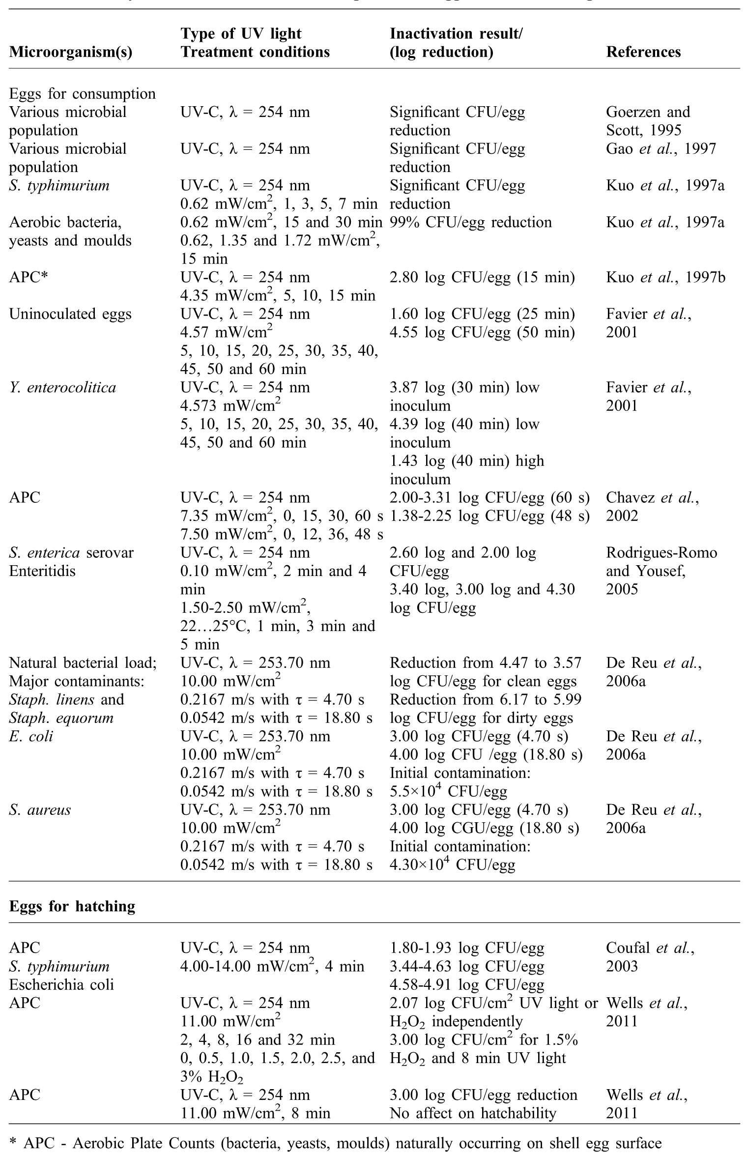
Table 2 summarises the most relevant studies on UV light inactivation of microorganisms on egg shells.
Summary of the inactivation of microorganisms on egg shells with UV light.
* APC - Aerobic Plate Counts (bacteria, yeasts, moulds) naturally occurring on shell egg surface
Conclusions
The importance of eggs for human consumption and hatching in conjunction with the high susceptibility of microbial contamination has led to the necessity of egg shell sanitation. Chemical sanitizers are prone to damage eggs cuticle layers and besides limited effectiveness in reducing pathogens the residues it could represent a chemical hazards to food safety. To avoid these negative aspects, new non-destructive, non-thermal methods were investigated, i.e. treatments with UV-C light and combined methods such as UV-light and ozone and UV-light and hydrogen peroxide.
From the review of the research conducted to date, the most relevant conclusions arising from the reviewed literature are:
1. UV light treatment achieved significantly greater reductions in bacterial population of clean and recently contaminated egg shells compared with methods in which chemical sanitizers were used;
2. UV light treatment ensured significant reductions of Salmonella spp., the most frequently present pathogen reported in eggs;
3. UV light treatment facilitates the hygiene control of the transportation system and enables rollers decontamination;
4. Elimination of microorganisms could be achieved at low temperatures and under relative dry conditions by using UV light treatments;
5. UV light treatment at high intensities and low time periods has the potential to reduce APC of the egg shells;
6. S. typhimurium treated with UV light on egg shells did not recover after subsequent incubation under either dark or light conditions;
7. Y. enterocolitica was more resistant to UV light than the natural aerobic microbiota of the egg shells;
8. UV light treatment of hatching eggs in a prototype cabinet can effectively reduce the aerobic and pathogenic bacteria on egg shells without affecting either egg shells conductance or eggs’ hatchability;
9. Salmonella was effectively inactivated on egg shells in a short time and at low temperature with the use of a combination of UV light and ozone treatment, or UV light and H2O2 treatment;
10. UV light treatment does not reduce the internal contamination of eggs due to the impermeability of egg shells to UV radiation.
All these recent advances have rendered UV light treatment a reliable, non-thermal alternative to the traditional treatments of egg shells and make it a suitable option in controlling microbial contamination of the egg shells surface.
Acknowledgements
The research was supported by the European Union funded FOODSEG Project (FP7 266061 contract) under the 7th RTD Framework.