Pulmonary atresia with intact ventricular septum is a rare congenital cardiac lesion, characterised by an imperforate pulmonary valve or atretic ventriculo-pulmonary junction, variable development of the right ventricular cavity in consequence of mural overgrowth, and potential ventriculo-coronary connections. The malformation is known to be associated with other cardiac and non-cardiac anomalies, but the combination of right- and left-sided obstructive lesions is extremely rare. We describe here our experience with an infant born with pulmonary atresia with intact ventricular septum in association with aortic coarctation.
Case report
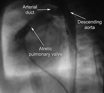
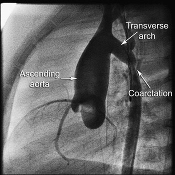
A newborn infant, born weighing 2.0 kg, presented with central cyanosis shortly after delivery. Investigations revealed the diagnosis of pulmonary atreisa with intact ventricular septum. The right ventricular cavity was reasonably well developed, and he underwent successful radiofrequency perforation of pulmonary valve and balloon angioplasty at 5 days of age without complications (Fig. 1). Prostaglandin was continued for few days, but as the saturations of oxygen were acceptable, it was withdrawn 5 days following the catheter intervention. When the arterial duct became insignificant, he was discharged home at 3 weeks of age. Initial follow up was very satisfactory, with Doppler velocities measured across the right ventricular outflow tract continuing to improve, and saturations of oxygen reaching normal values despite complete closure of the arterial duct. Echocardiographic assessment at 4 months of age, however, demonstrated aortic coarctation. He was otherwise well apart from slightly weak femoral pulses, but was normotensive. Review of the earlier echocardiograms failed to demonstrate any evidence of coarctation, even when the previously patent arterial duct became very constricted. The coarctation was confirmed angiographically, and was successfully relieved by balloon dilation (Fig. 2).
The angiogram taken prior to perforation of pulmonary valve. The catheter is below the imperforate pulmonary valve, the arterial duct is wide open, and there is no evidence of aortic coarctation.

The angiogram taken prior to balloon dilation shows discrete aortic coarctation.

Discussion
It is rare to find the combination of right- and left-sided obstructive lesions of the cardiac outflow tracts, with very few cases previously described. Those that have been documented include Fallot’s tetralogy and aortic coarctation,Reference Gunthard, Murdison, Wagner and Norwood1–Reference Yoshigi, Momma and Imai3 pulmonary valvar stenosis and coarctation,Reference Roguin and Shem-Tov4–Reference Wilson, Fonseka and Walker6 the combination of tricuspid and pulmonary atresia with coarctation,Reference Freedom, Silver and Miyamura7 and valvar pulmonary stenosis or atresia with aortic stenosis.Reference Peraira Moral, Burguens Valero and Garcia-Guereta Silva8, Reference Rivera, Moises, Siva, Andrade and Carvalho9 These combinations are difficult to explain in terms of embryology, but clearly do exist. Pulmonary atresia with an intact ventricular septum associated with left-sided obstructive lesions is extremely rare. To our knowledge, there has been but one previous report, with coarctation being the left-sided lesion as in our case.Reference Mackie, Lange and Powell10
Among the aetiological theories to explain aortic coarctation, one of the best documented is that, if ductal tissue extends into the descending aorta, then on closure of the duct this may cause coarctation of aorta. In our case, as well as in the other published case,Reference Mackie, Lange and Powell10 the small duct remain patent for several weeks, and on spontaneous closure of duct, coarctation became obvious. There had been no evidence of coarctation on aortic angiography undertaken at the time of radiofrequency perforation of the pulmonary valve. It was only during follow up, when the arterial duct finally closed, that we noted weak femoral pulses clinically, and found evidence of coarctation of aorta on echocardiography.
Thus, although the combination of pulmonary atresia with intact ventricular septum and left-sided obstructive lesions is exceedingly rare, it does happen. It should be excluded on closure of the arterial duct, as constriction of pre-existing ductal tissue in the descending aorta can clearly lead to coarctation.