Introduction
The transcanal approach to the middle ear is attractive because it avoids the necessity for mastoid surgery. The anterior middle ear, especially the antero-superior tympanum, is a difficult area to visualise using the conventional technique of tympanomeatal flap creation.Reference Wehrs1 We report a patient with congenital cholesteatoma and present a technique which we found useful for the management of congenital cholesteatoma extensively involving the middle ear.
Case report
A five-year-old boy was referred by his paediatrician for evaluation of a white mass on the right tympanic membrane. At the time of examination, the boy's mother denied any previous ear surgery, including myringotomy and tubes. The child had no history of chronic ear infections, although he had suffered the occasional ear infection.
On examination, a large, whitish mass was seen on the right tympanic membrane occluding most of its anterior half.
Audiography demonstrated speech reception threshold scores of 0 dB bilaterally and 100 per cent speech discrimination bilaterally. Computed tomography (CT) scanning demonstrated a normal mastoid with a soft tissue density in the right tympanum and anterior epitympanum (Figure 1).
Pre-operative high resolution computed tomography scans of the congenital cholesteatoma, showing a soft, rounded tissue mass in the anterior half of the tympanic cavity, extending into the protympanum, hypotympanum and anterior epitympanum.

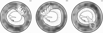
The patient underwent transcanal tympanotomy, beginning with a circular skin incision of the external auditory canal, 6 mm from the tympanic annulus out. The flap was elevated with a canal wall elevator until the fibrous annulus was identified. The tympanotomy approach was then extended by gently separating the tympanic annulus from its sulcus in a circular manner, and further by gently separating the pars tensa from the handle of the malleus. The firm attachment of the tympanic membrane at the umbo was not severed, in order to avoid lateralisation of the tympanic membrane (Figure 2c). Once the tympanic membrane was freed, the entire tympanic cavity, including the antero-superior part, was easily visualised.
Operative techniques for extended tympanotomy. (a) Extended tympanotomy using an inferiorly based flap.Reference Levenson, Parisier, Chute, Wenig and Juarbe2–Reference McGill, Rahbar, Nadol and McKenna4 (b) Extended tympanotomy using a superiorly based flap.Reference Sooy and Sooy5 (c) Extended tympanotomy using our modified sleeve technique.

The cholesteatoma was found to have occluded the anterior mesotympanum, protympanum, some of the hypotympanum and the lower part of the anterior epitympanum anterior to the mallear head. The sac extended beneath the malleus posteriorly, touching the anterior crus of the stapes and the long process of the incus. There were no actual attachments to the ossicles except the anterior surface of the mallear head, which was believed to be the origin of the cholesteatoma. Because the sac was too large and the margin of the sac was not able to be visualised, the sac in the mesotympanum was partially incised and the keratin debris removed. This deflating procedure was helpful in enabling direct visualisation of the margin of the cholesteatoma sac and removal of the cholesteatoma sac as a whole.
After removal of the sac, the middle ear was inspected with a 30° otoscope; no residual cholesteatoma was found. The tympanic membrane and canal wall skin were replaced and held in position with cotton wicks saturated in antibiotic solution.
Post-operatively, it was apparent that the procedure had been successful, with no damage or lateralisation of the tympanic membrane and no significant sensorineural hearing loss.
Histopathological analysis confirmed that the resected specimen was a cholesteatoma.
The patient showed no signs of recurrence after one year of follow up. At this time, CT scanning indicated no signs of recurrence. The patient was scheduled for another CT scan two years later.
Discussion
Congenital cholesteatoma of the middle ear classically presents as a white ‘pearl’ behind an intact tympanic membrane. The treatment for congenital cholesteatoma of the middle ear is removal. Surgical management depends on the extent of the lesion. Surgical removal can be performed via tympanotomy for lesions in the middle ear and via tympanomastoidectomy for lesions extending into the attic and mastoid air cells. Many lesions can be removed via an extended anterior tympanotomy approach (Figure 2a).Reference Levenson, Parisier, Chute, Wenig and Juarbe2–Reference McGill, Rahbar, Nadol and McKenna4
As the majority of congenital cholesteatomas are limited to the antero-superior quadrant of the middle ear, they can be accessed via a tympanomeatal flap created anterior to the malleus. The flap can be elevated medially and the anterior fibrous annulus can be lifted to access the middle ear in the antero-superior quadrant. This allows a more direct approach to cholesteatoma in this area while avoiding manipulation of the ossicular chain. If the sac occupies the entire tympanum including the protympanum and hypotympanum, it is prudent to remove the sac under direct vision. If further exposure is necessary, the tympanic membrane can be released from the umbo, thus affording exposure of the entire mesotympanum.Reference McGill, Rahbar, Nadol and McKenna4 However, this has a theoretical risk of post-operative lateralisation of the tympanic membrane if it is released from the umbo for exposure. A similar technique has been reported involving separation of the pars tensa from the malleus, leaving the entire eardrum and canal skin as a superiorly based flap (Figure 2b).Reference Sooy and Sooy5 Another reported technique involves removal of an eardrum–malleus–canal skin autograft (sleeve autograft technique) and then its replacement, with hearing reconstructed using a homograft notched incus.Reference Wehrs1 However, these techniques have a theoretical risk of post-operative conductive hearing loss.
Our modified sleeve tympanotomy (Figure 2c) approach enables similarly sufficient and direct visualisation of the entire middle ear, with a reduced possibility of lateralisation of the tympanic membrane and post-operative conductive hearing loss. If the congenital cholesteatoma has extended too far superiorly into the anterior epitympanum to be removable with this method, atticotomy should be added for further exposure.