Introduction
In this report, we present a patient with subtotal hearing loss, who had previously undergone a selective vestibular nerve section. Magnetic resonance imaging (MRI) studies confirmed the presence of a cochlear nerve on the operated side, enabling successful cochlear implantation.
Case report
A 70-year-old white man presented with subtotal hearing loss. In 1994, he had developed unilateral Ménière's disease for which he had undergone a right vestibular nerve section. Hearing had been preserved. However, over subsequent years his hearing had progressively deteriorated in both ears, until he re-presented in 2010 with subtotal hearing loss. Hearing was better in the left ear; however, hearing aid benefit was minimal and the patient reported that amplification caused distortion of sound.
It was decided that the patient was a suitable candidate for cochlear implantation.
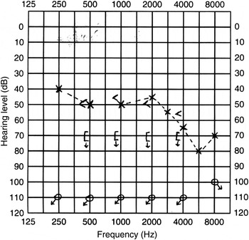
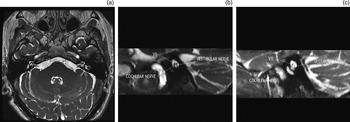
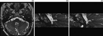
Pre-operatively, audiography and MRI examination were performed (Figures 1 to 3), the latter verifying that the right cochlear nerve was intact.
Pre-surgery audiogram using live-voice Bamford–Kowal–Bench sentences; results were 88 per cent correct. Only the left ear could be aided. With aiding of the left ear, the speech perception score for the prerecorded condition (65 dBHL) was 24 per cent. These results indicated the patient was within the current audiological criteria for cochlear implantation of either the right or left ear.

Images from a 1.5 T, three-dimensional, Fast Imaging Employing Steady-state Acquisition (‘FIESTA’) magnetic resonance imaging sequence through the internal auditory meatus. (a) Axial image showing location of the oblique sagittal reformatted images through the internal auditory meatus on the right (b) and left (c). Part (b) demonstrates post-operative attenuation of the right cochlear and vestibular nerves. To enable comparison, part (c) shows the normal-sized vestibular and cochlear nerves. As the vestibular nerve section was performed in the cerebellopontine angle, the attenuated right vestibular nerve contrasts markedly with the normal thickness, volume and size of the left vestibular nerve at the corresponding level. VII = facial nerve

Images from a 1.5 T, three-dimensional, Fast Imaging Employing Steady-state Acquisition (‘FIESTA’) magnetic resonance imaging sequence through the cerebellopontine angle. (a) Axial image showing location of the oblique sagittal reformatted images through the cerebellopontine angle on the right (b) and left (c). Part (b) demonstrates post-operative attenuation of the right vestibulocochlear nerve (VIII); subsequent events proved that this nerve consisted solely of the cochlear division. To enable comparison, part (c) shows a normal-sized left vestibulocochlear nerve. VII = facial nerve

Cochlear implantation was carried out on the right side using a Cochlear Nucleus CI512 Cochlear Implant with Contour Advance electrode (Cochlear Ltd, Sydney, New South Wales, Australia).
The device was activated three weeks later using a Nucleus CP800 Sound Processor. All 22 intracochlear electrodes were operating and within compliance. All 22 electrodes produced an auditory percept. At two weeks post-activation, two mid-array electrodes were disabled due to non-auditory sensation felt at the right upper side of the head.
Initial testing of open-set speech perception of live-voice Bamford–Kowal–Bench (‘BKB’) sentences resulted in a score of 78 per cent with the device activated and 66 per cent with the device inactivated. This was without a hearing aid.
Further follow up was done by telephone as the patient lived in a remote area; there was no further investigation. The patient reported significant benefit from his right cochlear implant during everyday conversation.
Discussion
A survey of the literature identified no other cases of cochlear implantation carried out after selective vestibular nerve section. Thus, we believe that the presented patient represents the first reported case. Lustig et al. have described cochlear implantation after acoustic tumour removal, which is in effect a vestibular nerve section.Reference Lustig, Yeagle, Niparko and Minor1 However, from our reading of this paper, it would seem that it was the contralateral ear that was implanted.
• Vestibular nerve section effectively controls vertigo in Ménière's disease
• If there is subsequent hearing loss, magnetic resonance imaging is needed to check for an intact cochlear nerve
• Recent advances in cochlear implantation mean that this may be possible in such patients
• Thus, vestibular nerve section should aim to spare the cochlear nerve
• A successfully implanted case is presented
Magnetic resonance imaging has been used to establish the presence of a cochlear nerve in cases of labyrinthine dysplasia and cochleovestibular malformation.Reference Zanetti, Guida, Barezzani, Campovecchi, Nassif and Pinelli2–Reference Govaerts, Casselman, Daemers, De Beukelaer, Yperman and De Ceulaer4 In most cases, modern MRI sequences should make it possible to establish the presence or absence of an adequate cochlear nerve.
Conclusion
If vestibular nerve section is to be carried out for Ménière's disease or for any other reason, it is essential that every effort be made to spare the cochlear nerve, as cochlear implantation may be a future possibility given recent advances in this field.
If hearing loss occurs after vestibular nerve section, MRI should be undertaken to investigate whether the cochlear nerve is intact.