Introduction
Chorioamnionitis is a polymicrobial infection – most often due to ascending genital bacteria – characterized by acute maternofetal inflammation.Reference Tita and Andrews1, Reference Sperling, Newton and Gibbs2 Chorioamnionitis is responsible for more than 50% of preterm prelabor rupture of membranes, which is a major risk factor for intrauterine growth retardation (IUGR).Reference Pugni, Pietrasanta and Acaia3, Reference Galinsky, Polglase, Hooper, Black and Moss4 IUGR is the main risk factor for cerebral palsy (CP) in preterm as well as term babies.Reference Ruff, Faulkner and Rumajogee5, Reference Tolcos, Petratos and Hirst6 Clinical evidence have linked perinatal inflammation, premature birth, IUGR and brain injuries, which may result in CP and other long-term neurological deficits.Reference Tita and Andrews1, Reference Spencer and Meyer7 Chorioamnionitis is associated with an estimated two-fold and four-fold increased risk for CP in, respectively, the preterm and term population.Reference Schendel, Schuchat and Thorsen8 The fetal inflammatory response syndrome (FIRS) arising from infected or inflamed placentas seems to be involved in the induction of abnormal fetal growth.Reference Romero, Gomez and Ghezzi9 Brain injuries associated with the FIRS and/or IUGR in preterm and term newborns are characterized by forebrain white matter injuries, including changes in glial cell population in addition to neuronal loss in gray matter structures.Reference Ruff, Faulkner and Rumajogee5, Reference Zhang, Lu and Wang10, Reference Gotsch, Romero and Kusanovic11 However, the causal link between pathogen-induced chorioamnionitis, IUGR and associated neurodevelopmental impairments has not been established yet.
Group B Streptococcus (GBS) is a gram-positive bacterium commonly colonizing the urogenital and/or gastrointestinal tracts of 20–30% of healthy pregnant women.Reference Patras and Nizet12 GBS is one of the most common bacterium detected in clinical and histological chorioamnionitis, being isolated in 15% of chorioamnionitis.Reference Tita and Andrews1 GBS colonization is also associated with preterm premature rupture of the membranes, low birth weight and with a higher risk for CP.Reference Schendel, Schuchat and Thorsen8, Reference Regan, Klebanoff and Nugent13 The most frequently isolated GBS serotypes from maternal or fetal tissues are serotype III (GBSIII, 25–53%) and Ia (GBSIa, 13–23%).Reference Teatero, Ferrieri and Martin14, Reference Lu, Wu and Chen15 Bacterial components modulate the microbial virulence between bacterial serotypes.Reference O’Riordan and Lee16 For instance, the capsular polysaccharide, that is, the outermost layer of bacterial surface, is serotype-specific and is a major GBS virulence factor.Reference Lemire, Houde, Lecours, Fittipaldi and Segura17 GBSIII is documented as more virulent than GBSIa, and is associated with worse clinical outcomes.Reference Joubrel, Tazi and Six18–Reference Andrade, Magalhães and Puga20
Using rat models of end-gestational active GBSIa infection and inactivated GBS-induced inflammation, we previously reported sex dichotomous effects in maternofetal inflammatory responses, chorioamnionitis and subsequent forebrain injuries as well as time-specific neurobehavioral impairments.Reference Allard, Bergeron and Baharnoori21–Reference Bergeron, Deslauriers and Grignon23 We hypothesized that end-gestational exposure to GBSIII will cause IUGR and CP-like neurobehavioral features. We based our experimental design on our established rat model of chorioamnionitis induced by killed GBSIa.Reference Bergeron, Deslauriers and Grignon23
Method
Experimental design
This study design was based on previous findings from our team, using a rat model of end-gestational exposure to inactivated GBSIa.Reference Bergeron, Deslauriers and Grignon23 The present study was conducted under the same ethical protocol (147-11R), animal housing condition and facility at Université de Sherbrooke.Reference Bergeron, Deslauriers and Grignon23 GBS A909 capsular serotype Ia sequence type (ST)-9 (GBSIa) and GBS BM110 capsular serotype III ST17 (GBSIII), both well-characterized isolates from human with invasive infections, were killed as previously described.Reference Périchon, Szili and du Merle19, Reference Bergeron, Deslauriers and Grignon23 This killing process using formaldehyde allows bacterial inactivation – preventing active infection and toxicity – while preserving most GBS components, as shown in other animal models of immunization.Reference Lee, Perez, Hopkins and Pier24, Reference Tobias, Svennerholm, Carlin, Lebens and Holmgren25
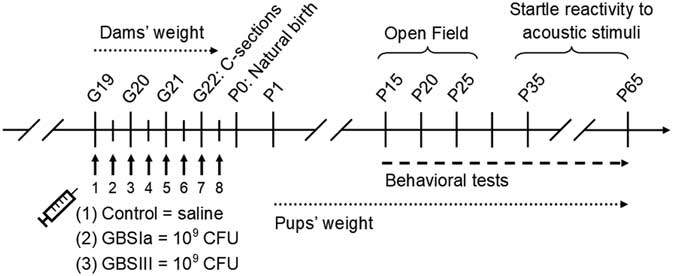
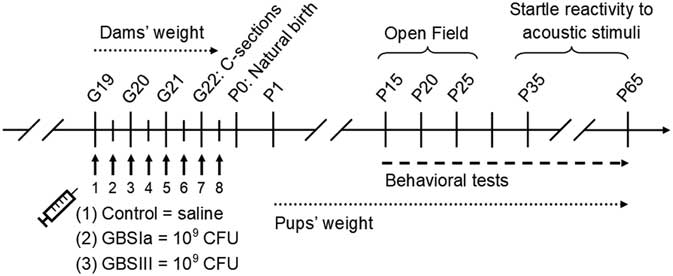
A total of 32 time-pregnant primiparous Lewis rats (Charles River Laboratories) were randomized into three groups and injected intraperitoneally every 12 h (8 am/pm) from gestational day (G) G19 to G22 with 100 µl of either: (Group 1) sterile 0.9% saline (control; CTL, number [n] = 8 dams), (Group 2) GBSIa (109 CFU suspended in saline, n = 8 dams) or (Group 3) GBSIII (109 CFU suspended in saline, n = 16 dams). Injection timing was chosen to mimic the time course of end-gestational human placental inflammation associated with known GBS urogenital colonization. Dams were weighed daily from G19 to G22, and were regularly monitored until delivery [at G23, or postnatal day (P) 0] to detect any sickness-related behavior. Cesarian (C)-sections were performed at G22 on two dams per experimental condition to confirm that GBSIa and GBSIII bacteria reached the placental tissue, inducing histological chorioamnionitis, as also described in our GBSIa rat model.Reference Bergeron, Gerges and Guiraut22 Pups born naturally were counted and weighed on P1 to prevent any stressful situation by handling dams on G23/P0. A total of six CTL litters and 14 GBSIII litters were used for behavioral testing (Fig. 1).
Experimental design and behavioral experiment scheme.
Motor, exploratory and anxiety-like behavior: Open Field test (P15, P20 and P25)
The Open Field apparatus (40 × 40 × 40 cm, divided into 16 equal virtual squares) was used to evaluate spontaneous locomotion, exploratory behavior and thigmotaxis (i.e., an index of anxiety in rodents reflected by a tendency to remain close to the walls) in a novel environment, as previously described.Reference Allard, Bergeron and Baharnoori21, Reference Simon, Dupuis and Costentin26 The tests were longitudinally performed using the same subjects at P15, P20 and P25. Automatic tracking and recording of freely moving animals (Any-Maze Video Tracking System) for 5 min was started as soon as the rat was placed in the corner, facing the central zone. Parameters analyzed were total distance traveled, duration of mobility, number of visited squares, number of lines crossed and time spent in the central zone (anxiety index). The number of rats tested in the Open Field was CTL males (n = 7 from four litters), GBSIII males (n = 14 from nine litters), CTL females (n = 13 from four litters), GBS females (n = 18 from nine litters).
Startle reactivity to acoustic stimuli: Startle response (P35, P65)
Startle reactivity to acoustic stimuli was measured in SR-LAB startle sound attenuated chambers (San Diego Instruments, Inc.) to assess sensorimotor gating longitudinally (same subjects) at P35 and P65 by performing the experiment as described.Reference Deslauriers, Racine, Sarret and Grignon27, Reference Porambo, Phillips and Marx28 Briefly, each rat was restrained in an inner Plexiglas cylinder fixed on an acrylic plate placed on a piezoelectric accelerometer transducing the force of animal startle response (vertical movements of the floor), to avoid locomotion artifacts. Before testing, the rat was acclimatized for a 5 min period to the continuous 71 dB background noise present throughout the session. The rat was then confronted to a first set of six 120 dB pulses (40 ms; habituation session), corresponding to startle signals. Subsequently, the subject was exposed to two sessions – separated by a 60 s delay – of 30 pseudo-random trials: five pulse-alone trials (120 dB for 40 ms), five null trials (no stimulation) and five prepulse plus pulse trials at four different intensities. The prepulse plus pulse trials consisted of a 20 ms prepulse of 75, 79, 83 and 87 dB (4, 8, 12 and 16 dB over background noise, respectively), followed by an 80 ms delay and a startle pulse (120 dB for 40 ms). At the end, the rat was confronted to a second set of six pulses of 120 dB (40 ms; habituation session). The cylinder containing the rat during the test was cleaned between each rat. The startle response amplitudes (mV) during the pre- and post-test pulse-alone (120 dB) trials were statistically analyzed to evaluate habituation throughout the testing period. The number of rats tested in the Open Field was: CTL males (n = 3 from three litters), GBSIII males (n = 4 from four litters), CTL females (n = 4 from four litters) and GBSIII females (n = 3 from three litters).
Histological procedures
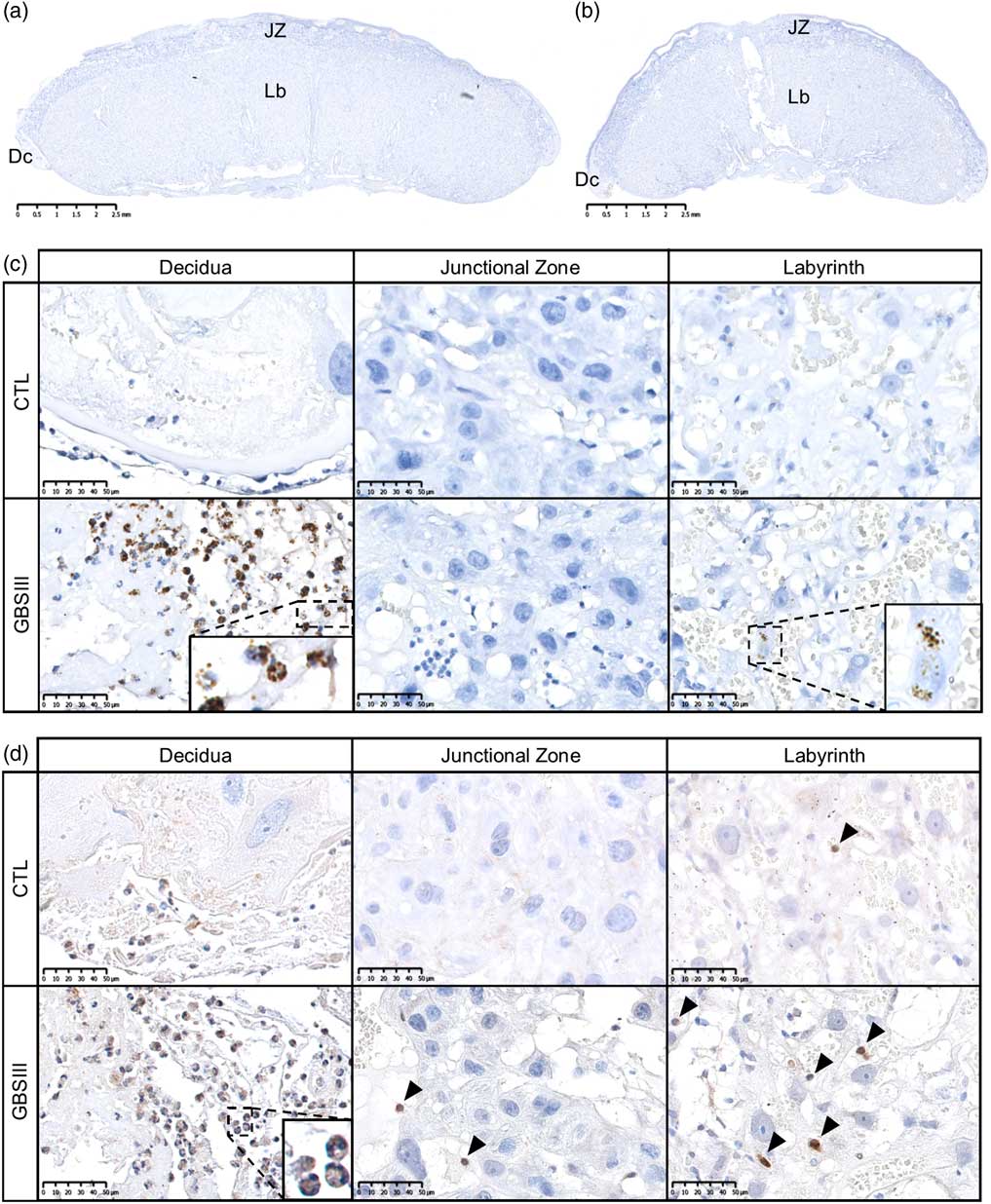
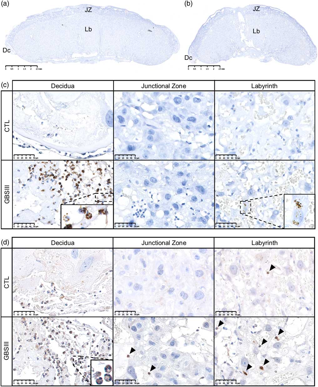
Six placentas per dam were removed by C-section on G22, and immediately fixed (4% paraformaldehyde, 0.1% glutaraldehyde, pH 7.4) as described.Reference Allard, Bergeron and Baharnoori21 Four coronal sections (5 µm thick section) per paraffin-embedded placenta were taken at the level of the umbilical cord insertion, and examined histologically. Rabbit GBS serotype Ia (1:500; Denka Seiken Co.) and serotype III (1:500; Denka Seiken Co.) antisera sets were used to detect GBSIa and GBSIII bacteria in placentas. Anti-PMN cell antibody (rabbit anti-PMN, 1:100; Cedarlane) was used to verify neutrophils’ infiltration throughout placental tissues, that is, a key marker for histological chorioamnionitis. Mouse horseradish peroxidase (HRP)-conjugated anti-rabbit (1:100; Santa Cruz Biotechnology) was used as secondary antibody for anti-PMN and anti-GBS experiments. We evaluated GBS and PMN infiltration within the maternal (decidua), maternofetal (junctional zone) and fetal (labyrinth) compartment (Fig. 2a and 2b).
Immunohistochemical analyses of GBS serotype III and PMN staining in GBSIII-exposed – or unexposed (CTL) – placentas. (a,b) Representative images of CTL (a) and GBSIII (b) placentas at G22, showing maternal (decidua; Dc), maternofetal (junctional zone; JZ) and fetal (labyrinth; Lb) compartments. (c) At G22, clusters of GBSIII-positive bacteria (brown staining) were disseminated in the decidua (left panel) and labyrinth (right panel) of GBSIII-exposed – but not unexposed – dams. (d) PMN infiltrations (brown staining, black arrow heads) in maternal (left panel), maternofetal (middle panel) and fetal (right panel) compartments were observed in GBSIII-exposed v. CTL placentas. Abbreviations: CTL, control; Dc, decidua; G, gestational day; JZ, junctional zone; Lb, labyrinth; GBS, group B Streptococcus; PMN, polymorphonuclear cells.
Rats assessed in the Open Field were euthanized at P40, and the whole brain was post-fixed (4% paraformaldehyde, 0.1% glutaraldehyde, pH 7.4). Paraffin-embedded forebrains were sectioned at a 5 μm thickness, and two consecutive slides were taken for histological analysis. Coronal sections between −2.86 and −3.11 mm posterior to Bregma, that is, at the level of dorsal hippocampus, for immunohistochemical experiments performed as previously described,Reference Allard, Bergeron and Baharnoori21, Reference Paxinos, Watson, Calabrese and Badea29 using a mouse anti-myelin basic protein (MBP) primary antibody (dilution 1:50; Chemicon) to detect nonphosphorylated neurofilament is a characteristic of abnormal axons.Reference Porambo, Phillips and Marx28 A goat anti-mouse-HRP (dilution 1:100; Santa Cruz Biotechnology) was used as secondary antibody. The slides were scanned with a NanoZoomer digital pathology (NDP) scanner (NanoZoomer 2.0-RS, Hamamatsu Photonics). Lateral ventricles’ area and thicknesses of corpus callosum (coronal plan, at the level of the interhemispheric sulcus) and primary (M1) and secondary (M2) motor cortices were measured by an experimenter blinded to conditions using the NDP viewing software. The average of two thickness measurements per anatomical area studied (i.e., the corpus callosum, M1 and M2 cortices) was used for statistical analysis. MBP density, that is, the HRP-positive area on the total corpus callosum area, was studied by colorimetric analysis as previously described.Reference Brochu, Girard, Lavoie and Sébire30 Densitometric measures from male CTLs were used as reference (mean adjusted to 1) to calculate fold differences of the other conditions (CTL females, GBSIII males, GBSIII females). Astrocytes glial fibrillary acidic protein (GFAP) and microglia/macrophages (ionized calcium-binding adapter molecule 1[Iba-1]) were double-labeled by immunofluorescence with, respectively, a rabbit anti-GFAP (dilution 1:100; Millipore) and a mouse anti-Iba-1 (dilution 1:250; Abcam) primary antibody. Anti-rabbit Alexa Fluor 488 conjugated (dilution 1:500; Invitrogen) and anti-mouse Alexa Fluor 594 conjugated (dilution 1:500; Invitrogen) were used as fluorescent secondary antibodies. Slides were mounted using Prolong® Gold Antifade Reagent with DAPI (Thermo Fisher Scientific, Inc.). Slides were examined and images were acquired at 200× magnification (Leica DM4000 B LED) by one investigator blinded to the groups. Counting of GFAP-positive and Iba-1-positive cells in M1 cortices and the corpus callosum by a blinded-to-conditions experimenter was performed using the Image J analysis software’s cell counter. The average number of GFAP- and Iba-1-positive cells per field in two areas in M1 cortices was used in the statistical analysis. The average value of GFAP- and Iba-1-positive cells per two consecutive areas measured in the corpus callosum, using the Image J analysis software, was used for statistical analysis. A total of 24 brains was analyzed [CTL females (n = 6), GBSIII females (n = 8), CTL males (n = 4), GBSIII males (n = 6)] from four CTL and six GBSIII litters. The same forebrains were used for all histological analysis.
Statistics
As treatments were administered prenatally and each pup belong to one specific treated dam, mates from the same litter are considered pseudo-replications of each other.Reference Spencer and Meyer7 Therefore, they should not be considered as totally independent in statistical analyses. Statistical analyses were performed using the statistical package IBM Statistics 25 (SPSS). Assessments of normality of distribution and homogeneity of variance for each data set were initially conducted by, respectively, a Shapiro–Wilk normality test and Levene’s test (P >0.05). Weight of dams at G19 was included as a covariate [two-way analysis of covariance (ANCOVA), general linear model in SPSS] to analyze the mean weight of dams between treatments (between-subjects; CTL, GBSIa and GBSIII) at three gestational timepoints (within-subject repeated measures; G20, G21 and G22). Sex was considered in our analysis design – given that we previously reported sex dichotomous effects in GBS-induced immune responses – forebrain injuries and behavioral impairments. To avoid the artificial sample size inflation caused by treating each rat from a same litter as an independent n, the average weight of pups was calculated for each sex in each litter, and treated as n = 1. Mean weight of offspring from each sex at P1 was analyzed by one-way between-subjects ANCOVA, using Treatment as fixed effect and the number of pups per litter (i.e., litter size) as covariate (general linear model in SPSS). All behavioral data were analyzed using linear mixed models (MIXED, SPSS), using Sex and Treatment (CTL, GBSIII) as fixed effects and litterID as a random effect. Sidak pairwise comparisons were applied in SPSS when there was a significant (P <0.05) effect of Treatment (three factors: CTL, GBSIa and GBSIII), or a significant (P <0.05) interaction between Sex and Treatment. All figures were constructed using GraphPad Prism software version 7.01 for Windows (Graph Pad Software, San Diego, CA, USA). Data are presented as the mean ± s.e.m. Specific values for test statistics and P-values are listed in Supplementary Table S1.
Results
Serotype-specific effects of GBS on gestation, placenta, growth and postnatal development
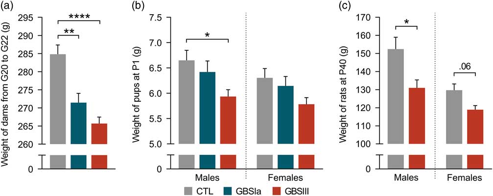
GBSIII induced a histological deciduitis and chorioamnionitis characterized by the presence of GBSIII within the decidua (maternal compartment, Fig. 2a–2c, left panel) and labyrinth (fetal compartment, Fig. 2a–2c, right panel), as well as a PMN infiltrates (Fig. 2d). No premature delivery was noticed in any group, that is, all dams gave birth naturally at G23/P0. No sickness behavior or mortality was observed in any groups. There was no difference for the mean weight of dams between experimental groups before the first injection at G19 (mean weight ± s.e.m.: CTL = 261.6 ± 17.1 g, GBSIa: 263.9 ± 8.0 g, GBSIII: 261.0 ± 11.1 g; P >0.05). Weight of dams at G19 was included as a covariate, adjusting means for the weight gain from G20 to G22. Treatment had an impact on the weight gain of dams during gestation (P <0.0001); dams exposed to GBSIa (adjusted mean: 271.5 ± 2.7 g, P <0.01, n = 8) and GBSIII (adjusted mean: 265.7 ± 1.8 g, P <0.0001, n = 16) gained less weight than CTLs (adjusted mean: 284.8 ± 2.6 g, n = 8) from G20 to G22 (Fig. 3a). There was no weight difference between dams exposed to GBSIa and GBSIII (P >0.05, Fig. 3a). No difference in litter size was detected between experimental conditions (mean number of pups per litter ± s.e.m.: CTL = 9.0 ± 0.9, GBSIa: 7.5 ± 0.5, GBSIII: 6.4 ± 0.8; P >0.05). There was no difference in the sex ratio (i.e., the percentage of male pups per litter) between experimental conditions (P >0.05, data not shown). Our results showed that end-gestational exposure to GBSIa and GBSIII and subsequent chorioamnionitis impacted negatively the maternal weight gain, without any change of mortality rate.
Differential effect of GBSI- and GBSIII-induced chorioamnionitis on maternal weight gain and offspring growth. (a) Dams exposed to GBSIa and GBSIII gained less weight than CTLs from G20 to G22. (b, c) The IUGR observed at P1 (b) persisted until P40 (c) in GBSIII-exposed offspring compared to CTLs. Adjusted means for weight of dams from G20 to G22 are presented, with covariate evaluated at G19 = 261.4 g (general linear model; SPSS). Abbreviations: CTL, control; G, gestational day; GBS, group B Streptococcus; IUGR, intrauterine growth retardation. Data are expressed as mean ± s.e.m. *P <0.05, **P <0.01, ****P <0.0001.
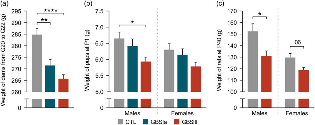
At P1, the mean weight of male (P <0.05) and female (trend, P = 0.07) offspring was different between treatments. Male (P <0.05) and female (trend, P = 0.09) offspring in utero exposed to GBSIII – but not to GBSIa (P >0.05) – weighted less than sex-matched CTLs at P1 (Fig. 3b). This decreased weight, compared to CTLs, was still present at P40 in GBSIII-exposed male (P <0.05) and female (trend, P = 0.06) rats (Fig. 3c). Taken together, GBSIII – but not GBSIa – induced chorioamnionitis, impacted negatively the pups’ growth at birth, and this IUGR was still present at P40.
Neuromotor impairments in IUGR rats in utero exposed to GBSIII
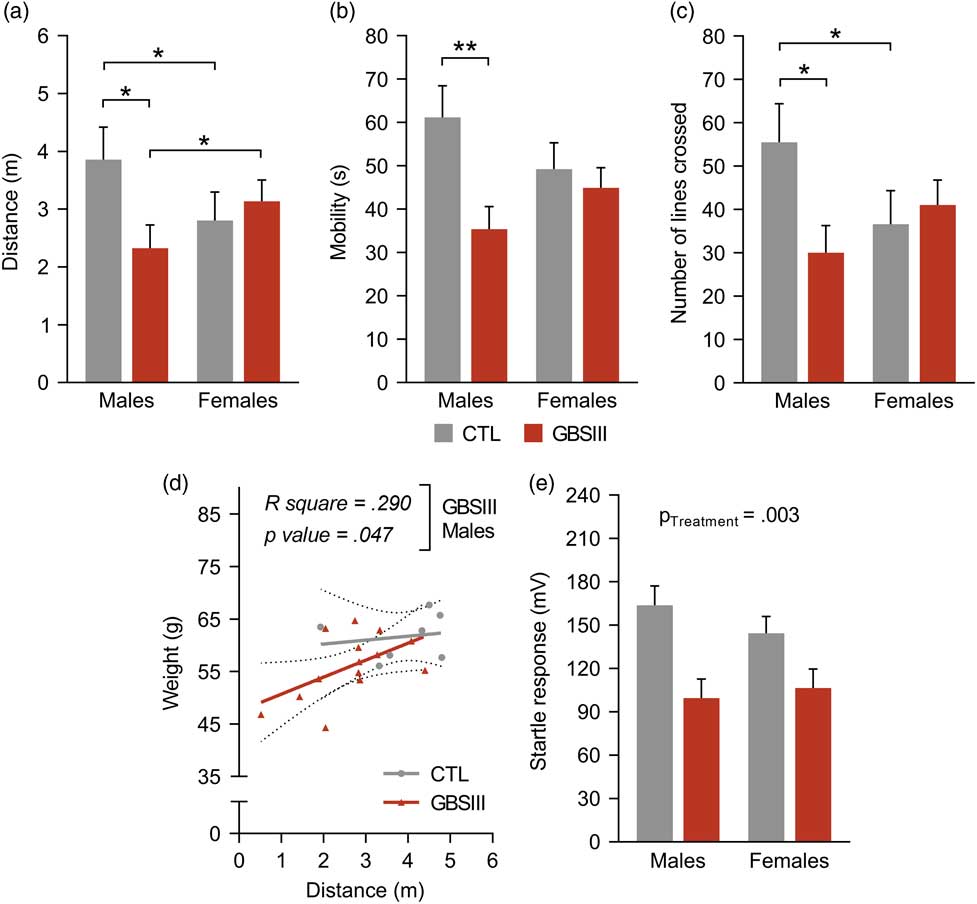
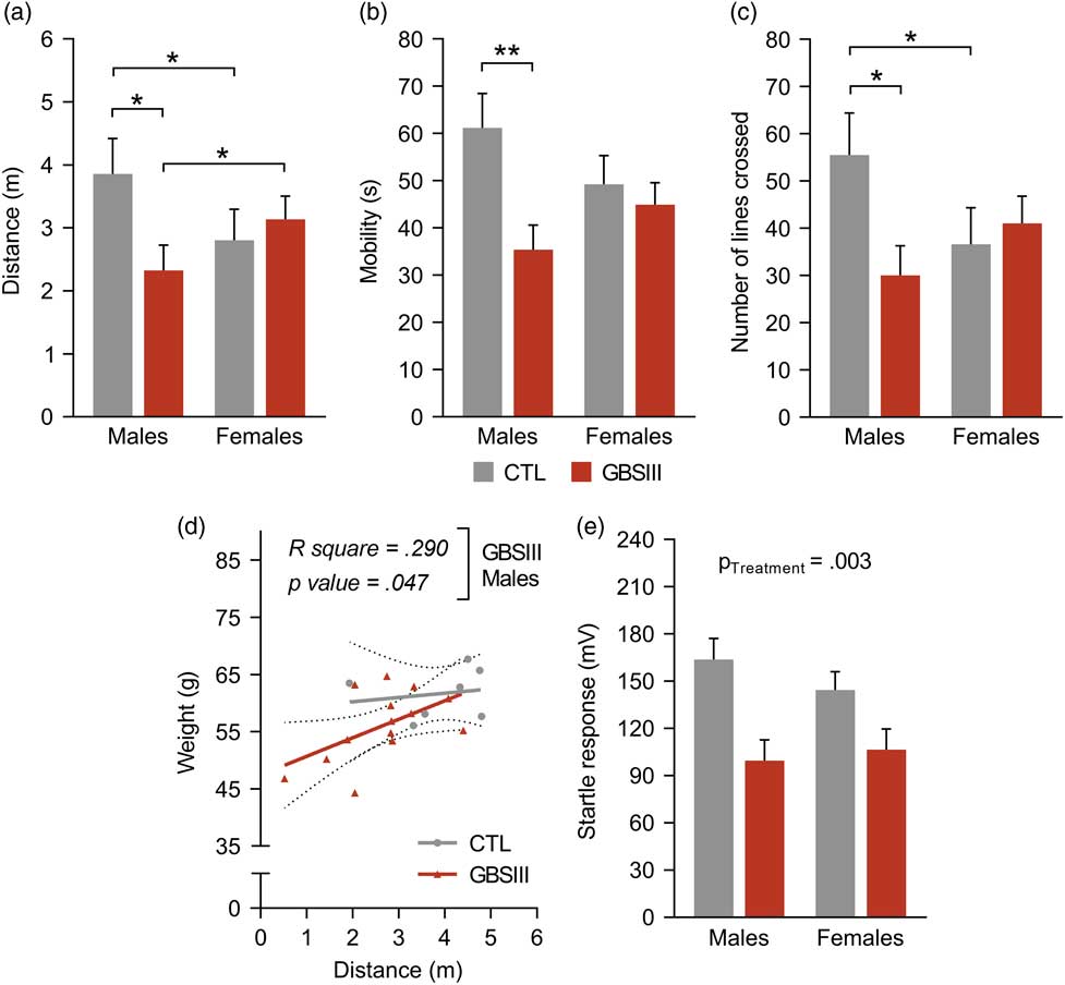
To analyze the impact of GBSIII-associated chorioamnionitis and growth deficit on neuromotor development, male and female rats were assessed longitudinally in the Open Field apparatus at three developmental time points (P15, P20 and P25). At P25, decreased total distance traveled (P <0.05, Fig. 4a), time of mobility (P <0.01, Fig. 4b) and total number of line crossed (P <0.05, Fig. 4c) were detected in GBSIII-exposed male – but not female – rats, compared to sex-matched CTLs. GBSIII-exposed males traveled a lower distance than GBSIII-exposed females (P <0.05, Fig. 4a). The opposite result was observed in CTLs: males traveled a higher distance and crossed more lines than females (P <0.05, Fig. 4a and 4c). These differences were not detected at P15 or P20 (P >0.05, data not shown). No difference in the number of squares visited, that is, an index of exploratory behavior, was detected between GBSIII-exposed and CTL rats at P15, P20 or P25 (P >0.05, data not shown). There was no difference in the time spent in the central zone, that is, an index of anxiety, between GBSIII-exposed versus CTL rats at P15, P20 or P25 (P >0.05, data not shown). These results showed impaired motor behavior in male – but not female – pups presenting IUGR following exposure to GBSIII chorioamnionitis. This impaired motor behavior was characterized by a decreased spontaneous locomotion, without anxiety-like behavior or impaired exploratory behavior. The level of IUGR (weight of rats) in GBSIII-exposed male – but not female – rats was positively correlating with the distance traveled in the Open Field apparatus (P <0.05, Fig. 4d). To investigate whether neuromotor deficits were persisting beyond P25, rats were assessed longitudinally (P35 and P65) for motor responses to acoustic stimuli. Between-subject analysis revealed that GBSIII-exposed rats from both sexes showed significantly reduced startle amplitudes compared to CTLs (P <0.01, Fig. 4e).
Sex-specific impact of GBSIII-induced chorioamnionitis on neurobehavioral parameters of the IUGR-affected offspring. (a–c) At P25, decreased total distance traveled (a), time of mobility (b) and number of lines (c) crossed in the Open Field apparatus were detected in male – but not female – IUGR rats in utero exposed to GBSIII. (d) At P25, weight of rats was positively correlating with the total distance traveled in the Open Fiel test. (e) Decreased startle responses to 120 dB acoustic stimuli were revealed in GBSIII-exposed rats from both sexes, compared to CTLs. Abbreviations: CTL, control; GBS, group B Streptococcus; IUGR, intrauterine growth retardation. Data are expressed as mean ± s.e.m. *P <0.05, **P <0.01.
Forebrain injuries in GBSIII-exposed rats
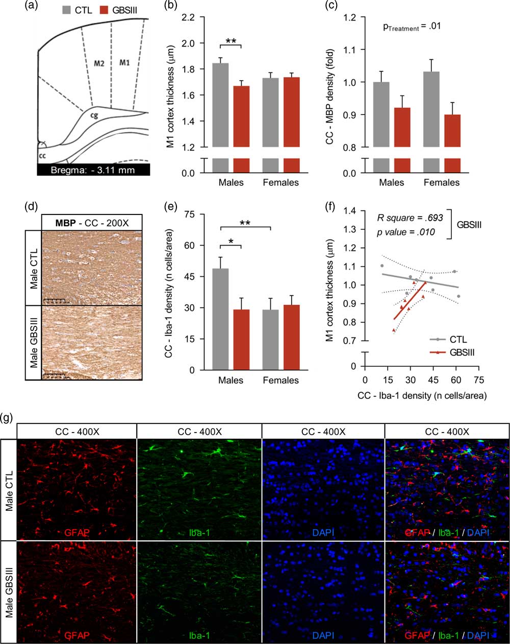
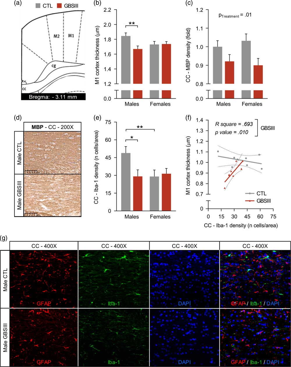
On the basis of our neuromotor results and data showing that IUGR is a major risk factor of CP in humans, neuroanatomical assessments performed on forebrains at P40 were focused on motor cortices and underlying white matter areas (Fig. 5a). Thinner primary motor (M1) cortices were detected in GBSIII-exposed males – but not females – compared to sex-matched CTLs (P <0.01, Fig. 5b). The densities of GFAP- and Iba-1-positive cells in M1 cortices were similar between all experimental conditions (P >0.05, data not shown). Although no difference in the corpus callosum thickness was observed (P <0.05, data not shown), a decreased density of MBP staining was detected in GBSIII-exposed rats from both sexes compared to CTLs (P <0.05, Fig. 5c and 5d). GBSIII-exposed males presented decreased density of microglial Iba-1-positive cells in the corpus callosum (Fig. 5e–5g), but no difference in astroglial GFAP-positive cells density was detected (P >0.05, data not shown). Densities of microglial cells were positively correlating with M1 cortex thicknesses of GBSIII-exposed rats (P <0.05, Fig. 5f and 5g).
Immunohistochemical (MBP) and immunofluorescent (GFAP, Iba-1) analyses of primary motor (M1) cortices and adjacent corpus callosum (CC) of rats’ forebrain at P40. (a,b) M1 cortices (a) of rats affected by IUGR were thinner (b) in GBSIII-exposed males, compared to sex-matched CTLs. (c,d) MBP staining density was reduced in GBSIII-exposed rats, compared to CTLs. (e) Reduced densities of Iba-1-positive cells in the CC were detected in GBSIII-exposed males and female CTLs, compared to male CTLs. (f) M1 cortices’ thicknesses were positively correlating with the densities of Iba-1-positive cells in the CC of GBSIII-exposed rats. (g) Representative images of cells positive for GFAP (astrocytes) and Iba-1 (microglia) in the CC. Abbreviations: CC, corpus callosum; CTL, control; GBS, group B Streptococcus; GFAP, glial fibrillary acidic protein; Iba-1, ionized calcium-binding adapter molecule 1; IUGR, intrauterine growth retardation; M1, primary motor cortex; MBP, myelin basic protein. Data are expressed as mean ± s.e.m. *P <0.05, **P <0.01.
Discussion
This study was the first to test in a preclinical model the effect of inflammation – but not infection – induced by the two most common GBS serotypes (i.e., Ia and III) on gestational parameters and neurobehavioral features in the progeny. The end-gestational exposure to inactivated GBS induced a deleterious impact on maternal and fetal weight gains in serotype- and sex-specific manners: GBSIa and GBSIII impacted negatively the maternal weight gain, although GBSIII – but not GBSIa – induced IUGR and persisting neuromotor impairments. Motor impairments detected in male rats in utero exposed to GBSIII were correlating positively with their level of IUGR. GBSIII-exposed rats presented decreased startle responses to acoustic stimuli beyond adolescent age. Altogether, our data showed that motor impairments observed in GBSIII-exposed rats were associated with a chorioamnionitis-induced IUGR, white matter injuries in the corpus callosum and thinner M1 cortices correlating with reduced microglial density in the adjacent white matter. The delayed appearance of motor impairments in GBSIII-exposed rats is in keeping with the delayed occurrence of CP symptoms in humans.
IUGR, occurring in 5–10% of pregnancy, is classically attributed to placental insufficiency or utero-placental dysfunction.Reference Wixey, Chand, Colditz and Bjorkman31 Restricted growth of the fetus is associated to long-term and severe neurological disorders, including CP.Reference Wixey, Chand, Colditz and Bjorkman31 A 30-fold increase in odds of CP has been reported in near-term and term newborns presenting IUGR.Reference Blair and Nelson32 To our knowledge, our results documented for the first time the link between GBSIII-induced chorioamnionitis, IUGR, and subsequent CP-like periventricular white matter injuries, and neuromotor behavioral impairments. Behavioral outcomes from the Open Field test – in which males performed worse than females – are in line with other preclinical models of IUGR showing worse neurobehavioral deficits in IUGR males than females, and human epidemiological studies documenting higher frequency of CP in boys than girls.Reference Wixey, Chand, Colditz and Bjorkman31 The startle response test performed at later developmental timepoints revealed that GBSIII rats from both sexes exhibited lower acoustic-induced startle responses than CTLs, suggesting sensorimotor deficits persisting beyond early ages.Reference Porambo, Phillips and Marx28 The diminished startle amplitude in IUGR rodents is of great clinical relevance since neurosensory deficits are common in the preterm population presenting white matter injuries, as well as an important feature in motor and psychiatric morbidities present in children born prematurely.Reference Dupin, Laurent, Stauder and Saliba33–Reference Paolicelli and Ferretti36
These neurobehaviorally impaired rats displayed thinner M1 cortices adjacent to a dysmyelinated corpus callosum containing fewer microglial cells than CTLs. Clinical neuroimaging and preclinical models showed forebrain injuries characterized by delayed cortical development, decreased cortical thickness and altered brain connectivity in preterm and term IUGR v. non-IUGR subjects.Reference Wixey, Chand, Colditz and Bjorkman31, Reference Tolsa, Zimine and Warfield37 Moreover, microglial cells were recently identified as having an active role in different processes within the developing brain, such as neuronal migration, axonal tract pathfinding and processes participating in the shaping of prenatal brain circuits.Reference Squarzoni, Thion and Garel35 Depletion of microglia, as observed in the white matter rats exposed to GBSIII, has been shown to interfere with brain wiring processes in the neocortex as well as related myelination.Reference Squarzoni, Thion and Garel35, Reference Paolicelli and Ferretti36, Reference Hagemeyer, Hanft and Akriditou38 In keeping with these findings, the decreased number of microglia we observed in our model might be a key pathological feature linking microglial alterations to abnormal cortical development and defective oligodendroglial myelination.Reference Hagemeyer, Hanft and Akriditou38
Our results also demonstrated a differential effect of GBS depending on its serotype: GBSIII – but not GBSIa – induced IUGR and neuromotor impairments, although both serotypes reached placental tissues. It has been shown that GBSIII adheres more than GBSIa to placental cell preparations, which suggested strain differences in number of receptors expressed and/or receptor types.Reference Périchon, Szili and du Merle19, Reference Winram, Jonas, Chi and Rubens39 On the other hand, GBS induces distinct inflammatory responses depending on serotypes and host’s sex.Reference Lemire, Roy and Fittipaldi40 Our results validate the pertinence to further investigate whether GBSIII triggers a differentiated inflammatory response compared to GBSIa, a potentially important question that has not been addressed yet. Further characterizing GBS serotype-specific maternofetal inflammatory responses will help to develop placento-protective strategies using personalized anti-inflammatory treatments.
This novel preclinical research has some limitations. Some severe complications involving GBSIII are due to live bacteria.Reference Russell, Seale and O’Driscoll41 However, many research groups showed that bacterial component exposures – without any active infection – induced FIRS, and led to fetal brain injuries.Reference Gilman-Sachs, Dambaeva and Salazar Garcia42–Reference Boveri, Kinsner and Berezowski45 Hence, the immune activation triggered by components of pathogens plays a critical role in the damaging consequences of infections, which provide a rational for our experimental design. In the present study, our goal was to investigate the inflammatory effects on the offspring of placental exposure to GBS serotype III on the offspring growth and brain development. Despite this above-mentioned limitation, the advantage of using inactivated bacteria limits active bacterial dissemination and infections of different organs in both the dams and fetuses, making more reproducible our experimental design and associated findings. Finally, we did not address in this study the specific mechanisms underlying sex-specific impairments. This is an important, but complex matter involving the interplay between several hormones and receptors as well as many other placental and brain mechanisms, opening new research avenues.
In sum, our results pave the way to further study interventions aiming to prevent or limit chorioamnionitis, subsequent IUGR and neurodevelopmental impairments. These studies should consider the pathogen(s) involved, as well as its serotype. The ongoing development of anti-GBS vaccine offers a promising way to prevent GBS infection during pregnancy and its deleterious effect on the progeny.
Supplementary material
To view supplementary material for this article, please visit https://doi.org/10.1017/S2040174418001083
Acknowledgments
This work was supported by grants from the Canadian Institutes of Health Research (CIHR), Fonds de Recherche du Québec – Santé (FRQ-S), and Foundation of Stars. We thank Dr Claire Poyart (Service de Bactériologie et Centre national de référence des Streptocoques, Hôpitaux Universitaires Paris Centre; INSERM, Équipe Barrières et Pathogènes, Institut Cochin, Paris, France) for providing the GBS strains (A909, BM110) used in this study.