INTRODUCTION
Harmful algal blooms (HABs) are well-known for their ecological and economic impacts in coastal areas. They are one of the major sources of contamination of the marine environment and can cause mass mortality of wild and cultivated animals due to the potent phycotoxins they produce (see reviews of Shumway, Reference Shumway1990; Landsberg, Reference Landsberg2002).
Among cultivated species, marine bivalves are of particular importance during HAB events in view of their sessile and filter-feeding habits that favour the accumulation of high levels of algal toxins in their tissues. As a consequence, the consumption of contaminated bivalves can be dangerous for human health and their commercialization is prohibited when harmful algae concentrations reach critical levels. These commercial embargos are becoming more and more frequent worldwide, including in Brazil, where they are causing important economic losses to fisheries and aquaculture-based industries (see http://www.algasnocivas.pro.br/monitoramento.php).
Most of the studies on algal toxins are focused on their impact on human health and only few of them examine their potential negative effects on bivalve physiology and survival. Even though mortalities are uncommon among bivalves during HAB events since they seem to be particularly resistant to phycotoxins, recent studies have shown that microalgal toxins may indeed have damaging or stressful effects on bivalve metabolism and make them more susceptible to infections (Hégaret et al., Reference Hégaret, da Silva, Wikfors, Lambert, De Bettignies, Shumway and Soudant2007a, Reference Hégaret, Smolowitz, Sunila, Shumway, Alix, Dixon and Wikfors2010).
Diarrheic shellfish poisoning (DSP) is a symptom caused in humans by the consumption of contaminated bivalves, which have accumulated toxins from certain species of dinoflagellates belonging to the genera Dinophysis and Prorocentrum (Yasumoto et al., Reference Yasumoto, Murata, Oshima, Sano, Matsumoto and Clardy1985; Lee et al., Reference Lee, Igarashi, Fraga, Dahl, Hovgaard and Yasumoto1989; Bravo et al., Reference Bravo, Fernandez, Ramilo and Martinez2001). The major DSP toxins are okadaic acid (OA) and its structural derivatives, the dinophysistoxins -1 and -2 (DTX1 and DTX2). After consumption of contaminated shellfish, these lipophilic toxins produce gastrointestinal symptoms such as nausea, vomiting, diarrhoea and abdominal pain (Yasumoto et al., Reference Yasumoto, Oshima and Yamaguchi1978). In mammals, OA toxins are known to inhibit serine-threonine protein phosphatases (PP-1 and especially PP-2A) (Biolojan & Takai, Reference Biolojan and Takai1988), which are key components of cell signalling and cell regulation pathways that underlie a myriad of essential physiological processes. Interestingly, mussel phosphatases 1 and 2A are not apparently affected by OA (Svensson & Förlin, Reference Svensson and Förlin1998). More recently, OA and derivatives also have been shown to be potent tumour promoters in mammals (Fujiki & Suganuma, Reference Fujiki, Suganuma, Fusetani and Kem2009) and implicated in micronucleus formation (Carvalho Pinto-Silva et al., Reference Carvalho Pinto-Silva, Ferreira, Costa, Belli Filho, Creppy and Matias2003, Reference Carvalho Pinto-Silva, Creppy and Matias2005), cell apoptosis (Lago et al., Reference Lago, Santaclara, Vieites and Cabado2005; Prado-Alvarez et al., Reference Prado-Alvarez, Florez-Barros, Mendez and Fernandez-Tajes2013) and genotoxicity (Valdiglesias et al., Reference Valdiglesias, Mendez, Pasaro, Cemeli, Anderson and Laffon2010; Gonzalez-Romero et al., Reference Gonzalez-Romero, Rivera-Casas, Fernandez-Tajes, Ausio, Mendez and Eirin-Lopez2012) in mammals and bivalves.
Bivalve immune responses against pathogens and/or toxicants are carried out by the circulating blood cells or haemocytes and by a variety of soluble molecules found in their haemolymph (Hine, Reference Hine1999; Roch, Reference Roch1999). Recent reports have shown that some immune functions might be affected in bivalves upon experimental exposure to toxic microalgae or their purified toxins (Hégaret & Wikfors, Reference Hégaret and Wikfors2005a; da Silva et al., Reference da Silva, Hegaret, Lambert, Wikfors, Le Goic, Shumway and Soudant2008; Galimany et al., Reference Galimany, Sunila, Hegaret, Ramon and Wikfors2008a, Reference Galimany, Sunila, Hegaret, Ramon and Wikforsb; Malagoli et al., Reference Malagoli, Casarini and Ottaviani2008; Haberkorn et al., Reference Haberkorn, Lambert, Le Goic, Gueguen, Moal, Palacios, Lassus and Soudant2010a; Bricelj et al., Reference Bricelj, Ford, Lambert, Barbou and Paillard2011; Hégaret et al., Reference Hégaret, da Silva, Wikfors, Haberkorn, Shumway and Soudant2011; Mello et al., Reference Mello, da Silva, Barracco, Soudant and Hégaret2013; Prado-Alvarez et al., Reference Prado-Alvarez, Florez-Barros, Mendez and Fernandez-Tajes2013). However, very few studies (Hégaret & Wikfors, Reference Hégaret and Wikfors2005b; Mello et al., Reference Mello, Proença and Barracco2010; Prado-Alvarez et al., Reference Prado-Alvarez, Florez-Barros, Sexto-Iglesias, Mendez and Fernandez-Tajes2012) have examined the effect of natural harmful algal blooms on the immune functions of cultivated or wild shellfish.
The coastal area of Santa Catarina (southern Brazil) is by far the major producer of cultured bivalves in Brazil (more than 95% of the total national production). In the last few decades, blooms of harmful algae have been reported along the Brazilian coast, such as diatoms of the genus Pseudo-nitzschia that cause amnesic shellfish poisoning (ASP) and the dinoflagellates Alexandrium tamarense and Dinophysis acuminata Claperede & Lachman (1859) that cause paralytic shellfish poisoning (PSP) and diarrhoeic shellfish poisoning (DSP) respectively (Proença et al., Reference Proença, Schramm, Tamanaha and Alves2007; Schramm & Proença, Reference Schramm and Proença2008). In Santa Catarina coast, blooms of D. acuminata are currently becoming more and more frequent (Proença et al., Reference Proença, Schramm, Tamanaha and Alves2007) and besides the ecological impact and risk to human health, these toxic blooms are also threatening the malacoculture activity.
Recently, Mello et al. (Reference Mello, Proença and Barracco2010) reported that natural blooms of D. acuminata may affect some haemato-immunological parameters in oysters (Crassostrea gigas Thunberg, 1793), clams (Anomalocardia brasiliana Gmelin, 1791) and especially mussels (Perna perna Linnaeus, 1758) cultivated in the south bay of Santa Catarina Island. The aim of this study was to expand the observations of Mello et al. (Reference Mello, Proença and Barracco2010) to other regions of Santa Catarina coast, where bivalves are being intensely cultivated, in order to validate the results of these authors and to better understand the potential toxic effects of D. acuminata toxins on bivalve immunity. In this study, we report the modulation of different haemato-immunological parameters in C. gigas and P. perna during two blooms of D. acuminata that took place in different localities of Santa Catarina coast.
MATERIALS AND METHODS
Animals and experimental design
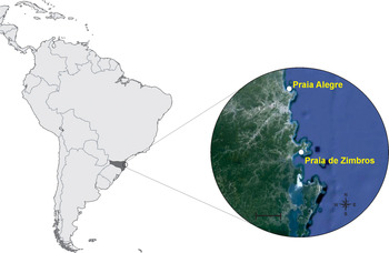
Adult Pacific oysters Crassostrea gigas (shell height, 90–100 mm, N = 60) and brown mussels Perna perna (60–70 mm, N = 80) were obtained from commercial marine farms in Santa Catarina coast during two natural blooms of D. acuminata (lasting from 6–9 days). The first (2950 microalgal cells l−1) occurred in November 2009 at Praia Alegre (PA – 26°46′S; 48°39′08″W) and the second (4150 cells l−1) in March 2010 at Praia de Zimbros (PZ – 27°11′S; 48°32′31″W) (Figure 1). At PZ both bivalves were collected, whereas at PA only mussels were obtained because oysters were not available. Animals were sampled and used in different analyses (immunological and histological assays) during the algal blooms and also after 30 days from the end of the bloom (reference animal group).
Sites of oysters and mussels samplings at Santa Catarina coast during blooms of Dinophysis acuminata. Bar: 20 km.

Seawater samples were preserved in lugol (1%) for algal cell counts. Water salinity, temperature and dissolved oxygen were measured during the samplings.
Microalgal cell counts, concentration of okadaic acid (OA) and mouse bioassays
The number of D. acuminata cells in seawater samples was estimated under an inverted phase-contrast microscope according to the protocol of Utermöhl (Reference Utermöhl1958).
The concentration of OA was determined in mussel and oyster digestive glands (N = 12, for each animal group) by liquid chromatography (LC) coupled to mass spectrometry (LC-MS/MS). The gland extracts (2 g) were prepared by alcohol extraction (absolute methanol), centrifuged and filtered (0.2 µM nylon filters). Chromatography was performed on Agilent 1200 LC system RR equipped with a fast Zorbax Eclipse XDB-C18, 4.6 × 50 mm chromatography column at 35°C. The identification and quantification of OA and its derivatives was performed using an API 3200 QTrap MS/MS detector calibrated with pure standards from NRC Canada, following the settings obtained from Villar-González et al. (Reference Villar-González, Rodríguez-Velasco, Botana and Gilbert2008).
Mouse bioassays (MB) were carried out by injecting 1 ml of the digestive gland extracts of both bivalves in three mice (weight 18–20 g) intraperitoneally. A reaction was considered positive when at least two mice died within 24 h.
Haemolymph preparation
Haemolymph was withdrawn from animal adductor muscle with the aid of a needle (21 G) coupled to a 1 ml syringe (kept on ice). Haemolymph pools (3 pools of 10 animals from each species and from each locality) were separated in two subgroups. The first was fixed in 4% formaldehyde diluted in modified Alsever solution or MAS (27 mM sodium citrate, 336 mM sodium chloride, 115 mM glucose, 9 mM EDTA, pH 7.0) (2:1 v/v) and used to determine the haemograms and the percentage of apparent apoptotic cells. The second subgroup was used to prepare total haemolymph (TH). TH was obtained by lysing the haemocytes through sonication (3 cycles of 7 s each, at 22.5 kHz/50 W, at 4°C). The disrupted cell suspension was centrifuged (12 000 g for 30 min at 4°C) and the supernatant or TH (exocytosed cell products + plasma) was separated and stored at −20°C until use (determination of AT, PO and PC).
Haemograms: total (THC) and differential (DHC) haemocyte counts
Total haemocyte counts (THC) were determined from fixed haemolymph pools with the aid of a Neubauer chamber (in duplicates). The relative percentage of the different haemocyte populations (DHC) was estimated by counting 200 cells from each fixed blood sample under a phase-contrast microscope. Results were expressed as the relative percentage of granular haemocytes (GHs). The remaining percentage (complementary) corresponds to the hyaline haemocytes (HHs).
Percentage of apoptotic haemocytes (AH)
The percentage of apparent apoptotic haemocytes was determined by using Hoechst 33258 staining (SIGMA). Fixed haemocyte smears were immersed in McIlvane buffer (0.1 M citric acid, 0.4 M disodium hydrogen phosphate, pH 5.5) for 5 min and then incubated for 5 min in a solution of bisbenzimida fluorophore (1 mg ml−1) in McIlvane buffer for 5 min. The slides were then mounted with coverslips and observed under a fluorescence microscope (365 nm). The percentage of apoptotic cells was estimated by examining 200 cells per sample and counting the morphologically altered nuclei characteristic of apoptotic cells.
Haemagglutinating activity
Samples of 50 µl of TH from the different pools were serially diluted in TBS-1 (50 mM Tris, 150 mM NaCl, 10 mM CaCl2, 5 mM MgCl2, pH 7.4) in 96-well microplates (U-shaped bottom) and incubated with the same volume of a suspension of dog erythrocytes (2% in TBS-1) for 2 h at 20°C in a humid chamber. In controls, TH was replaced by TBS-1. The agglutinating titre (AT) was expressed as the reciprocal of the highest TH dilution showing positive agglutination. The titres were converted to log2 and the assays were performed in triplicate.
Determination of phenoloxidase (PO) activity
PO activity was determined spectrophotometrically through the formation of DOPA-chrome (red pigment) by the oxidation of the enzyme substrate, L-dihydroxyphenylalanine (L-DOPA). TH samples (50 µl) from the different pools were diluted (v/v) in TBS-2 (50 mM Tris, 400 mM NaCl) and incubated with 50 µl of L-DOPA (3 mg ml−1) in 96-well plates (flat-shaped bottom) at 20°C. The reaction was carried out at pH 9.0, since alkaline pH is a strong enzyme inducer. The formation of DOPA-chrome was recorded on a microplate reader (A490), every 30 s, for 20 min. In controls, TH was replaced by TBS-2. One enzyme unit (1 U) corresponded to an increase of 0.001 in the absorbance per min, per mg of protein at 20°C (Söderhäll & Häll, Reference Söderhäll and Häll1984). All assays were carried out in triplicate.
Total protein concentration (PC)
PC was determined in the different TH pools according to the method of Bradford (Reference Bradford1976), using bovine serum albumin (BSA) as a standard. Assays were carried out in triplicate.
Histopathological analysis
Whole oysters from PZ (N = 15 for each group) were removed from the shells and preserved in a Davidson solution for 24 h. Mussels were not preserved for histology. The fixed animals were then transferred to 70% ethanol and sectioned diagonally to enable the exposure of the mantle (and gonads), gills and digestive gland. Tissues were embedded in paraffin, sectioned to 5 µm and stained in haematoxylin-eosin (HE). Permanent slides were examined under a light microscope and the histological lesions were recorded with digital micrographs.
Statistical analyses
The results were first subjected to Bartlett's test to evaluate variance homogeneity. Then, the results of each immune parameter were compared by one-way ANOVA followed by Tukey's post-test (mean comparison). For DHC and AH (percentages), the data were arcsin transformed. The results were considered significant at P < 0.05. Statistical analysis was performed using GraphPad Prism® software, version 5.0.
RESULTS
Abiotic parameters
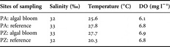
Water salinity, temperature and concentration of dissolved oxygen (DO) were very similar in both sampling sites during the toxic algal blooms (PA: November; PZ: March) and 30 days after the bloom-end (reference groups) (Table 1). The sole exception was the water temperature at PZ (reference, 20°C) that was considerably lower than during the algal bloom (27°C), probably due to the beginning of autumn.
Abiotic parameters at the sampling sites during blooms of Dinophysis acuminata.

DO, dissolved oxygen; PA, Praia Alegre; PZ, Praia de Zimbros.
Number of algal cells, okadaic acid (OA) concentration and mouse bioassays
Dinophysis acuminata concentration in seawater reached 2950 cells l−1 at PA in November 2009, and 4150 cells l−1 at PZ in March 2010. In Santa Catarina coast, a concentration of 500 cells l−1 of D. acuminata in seawater is already considered critical and bivalve consumption is unsafe. Mouse bioassays gave positive results in both natural microalgal blooms. Bivalve mortalities were not recorded during both HABs. Following 30 days of the bloom period the concentration of D. acuminata in seawater dropped to 0 cells l−1 at both sites.
The concentration of OA in the digestive gland extracts was measured only in animals from PZ blooms and was 10× higher in mussels (60.1 µg kg−1) than in oysters (5.9 µg kg−1). Following 30 days of the end of the blooms there was a marked decrease of the OA concentration in mussel tissue extracts (0.9 µg kg−1), whereas no traces of toxins were found in oysters.
Haemograms
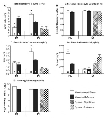
The total haemocyte count (THC) of mussels varied significantly during the algal blooms when compared with the reference groups, even though in a contrasting manner (Figure 2A). At PA, there was a decrease of THC (56%) during the D. acuminata bloom (2.9 ± 0.5 × 10⁶ cells ml−1) when compared with the reference group (5.2 ± 1.8 × 10⁶ cells ml−1), whereas at PZ it increased about 44% (algal bloom: 6.9 ± 1.2 × 10⁶ cells ml−1 and reference group: 4.8 ± 0.4 × 10⁶ cells ml−1). On the other hand, in oysters from PZ, the THC was similar in both algal-exposed and reference animals (about 2.5 × 10⁶ cells ml−1).
Immunological parameters of mussels and oysters sampled during (algal bloom) and after (reference) the bloom of Dinophysis acuminata. PA: Praia Alegre; PZ: Praia de Zimbros. *Represent significant differences (P < 0.05) between the exposed (HAB) and reference group at the same site. Letters represent significant differences (P < 0.05) between sites (mussels).

The differential haemocyte count (DHC) was presented here as the percentage of granular haemocyte populations (GHs). The complementary percentage represented by the hyaline haemocyte (HHs) population is not shown. The percentage of GHs did not change significantly in both bivalve species during the algal blooms (Figure 2B). This haemocyte population (GHs) was always predominant (more than 80%) over the hyaline haemocytes (HH) in both bivalve species.
Percentage of apoptotic haemocytes (AH)
The number of haemocytes showing altered nuclei suggesting apoptosis was very low in both bivalves (less than 1%) during both algal blooms and no significant differences were observed with the reference groups.
Total protein concentration (PC) in haemolymph
The PC of mussel haemolymph increased significantly (63%) only during the algal bloom at PZ (3.1 ± 0.5 mg ml−1) when compared with the reference group (1.9 ± 0.1 mg ml−1) (Figure 2C). No variation was observed on the PC of oyster haemolymph. Curiously, the PC of oyster haemolymph was considerably lower (less than 1 mg ml−1) than that of mussels.
Phenoloxidase activity (PO)
The PO activity varied significantly in both mussels and oysters during the algal blooms, but in contrasting manners (Figure 2D). In mussels, the enzyme activity increased strongly (about 100%) during the dinoflagellate bloom at PA (bloom: 575.4 ± 66.7 U min−1 mg−1, reference group: 287.2 ± 6.7 U min−1 mg−1) but remained unaltered at PZ and the values were about half (120–140 U min−1 mg−1) of those found at PA (reference group). On the other hand, PO activity in oysters dropped significantly (about 60%) during the algal bloom at PZ (algal bloom: 298.0 ± 8.1 U min−1 mg−1, reference: 478. 8 ± 68.0 U min−1 mg−1).
Haemagglutinating activity
The agglutinating titres (AT) of the TH of both bivalves against dog erythrocytes did not change significantly during both toxic algal blooms (Figure 2E). However, there was a tendency to higher values in mussels at PA (AT = 512) when compared with the reference group (AT = 256). The AT of both bivalve species was very similar.
Histopathological analysis
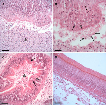
Histological analysis was carried out only in oysters. Animals exposed to the bloom of D. acuminata displayed an intense migration and infiltration of haemocytes into the lumen of the stomach, intestine, and to a lesser extent into the digestive primary or secondary tubules (diapedesis through the epithelium) (Figure 3). Tissue alteration was observed in the stomach epithelium. Oysters from the reference group did not exhibit such abnormalities.
Histological alterations of oysters Crassostrea gigas sampled during the bloom of Dinophysis acuminata at Praia de Zimbros. (A) Intense haemocytic infiltration at the stomach lumen (asterisk). Bar: 50 µm. (B) Magnification of figure A showing the altered stomach epithelium with haemocytes undergoing diapedesis (arrows). Bar: 20 µm. (C) Haemocytic infiltration at the lumen (asterisk) and epithelia (arrows) of the digestive tubule. Bar: 50 µm. (D) Undamaged stomach epithelium from oysters of reference group. Bar: 20 µm.

DISCUSSION
In the last two decades, blooms of Dinophysis acuminata have been increasing dangerously in the Santa Catarina coast and the presence of okadaic acid (OA) and derived toxins have been recorded in mussel and oyster tissues (Proença et al., Reference Proença, Schramm, Tamanaha and Alves2007). In 2010, Mello et al. reported that a severe natural bloom of D. acuminata in Santa Catarina Island resulted in the modulation of some haemato-immunological parameters in oysters (C. gigas), clams (Anomalocardia brasiliana) and especially in mussels (P. perna). The aim of this study was to extend these previous observations to other localities of the Santa Catarina coast, where shellfish are intensely cultivated, in order to validate the potential noxious effect of DSP toxins on bivalve immunity. As stated before, two natural blooms of D. acuminata occurred in Santa Catarina coast during this study: one at PA (November 2009–2950 cells l−1) and a more intense one at PZ (March 2010–4150 cells l−1). Both algal blooms lasted about 6–9 days and bivalve commercialization was prohibited until natural depuration was ascertained.
Various haemato-immunological parameters were examined in farmed P. perna and C. gigas during both dinoflagellate blooms as well as the presence of histological lesions in oysters. The number and type of circulating haemocytes (haemograms) are major immune parameters in the assessment of bivalve health status. In this study, the number of total circulating haemocytes (THC) was indeed altered in mussels, but not in oysters, during both D. acuminata blooms. Curiously, at PA mussel THC decreased significantly (56%), but at PZ it augmented greatly (44%) (more intense bloom). Possibly, the rise of THC was related to the activation of the mussel immune system triggered by the microalgal phycotoxins. On the other hand, the reduction of the circulating haemocyte number could be related to the migration of these cells to the tissues in contact with the microalgae in order to assist dinoflagellate clearance (Galimany et al., Reference Galimany, Sunila, Hegaret, Ramon and Wikfors2008b; Estrada et al., Reference Estrada, Rodríguez-Jaramillo, Contreras and Ascencio2010; Escobedo-Lozano et al., Reference Escobedo-Lozano, Estrada, Ascencio, Contreras and Alonso-Rodriguez2012). Alternatively, these contrasting responses could be related to the different concentration/lasting of the algal blooms or to different environmental variables underlying field experiments. Interestingly, the significant increase of THC observed in P. perna at PZ was very similar to that reported by Mello et al. (Reference Mello, Proença and Barracco2010) during the severe D. acuminata bloom (17 600 cells l−1) at the south bay of Santa Catarina Island.
The THC of C. gigas was determined only during the second bloom (PZ), since this species was not available at PA. In contrast to the mussels, oyster THC did not vary during the algal bloom and this result was in agreement with the report of Mello et al. (Reference Mello, Proença and Barracco2010). Similar results were also described in C. virginica exposed to A. fundyense and A. catenella (which cause PSP), where the THC remained unchanged (Hégaret et al., Reference Hégaret, Wikfors, Soudant, Lambert, Shumway, Berard and Lassus2007b).
The proportion of blood cell types or differential haemocyte counts (DHC) did not vary in both bivalves during both toxic blooms. This result differed from the observations of Mello et al. (Reference Mello, Proença and Barracco2010) where a decrease (12%) in the percentage of granulocytes (GHs) was observed in mussels, but not in oysters, during the toxic bloom. These differences might be due to the much higher D. acuminata concentration (4× higher than at PZ) that occurred in the study of Mello et al. (Reference Mello, Proença and Barracco2010). Changes in the percentage of haemocyte populations may result from the recruitment of a specific cell type, such as granulocytes, into the tissues in direct contact with the harmful microalgae, as are the digestive system and gills. Haberkorn et al. (Reference Haberkorn, Lambert, Le Goic, Gueguen, Moal, Palacios, Lassus and Soudant2010a) reported a drastic increase of GHs in C. gigas fed Alexandrium minutum. On the other hand, the DHC of C. virginica and C. gigas exposed to A. catenella and A. fundyense did not vary (Hégaret et al., Reference Hégaret, Wikfors, Soudant, Lambert, Shumway, Berard and Lassus2007b).
The exposure of bivalves to toxic compounds may stimulate cell death through apoptosis (Sokolova et al., Reference Sokolova, Evans and Hughes2004, Reference Sokolova, Foster, Grewal, Graves and Hughes2011; Marcheselli et al., Reference Marcheselli, Azzoni and Mauri2011). In this study, however, the percentage of altered haemocyte nuclei exhibiting morphological features of apoptosis was very low (<1%) in both bivalves during both algal blooms. Mello et al. (Reference Mello, Proença and Barracco2010) also found insignificant levels of apoptosis in clam, oyster and mussel haemocytes during the D. acuminata bloom. These results suggest that OA and derivatives do not trigger cell apoptosis in bivalve haemocytes. Galimany et al. (Reference Galimany, Sunila, Hegaret, Ramon and Wikfors2008b) also found very low levels of apoptosis in the haemocytes of M. edulis exposed to Prorocentrum minimum. Curiously, Prado-Alvarez et al. (Reference Prado-Alvarez, Florez-Barros, Sexto-Iglesias, Mendez and Fernandez-Tajes2012) reported an unexpected decrease in haemocyte apoptosis in Mytilus galloprovincialis (from field) highly contaminated by OA and other DSP toxins. The authors observed the same effect after incubating mussel haemocytes in vitro with high concentrations of purified OA toxin. They suggested that OA may inhibit the apoptosis pathway in haemocytes (e.g. inhibition of caspases). In contrast, in a more recent study, Prado-Alvarez et al. (Reference Prado-Alvarez, Florez-Barros, Mendez and Fernandez-Tajes2013) observed an increase in haemocyte apoptosis when the clam Ruditapes decussatus was exposed to Prorocentrum lima (OA producer) and when its haemocytes were incubated in vitro to the OA toxin. It should be emphasized that the recent genome sequencing of C. gigas (Zhang et al., Reference Zhang, Fang, Guo, Li, Luo, Xu, Yang, Zhang, Wang, Qi, Xiong, Que, Xie, Holland, Paps, Zhu, Wu, Chen, Wang, Peng, Meng, Yang, Liu, Wen, Zhang, Huang, Zhu, Feng, Mount, Hedgecock, Xu, Liu, Domazet-Loso, Du, Sun, Zhang, Liu, Cheng, Jiang, Li, Fan, Wang, Fu, Wang, Wang, Zhang, Peng, Li, Li, Chen, He, Tan, Song, Zheng, Huang, Yang, Du, Chen, Yang, Gaffney, Wang, Luo, She, Ming, Huang, Huang, Zhang, Qu, Ni, Miao, Wang, Steinberg, Wang, Qian, Liu and Yin2012) revealed that this bivalve has a particularly powerful anti-apoptosis system with more than 45 genes encoding for protein apoptosis inhibitors (IAPs), which is much higher than in other organisms such as sea urchins and humans that possess only 7 and 8 IAP coding genes, respectively.
Phenoloxidase (PO) activity is also an important immune parameter commonly used to assess invertebrate health, particularly in crustaceans and insects (Liu et al., Reference Liu, Jiravanichpaisal, Cerenius, Lee, Söderhäll and Söderhäll2007; Cerenius et al., Reference Cerenius, Kawabata, Lee, Nonaka and Soderhall2010). In bivalves, however, the activity of this enzyme has not yet been clearly related to the immune system. Nonetheless, several authors have been using this enzyme activity to express shellfish health status (Thiagarajan et al., Reference Thiagarajan, Gopalakrishnan and Thilagam2006; Aladaileh et al., Reference Aladaileh, Nair and Raftos2007; Schleder et al., Reference Schleder, Kayser, Suhnel, Ferreira, Rupp and Barracco2008). In this study, the level of PO activity was altered in both species during the algal blooms, but in contrasting manners. In mussels, PO activity increased by about 50% during the toxic bloom at PA but did not vary at PZ. In contrast, in oysters, PO activity decreased by about 40% in oysters during the PZ bloom. These results differ from those of Mello et al. (Reference Mello, Proença and Barracco2010), where PO activity decreased (30%) in P. perna, but did not vary in C. gigas. Haberkorn et al. (Reference Haberkorn, Lambert, Le Goic, Moal, Suquet, Gueguen, Sunila and Soudant2010b) also referred to contrasting results in C. gigas fed A. minutum (which causes PSP). In a first experiment, the authors observed a reduction on PO activity, but in a second one, the enzyme activity augmented. The authors suggested that these differences could be due to the different gonad maturation stages of the animals from both experiments.
Naturally occurring lectins may function as pattern recognition proteins (PRPs) that recognize molecular sugars on pathogen surface. It was already shown that the concentration of lectins may be modulated under stressful conditions (Schleder et al., Reference Schleder, Kayser, Suhnel, Ferreira, Rupp and Barracco2008; Chikalovets et al., Reference Chikalovets, Chernikov, Shekhova, Molchanova and Lukyanov2010; Song et al., Reference Song, Zhang, Zhao, Wang, Qiu, Mu, Liu and Song2010). In this study, the agglutinating titre (AT) of mussel and oyster haemolymph did not vary during both D. acuminata blooms. Only a tendency to higher values was observed in P. perna during the PA bloom. These results are consistent with those of Mello et al. (Reference Mello, Proença and Barracco2010) who also did not observe significant differences in the AT of both bivalves during the intense dinoflagellate bloom. Similarly, Haberkorn et al. (Reference Haberkorn, Lambert, Le Goic, Moal, Suquet, Gueguen, Sunila and Soudant2010b) did not find changes in the AT of C. gigas fed A. minutum, as well as da Silva et al. (Reference da Silva, Hegaret, Lambert, Wikfors, Le Goic, Shumway and Soudant2008) and Hégaret et al. (Reference Hégaret, da Silva, Sunila, Shumway, Dixon, Alix, Wikfors and Soudant2009) in the cockle Ruditapes philippinarum exposed to different harmful microalgae. Altogether these results, although still limited, suggest that the concentration of lectins from bivalve haemolymph is not influenced by toxic microalgae blooms.
The total protein concentration (PC) in bivalve haemolymph may be affected during stress situations such as physiological changes (Schleder et al., Reference Schleder, Kayser, Suhnel, Ferreira, Rupp and Barracco2008) or in the presence of xenobiotics (Auffret et al., Reference Auffret, Rousseau, Boutet, Tanguy, Baron, Moraga and Duchemin2006). In this study, only the PC of mussels from PZ increased significantly (63%) during the toxic bloom. Interestingly, the increase of PC was not the result of an increase of immune proteins, such as agglutinins and phenoloxidase. Similar results were also reported by Mello et al. (Reference Mello, Proença and Barracco2010) in P. perna.
Histopathological analysis was carried out only in oysters. The results revealed an intense migration and diapedesis of haemocytes through the digestive epithelium of intestine, digestive tubules and especially stomach, whose epithelium was clearly altered in oysters exposed to the algal bloom. Curiously, the THC and DHC did not fall in this oyster group. Maybe, when the animals were sampled, the production of new haemocytes had already been stimulated and the new cells replaced the infiltrated ones. Several studies pointed out the occurrence of lesions in different tissues of bivalves exposed to HABs. Juvenile scallops Nodipecten subnodosus (Ascencio et al., Reference Ascencio, Estrada, Romero, Campa-Cordova and Luna2007) and Argopecten ventricosus (Escobedo-Lozano et al., Reference Escobedo-Lozano, Estrada, Ascencio, Contreras and Alonso-Rodriguez2012) exposed to the dinoflagellate Gymnodinium catenatum (PSP producer) exhibited epithelial melanization (gills and mantle), and haemocyte infiltration and aggregation in several scallop tissues. Also in C. gigas seeds, abnormalities such as scrubs and erosions of the digestive tubules were reported after exposure to P. minimum (Imojen et al., Reference Imojen, Handlinger and Hallegraeff2005). Similarly, in mussels M. edulis exposed to P. minimum, haemocytes migrated through diapedesis into the stomach and intestine (Galimany et al., Reference Galimany, Sunila, Hegaret, Ramon and Wikfors2008b).
As previously pointed out by Mello et al. (Reference Mello, Proença and Barracco2010) in accordance with this study, the immune parameters of P. perna were more affected than those of C. gigas during the blooms of D. acuminata. These results might be due to the higher toxin accumulation (OA and derivatives) in mussel (10× more) than in oyster tissues. Other authors have also established that mussels accumulate larger quantities of phycotoxins than oysters and scallops (Reizopoulou et al., Reference Reizopoulou, Strogyloudi, Giannakourou, Pagou, Hatzianestis, Pyrgaki and Graneli2008; Lindegarth et al., Reference Lindegarth, Torgersen, Lundve and Sandvik2009; Kacem et al., Reference Kacem, Bouaïcha and Hajjem2010). In addition to the higher toxin accumulation, mussels may also have a lower toxin clearance rate than oysters and other bivalves. In effect, Kacem et al. (Reference Kacem, Bouaïcha and Hajjem2010) showed that OA clearance in C. gigas was faster than in M. galloprovincialis. Similarly, Vale (Reference Vale2004) reported that the clam Donax spp. has a higher OA clearance rate than M. edulis.
In conclusion, even though the obtained results did not express a consistent pattern of immunological response to harmful algal bloom, they support the observations of Mello et al. (Reference Mello, Proença and Barracco2010). We can thus infer that mussels are indeed more immunologically susceptible to blooms of D. acuminata than oysters. It is also of particular significance to determine if DSP toxins, even though non-lethal to bivalves, may work synergistically with other stress factors and reduce their resistance to infections, thus putting the local shellfish production at risk.
FINANCIAL SUPPORT
This study was supported by the Brazilian Research Council, CNPq (research project No. 474539/2008-3). The authors are also indebted to the scholarships assigned to Erik Simões (CNPq), Renato C. Vieira (CNPq) and Danielle F. Mello (REUNI/UFSC).