Introduction
A tracheal diverticulum is an outpouching of the tracheal lumen epithelium, typically occurring around the level of the thoracic inlet at the right posterolateral aspect of the trachea. They are rare, occurring in only 2.4 per cent of the population.Reference Tanrivermis Sayit, Elmali, Saglam and Celenk1 Tracheal diverticula are usually asymptomatic. If symptoms are present, they include cough, recurrent tracheobronchitis, dyspnoea, stridor, dysphagia and hoarseness. In rare cases, they can become infected and lead to abscess formation. We present the case of an abscess due to an infected tracheal diverticulum.
Case report
A 43-year-old woman presented to her general practitioner feeling generally unwell and lethargic. Her symptoms were attributed to an upper respiratory tract infection, and she was prescribed a course of oral amoxicillin and clavulanic acid.
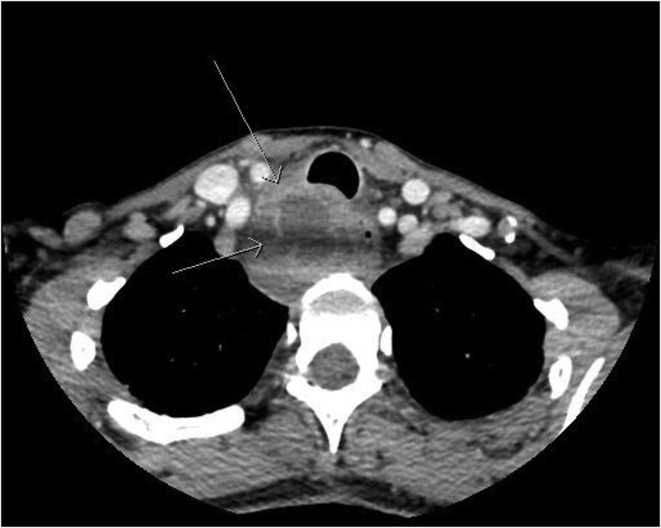
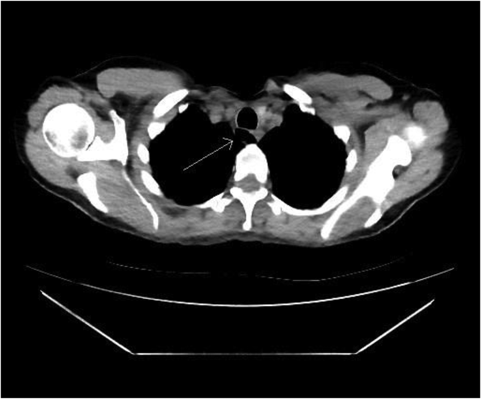
Her symptoms persisted and she developed globus sensation. Therefore, a computed tomography (CT) scan of her neck was arranged. This showed a 3.5 cm multiloculated right neck abscess, posterolateral to the trachea at the level of the thoracic inlet, causing mild airway narrowing, related to a known tracheal diverticulum (Figures 1 and 2). A CT scan of the chest conducted three months prior for investigation of shortness of breath and wheeze revealed an incidental finding of a posterior tracheal diverticulum at the level of the thoracic inlet (Figure 3).
Axial computed tomography scan of the neck with contrast (soft tissue window) showing the abscess of the posterior tracheal diverticulum (arrows), causing mild airway narrowing.

Coronal computed tomography scan of the neck with contrast (soft tissue window) showing the abscess to the right of the trachea and oesophagus (arrows) at the level of the thoracic inlet.

Axial computed tomography scan of the chest (soft tissue window) showing the posterior tracheal air-filled diverticulum (arrow) at the level of C7/T1.

The patient was referred acutely to the ENT team for management. On review, she was afebrile, and her blood pressure, heart rate, respiratory rate and oxygen saturations were within normal limits. The examination findings were unremarkable, with no increased work of breathing, no stridor and no dysphonia. The abscess was not palpable and she had a full range of neck movement. The supraglottis, glottis and visible subglottis were normal on fibre-optic nasal endoscopy, with normal vocal fold mobility. She had a raised C-reactive protein level of 80 mg/l and a normal white blood cell count of 9 × 10⁹/l.
The patient was commenced on intravenous tazobactam and piperacillin after consultation with the infectious disease physicians, and was admitted under the ENT team. Given the large size and multiloculation of the abscess, the decision was made to proceed with incision and drainage.
The surgical procedure included rigid oesophagoscopy to ensure there was no communication with the oesophagus. The oesophageal examination findings were normal. An anterior transcervical approach was then used, with a right hemicollar incision. A thick-walled abscess was exposed by dissection medial to the carotid sheath and lateral to the trachea. The lesion was confirmed to be the abscess by needle aspiration of pus. The abscess was incised, drained and washed out with normal saline. Samples of the purulent material were sent for microscopy, and cell culture and fungal growth testing. The recurrent laryngeal nerve was not seen. A non-suction drain was inserted.
The drain was removed and the patient was stepped down to oral antibiotics (amoxicillin and clavulanic acid) on day 1 post-operatively. She was discharged on day 2 on a 10-day course of oral antibiotics. At two weeks follow up, she had no ongoing infective symptoms, no subcutaneous emphysema and the globus sensation had resolved. The intra-operative samples yielded no bacterial growth on microscopy and culture testing. Fungal cultures grew Aspergillus fumigatus; however, on consultation with the infectious disease physicians, this was not thought to be clinically significant. The patient is scheduled to undergo surgical excision of the diverticulum and repair of the tracheal defect.
Discussion
A tracheal diverticulum is an outpouching of the tracheal ciliated columnar epithelium, usually occurring around the level of the thoracic inlet and at the right posterolateral aspect of the trachea (97.1 per cent).Reference Tanrivermis Sayit, Elmali, Saglam and Celenk1,Reference Monroe, McLean and Gross2 Tracheal diverticula are rare, occurring in only 2.4 per cent of the population.Reference Tanrivermis Sayit, Elmali, Saglam and Celenk1 They are usually asymptomatic and identified incidentally on cross-sectional imaging.Reference Tanrivermis Sayit, Elmali, Saglam and Celenk1 Chronic cough, dyspnoea, recurrent tracheobronchitis, stridor, dysphagia and dysphonia may be seen in symptomatic patients.Reference Tanrivermis Sayit, Elmali, Saglam and Celenk1 Tracheal diverticula can be congenital or acquired (iatrogenic or due to prolonged increased intraluminal pressure).Reference Tanrivermis Sayit, Elmali, Saglam and Celenk1,Reference Monroe, McLean and Gross2 A diagnosis of tracheal diverticula is usually made on a CT scan of the neck or chest. Tracheal diverticula may also be identified by direct bronchoscopic examination, though this is an invasive and less reliable method of diagnosis.Reference Tanrivermis Sayit, Elmali, Saglam and Celenk1
Tracheal diverticula can become infected and lead to recurrent tracheobronchitis.Reference Tanrivermis Sayit, Elmali, Saglam and Celenk1 In very rare cases, the infection can lead to abscess formation.Reference Tanrivermis Sayit, Elmali, Saglam and Celenk1 There are only three other similar reported cases in the literature, described below.Reference Monroe, McLean and Gross2–Reference Akabane, Kawachi, Fukai, Shimoyama, Kashiwagi and Ogino4
• A tracheal diverticulum is an outpouching of the tracheal epithelium, usually occurring at the thoracic inlet level and right posterolateral aspect of the trachea
• They are rare, occurring in only 2.4 per cent of the population, and can be congenital or acquired
• Tracheal diverticula are typically asymptomatic; symptoms include chronic cough, dyspnoea, recurrent tracheobronchitis, stridor, dysphagia and hoarseness
• Diagnosis is made based on computed tomography of the neck and/or chest with contrast
• Tracheal diverticula can rarely become infected and form an abscess; surgical drainage and excision may be performed
• To our knowledge, this is the fourth ever reported case of abscess formation related to an infected tracheal diverticulum
A 65-year-old Japanese man presented with airway distress due to a large paratracheal abscess causing airway compression.Reference Akabane, Kawachi, Fukai, Shimoyama, Kashiwagi and Ogino4 He required emergent intubation and surgical drainage. The patient was extubated on day 4 post-operatively, and was discharged home on day 7 with no major complications.
Charest et al. describe the case of an infected tracheal diverticulum with abscess formation that presented as a fluorodeoxyglucose-avid lesion in a 74-year-old woman with a past history of a lymphoproliferative disorder.Reference Charest, Sirois, Cartier and Rousseau3 She underwent mediastinoscopy to confirm the diagnosis. The 15 mm tracheal diverticulum was identified retrospectively, on a CT scan of the chest conducted five months previously. She underwent surgical drainage of the abscess and recovered well.
The third case in the literature was a 51-year-old man who presented with dyspnoea and an enlarging right neck mass.Reference Monroe, McLean and Gross2 A CT scan showed a complex air- and fluid-filled mass posterolateral to the trachea. The patient underwent incision and drainage of the abscess, but it re-collected. At repeat surgery, the mass was excised and a communication with the posterior trachea was noted and oversewn. The histopathology findings were consistent with a tracheal diverticulum.
As illustrated by this case, a tracheal diverticulum may become infected and present as a cervical abscess. Although this is rare, it is important to consider in the differential diagnosis of a lower neck or mediastinal mass. A CT scan is helpful in diagnosis and a communication with the digestive tract can be excluded with contrast swallow studies. To our knowledge, this is the fourth ever reported case of abscess formation related to an infected tracheal diverticulum.
Competing interests
None declared