INTRODUCTION
Clytia (Lamouroux, 1812) is the genus within Campanulariidae (Cnidaria, Leptomedusae) with the highest number of species and it is widely distributed throughout the world. Approximately 60 species of Clytia are currently recognized (Bouillon & Boero, Reference Bouillon and Boero2000; Calder et al., Reference Calder, Mallinson, Collins and Hickman2003; Schuchert, Reference Schuchert2003, Reference Schuchert2012; Bouillon et al., Reference Bouillon, Gravili, Pagés, Gili and Boero2006; Govindarajan et al., Reference Govindarajan, Boero and Halanych2006; Lindner et al., Reference Lindner, Govindarajan and Migotto2011), and because they are usually the most abundant hydromedusae, they play an important ecological role in the plankton and shallow-water benthic environments, acting as both competitor and predator (Madin et al., Reference Madin, Bollens, Horgan, Butler, Runge, Sullivan, Klein-Macphee, Durbin, van Keuren, Plourde, Bucklin and Clarke1996; Avent et al., Reference Avent, Bollens, Butler, Horgan and Rountree2001; Boero et al., Reference Boero, Camillo and Gravili2005; Adamík et al., Reference Adamík, Gallager, Horgan, Madin, McGillis, Govindarajan and Alatalo2006). Furthermore, the ubiquitous Clytia hemisphaerica (Linnaeus, 1767) has emerged as an important model organism in developmental biology and evolutionary studies (Amiel et al., Reference Amiel, Chang, Momose, Houliston, Verlhac and Villeneuve2010; Houliston et al., Reference Houliston, Momose and Manuel2010).
Clytia has a life history consisting of medusa and hydroid stages. Clytia medusae are commonly found in coastal waters, and their hydroids are either benthic or free-living planktonic colonies (Cornelius, Reference Cornelius, Barnes and Crothers1995; Lindner et al., Reference Lindner, Govindarajan and Migotto2011). However, taxonomy is particularly challenging in Clytia because of the phenotypic plasticity exhibited by its species. At either the medusa or hydroid level, most of the morphological characters used to distinguish the different species, have little or no taxonomic value because these characters have been observed to vary within a single species (Bouillon & Boero, Reference Bouillon and Boero2000). Furthermore, even among the few taxonomically relevant characters, it is unclear whether the differences are environmentally or genetically based, which presents a major limitation to their use in the identification of new species (Cornelius, Reference Cornelius1990). An additional obstacle is that most species of Clytia have been examined only at the hydroid or medusa stage (Reference Cornelius1982, Reference Cornelius, Bouillon, Boero, Cicogna and Cornelius1987; Calder, Reference Calder1991) and potentially distinguishing features of other life cycle stages are not yet recognized. In fact, specific identification is also hindered because the description of species from the plankton may have been based on developmental stages of other species (Lindner & Migotto, Reference Lindner and Migotto2002). Consequently, taxonomic homonyms and synonyms are common within Clytia, in particular for the nearly cosmopolitan C. hemisphaerica and C. gracilis (Sars, 1850). Elucidation of the complete life cycles of the known Clytia species is therefore important as it is likely to bring about important revisions (Bouillon & Boero, Reference Bouillon and Boero2000). Yet, for most Clytia species, life cycle studies may also be problematic since the developmental relationship between polyp and medusa stages is poorly understood. As reviewed by Lindner et al. (Reference Lindner, Govindarajan and Migotto2011), only ten species have had their life cycles investigated.
Most species of Clytia inhabiting the coastal waters of China have been described solely on the basis of their medusae (Huang, Reference Huang2008), with only a few reports in which hydroids were studied (Gao, Reference Gao1956). Recently, we used DNA barcoding based on two partial mitochondrial genes sequences, those of cytochrome c oxidase subunit I (COI) and the large subunit of ribosomal RNA (16S) (Zheng et al., unpublished data), to examine hydromedusae from the coastal waters of China. These efforts led to the identification of highly distinct lineages for a species of Clytia presented in the plankton samples from Xiamen Bay. Although this species is usually identified as C. hemisphaerica (Xu et al., Reference Xu, Huang and Wang1985; Cheng et al., Reference Cheng, Wang, Wang, Zhang, Li and Sun2012), in the phylogenetic tree it was determined to be the same species as Clytia cf. gracilis sp., from the coastal north-eastern United States (see Lindner et al., Reference Lindner, Govindarajan and Migotto2011). We therefore re-examined the morphology of the Xiamen Bay species and investigated its complete life cycle. The results showed that it differed from both C. hemisphaerica and C. gracilis based on the distinct morphological characters of its hydroid and medusae stages. Molecular and morphological evidence supported the valid placement of this species within the genus Clytia, and the name C. xiamenensis sp. nov. was chosen.
MATERIALS AND METHODS
Molecular methods
Total DNA was extracted from whole medusae (four field individuals and four individuals released from colonies) by SDS-proteinase K/phenol-chloroform extraction (Zheng et al., Reference Zheng, Lin, Li, Cao, Xu and Huang2009). The DNA was preserved in TE buffer and stored at −20 °C. The COI was amplified using the procedure and primers described by Zheng et al. (Reference Zheng, Lin, Li, Cao, Xu and Huang2009). 16S was partially amplified using the published primers (16S-L 5′GAC TGT TTA CCA AAA ACA TA3′ and 16S-H 5′CAT AAT TCA ACA TCG AGG3′) (Ender et al., Reference Ender and Schierwater2003). Amplification for 16S was achieved with five cycles of 94oC/1 min, 45oC/50 s, 72oC/1 min, followed by 30 cycles of 94°C/50 s, 50°C/1 min, 72°C/1 min, and a final elongation step at 72°C for 5 min. All PCR products were sequenced directly in both directions on an ABI PRISM 3730 Genetic Analyzer using BigDye® Terminator v.3.1 (Shanghai Sangon Biological Engineering Technology & Service Co., Ltd, China).
The COI and 16S sequences were aligned using ClustalX v.2 (Larkin et al., Reference Larkin, Blackshields, Brown, Chenna, McGettigan, McWilliam, Valentin, Wallace, Wilm, Lopez, Thompson, Gibson and Higgins2007) and then edited manually, using EditSeq v.7.1, to ensure the correct alignment and placement of insertion/deletion events. GenBank BLAST searches were performed to confirm the accuracy and validity of all sequences, which were then deposited in GenBank (Accession numbers for field samples: JQ716198–JQ716201 for COI and JQ716037–JQ716040 for 16S; Accession numbers for sequences derived from medusae liberated from the colonies: JQ7161202–JQ716205 for COI and JQ716041–JQ716044 for 16S). These sequences along with the sequences of Clytia folleata (McCrady, 1859) collected from the coastal waters of China (COI: JQ716211; 16S: JQ716051-JQ716055), and other species in Campanulariidae which have already been deposited in Genbank (Table S1, Supplementary Information) were used to infer phylogenetic trees using neighbour-joining (NJ, based on K2P model) and maximum-likelihood (ML, based on GTR + G model) algorithms in MEGA V5 (Tamura et al., Reference Tamura, Peterson, Peterson, Stecher, Nei and Kumar2011). Node support for the two approaches was inferred with a bootstrap analysis (1000 replicates). Two Leptothecata species were chosen as outgroups: Aequorea conica (Browne, 1905) and Gangliostoma guangdongensis (Xu, 1983).
Sample collection
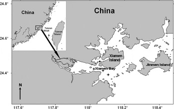
Medusae of Clytia xiamenensis sp. nov. were collected using a plankton net (mesh size: 500 μm) from Xiamen Bay, China, in March 2011 (Figure 1). Individuals used in DNA barcoding studies were acclimated in filtered seawater (FSW) for at least 24 h before they were photographed, and then preserved in 95% ethanol. The remaining individuals were kept alive and cultured together in FSW for morphology and life cycle observations.
Collecting sites of Clytia xiamenensis sp. nov.

Culturing and morphological description
Medusae collected from the sea were reared in covered glass vessels with a roughened glass slide placed at the bottom which facilitates colony settlement. Zygotes were collected and cultured in small crystallizing dishes to observe embryonic development. The glass slides with the newly settled colonies of C. xiamenensis sp. nov. were subsequently transferred to crystallizing dishes for further observation. Newly released medusae were collected and reared through maturity in the laboratory for morphological and DNA barcoding analyses. The hydroids and medusae were fed 2–3 times per day with Artemia sp. nauplii; the FSW was changed every second day. The temperature of the water was kept below room temperature (17–25°C) and the salinity was 30–32‰.
Planulae, hydroids, and medusae were examined under a stereomicroscope and by biological microscopy (Zeiss V12 and Nikon SMZ1000). Prior to their observation, hydranths and medusae were acclimated in a 10% MgCl2 solution. Artemia sp. nauplii juice and distilled water were used to induce the nematocysts to discharge in squash preparation (Östman, Reference Östman, Bouilon, Boero, Cicogna and Cornelius1987; Wang & Xu, Reference Wang and Xu1990). Nematocyst types and their distribution were determined using a light microscope equipped with differential-interference-contrast optics (Olympus BX51). Nematocyst nomenclature followed that of Weill (Reference Weill1934), Mariscal (Reference Mariscal, Muscatine and Lenhoff1974) and Östman (Reference Östman1979a, Reference Östmanb, Reference Östman and Stepanjants1999).
RESULTS
DNA barcoding
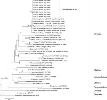
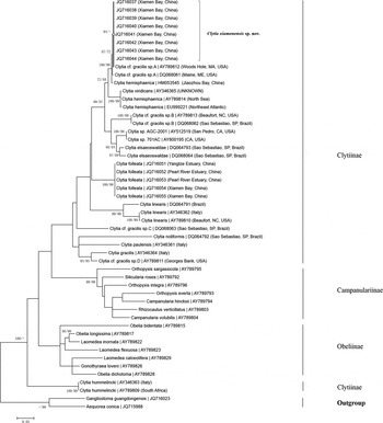
The COI and the 16S trees strongly supported Clytia xiamenensis sp. nov. as a member of the genus Clytia (Figures 2 & 3). In both trees, C. xiamenensis sp. nov. formed a strongly supported cluster with ‘C. hemisphaerica’ from the China Sea (Wand et al., 2010, sequence obtained from GenBank) and ‘Clytia cf. gracilis sp. A’ from the coastal north-eastern United States (see Lindner et al., Reference Lindner, Govindarajan and Migotto2011). All three were clearly separated from other Clytia species. For both the partial COI and the 16S sequences, genetic divergence was zero within individuals of C. xiamenensis sp. nov., and low within the clade comprising C. xiamenensis sp. nov., ‘C. hemisphaerica’ (from the China Sea), and ‘Clytia cf. gracilis sp. A’ (COI: 0–0.007; 16S: 0–0.006) (Tables S2 & S3; Supplementary Information), which supported the common lineage of its members. And there were obvious genetic divergences between C. xiamenensis sp. nov. and other species of Clytia (COI/16S: 0.093-0.315/0.055-0.241). According to the tree-based identification, C. xiamenensis sp. nov. is most closely related to the clade containing C. hemisphaerica (two individuals, collected from the North Sea and the north-east Atlantic; see Lindner et al., Reference Lindner, Govindarajan and Migotto2011 and Licandro et al., Reference Licandro, Conway, Yahia, de Puelles, Gasparini, Hecq, Tranter and Kirby2010, respectively).
Maximum-likelihood (ML) topology based on mtCOI. Bootstrap values higher than 70 were shown above the branches. First number along the branches refers to ML bootstrap values, second number refers to neighbour-joining bootstrap values, Clytia xiamenensis sp. nov. and subfamily lineages are indicated.

Maximum-likelihood (ML) topology based on 16S rRNA. Bootstrap values higher than 70 were shown above the branches. First number along the branches refers to ML bootstrap values, second number refers to neighbour-joining bootstrap values, Clytia xiamenensis sp. nov. and subfamily lineages are indicated.

RESULTS
SYSTEMATICS
Order LEPTOTHECATA Cornelius, 1992
Family CAMPANULARIIDAE Johnston, 1836
Genus Clytia Lamouroux, 1812
Clytia xiamenensis sp. nov.
(Figures 4–7; Tables 1–3)
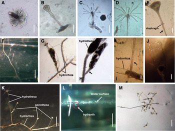
The hydranth of Clytia xiamenensis sp. nov.: (A–C) primary hydranth from the settled planulae; (D) distal portion of hydranth; (E) hydrotheca, arrow: annuli; (F) unbranched stem; (G–H) gonangium with medusa buds (G) gonothecae on the stolon; (H) gonothecae on the hydrocaulus, arrow: a short stalk with annuli; (I) pedicle with annuli (arrow); (J) hydrothecal margin with cusps; (K) a part of colony with a branch and gonothecae; (L–M) the floating colony. Scale bars: A,B,E,G–J, 100 μm; C, 250 μm; D, 125 μm; F,K,M, 1 mm; L, 2 mm

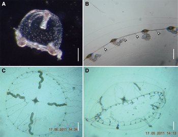
The medusae of Clytia xiamenensis sp. nov.: (A) newly liberated medusa; (B–D) mature medusa: (B) marginal structure of mature medusa; (C) male species; (D) female species. Note: Swallow-tailed arrow: the statocysts between tentacle (or marginal wart). Scale bars: A, 100 μm; B, 300 μm; C–D, 1 mm.

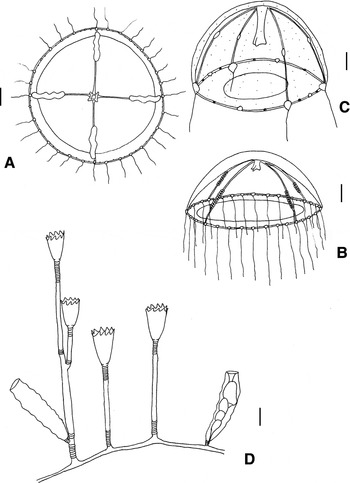
The line drawings of Clytia xiamenensis sp. nov.: (A) mature male individual; (B) mature female individual; (C) newly liberated medusa; (D) a part of colony with a branch and gonothecae. Scale bars: A,B, 1 mm; C,D, 100 μm.

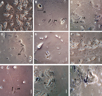
Interference contrast micrographs of the nematocyst from Clytia xiamenensis sp. nov.: (A–E) nematocysts form medusa: (A–B) A-type; (C) LA-type; (D) C-type; (E) isorhizas; (F–I) nematocysts form hydroid: (F–G) A-type; (H–I) B-type. Note: swallow-tailed arrow: the small protruding tip; arrow: the direction of discharge in nematocyst; pa: the proximal armature; dt: the distal tube; A: A-type; LA: LA-type; B: B-type. Scale bars: 5 μm.

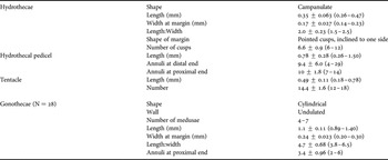
Measurements (mean ± SD (range)) of colonies of Clytia xiamenensis sp. nov. (N = 30 unless otherwise mentioned).

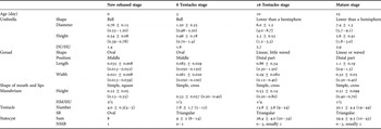
Comparison of morphology (mean ± SD (range)) of Clytia xiamenensis sp. nov. at successive developing medusae stages, in mm (N = 25).

DU/HU, diameter of umbrella/height of umbrella; HM, height of manubrium; SB, shape of bulbs; NSSB, number of statocysts between successive bulbs.
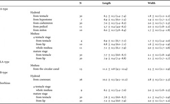
Measurements (mean ± SD (range)) of microbasic mastigophore nematocysts of Clytia xiamenensis sp. nov., in μm.

MATERIAL EXAMINED
Holotype: CXXB 11, female, 9.0 mm.
Paratype: CXXB 12, male, 8.6 mm. Both were collected in Xiamen Bay, China, 24.3871°N 118.1430°E, March 2011. The type specimens are deposited in the Department of Oceanography, Xiamen University, China.
ETYMOLOGY
Clytia xiamenensis sp. nov. is named after Xiamen Bay where the species was collected.
DIAGNOSIS
Clytia xiamenensis sp. nov. is distinguished from its congeners and other Campanulariidae based on a combination of the following characters:
Polyp: the stem is monosiphonic, stolonal, branching rarely. Hydrothecal cusps usually incline to one side. Gonothecae are borne on both hydrorhiza and pedicel, with a short annulated pedicel and undulated walls. B-type microbasic mastigophores are 9.5–11.2 μm long in vivo.
Adult medusa: the bell is 5.7–9.1 mm in its marginal diameter, with up to 44 tentacles and 0–3, but usually 1, statocysts between the hollow tentacles. In male medusae, green pigments are present in gonads, tentacle bulbs, and warts. LA-type microbasic mastigophores (9.5–12.4 μm, it shared similar shape with A-type, but much larger than A-type, so named it as LA-type) were also observed.
Hydroid
Colonies are stolonal, and the pedicels branch only rarely (Figures 4F, K & 6D). The pedicel is short (0.26–1.5 mm), with 4–29 distal and 7–14 proximal annuli (Figure 4D, E, I); several pedicels are thoroughly covered with annuli.
The hydrotheca is campanulate in shape, with 6–12 triangular, pointed cusps inclining to one side (Figures 4J & 6D), and variable in size: 0.26–0.47 mm long and 0.14–0.23 mm wide at the margins (Figure 4D, E). The hydrothecal diaphragm is thin, transverse, and located at the base of the hydrotheca (Figure 4E); the basal chamber is cup-like. The hydranth is elastic, reaching a height of up to 0.88 mm; the pedunculated hypostome is spherical in oral view. There are 12–18 filiform tentacles and two amphicoronate whorls, with an average length of 0.49 mm (Figure 4D). Nematocysts distribute on tentacles circularly. Hydrothecal length:width ratio: 1.5–2.5 (Table 1).
The gonothecae arise on stolons, sometimes on the pedicel or directly on a branch (Figures 4G, H, K & 6D). They have distinctly undulated walls (Figure 4G) and short pedicels, each with 2–6 annuli (Figure 4H); 4–7 buds are present (Figure 4G, H). Gonothecae are cylindrical, 0.89–1.4 mm long and 0.2–0.3 mm wide at their margins. Their length: width ratio is 3.8–6.5 (Table 2).
Most areas of the hydroid body contain A-type nematocysts with a spindle-shaped capsule (Figure 7F, G; details in Table 3). The tube is discharged from the tip in a direction almost coinciding with that of the long axis of the capsule (Figure 7G). B-type microbasic mastigophore nematocysts are 10.2 ± 0.5 μm (9.5–11.2 μm, N = 26) long and 2.8 ± 0.3 μm (2.1–3.3 μm, N = 26) wide in vivo. They are along coenosarcs and are much larger and more curved than other types of nematocysts (Figure 7H, I; details in Table 3).
Newly released medusa
The umbrella is bell-shaped, 0.55–1.2 mm wide and 0.39–0.78 mm in height. The jelly is comparatively thin. D-type nematocysts are scattered over the exumbrella (Figures 5A & 6C). There are four radial canals and one circular canal. The stomach is small and the manubrium is quadrate, with four simple lips. The ovate and transparent gonads are located in the middle of the radial canals. The velum is broad. Four hollow tentacles contain A- and C-type nematocysts. Additional features: four developing marginal warts; eight adradial statocysts, with closed marginal vesicles, containing one statolith (Figures 5A & 6C).
Development
Medusae attained up to eight tentacles within 3 d and 16 tentacles within about 10 d. During this period, D-type nematocysts on the exumbrella disappeared; the umbrella became flat and the lips recurved (details in Table 2).
Mature medusa
When the medusae are 25 d old, the umbrella is flattened, 5.7–9.1 mm wide and 1.8–3.0 mm in height (Figures 5C, D & 6A, B). The stomach is small; the manubrium is short, with undulated lips. There are four radial canals. The gonads are usually elongated and may be slightly wavy, about 0.9–1.5 mm long and 0.30–0.40 mm wide, occupying the distal part of the radial canal. The velum is narrow. There may be 19–44 well-developed hollow tentacles, but generally about 30. The tentacle bulbs are oval or triangular, without papilla. There are 10–43 statocysts in a medusa but the number is usually less than that of the total tentacles. Between 0 and 3 statocysts, but usually only one, are located between successive tentacles and margin warts. The statocysts contain one statolith, rarely two (Figure 5B). In male medusae, green pigments are present in the gonads, tentacle bulbs, and warts (Figure 5C).
A-type nematocysts are scattered throughout most of the body (Figure 7A, B). The nematocysts of mature medusae are larger than those of immature forms (Table 3). LA-type microbasic mastigophores are 11.2 ± 0.8 μm (9.5–12.4 μm, N = 15) long and 2.5 ± 0.2 μm (2.1–2.9 μm, N = 15) wide in vivo and are much larger than A-type (Figure 7C), scattering only at the level of the circular canal. C-type nematocysts are found only in the tentacles of the medusae, are relatively few in number, and their tubules roughly form a right angle with the long axis of the capsule when discharged (Figure 7D). Atrichous isorhiza nematocysts are present in the tentacles (Figure 7E) and in the lips (Table 3).
Biological notes
The mature medusae released eggs and sperm mainly at dawn, although occasionally at other times of the day. The eggs were variable in diameter, 158 ± 12.8 μm (130–182 μm), but the sperm were much smaller. The head of the sperm was 3.3 ± 0.28 μm (2.5–3.8 μm) long and 2.0 ± 0.25 μm (1.7–2.5 μm) wide. Embryonic development lasted only for several hours. Fertilized eggs developed into blastula within 3–4 h; the blastula became free-swimming hollow planulae after 1–3 h and then solid planulae during the following 3 h.
Planulae are small, milky white, 0.14–0.36 mm long and 0.060–0.12 mm wide. They can change direction by rotation during swimming. The planulae observed in this study settled mainly along the bottom and walls of the rearing jars, where they underwent typical development into hydroids (Figure 4A–C). However, some planulae settled on the underside of the surface film of the water, where they gave rise to floating colonies that developed for a few days (Figure 4L, M) before sinking to the bottom of the jar, where they continued to develop. We did not observe any morphological differences between free and attached hydranths.
The hydroids were kept at 17–25oC, as higher temperatures were lethal. The formation of a considerable number of gonothecae was observed in spring (17–20oC), while fewer than 20 gonothecae were counted in summer (24–25oC).
DISCUSSION
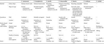
The similarities in shape and size of medusae of Clytia species, especially immature medusae, are greater than those of the polyps of this genus (e.g. Table 4). As a result, it is difficult to describe species unequivocally based on medusae collected in the plankton. This has frequently led to the synonymization of species within the genus Clytia at the medusa level (Bouillon & Boero, Reference Bouillon and Boero2000). For example, Millard (Reference Millard1966) combined C. johnstoni (Alder, 1856) and C. gracilis and established C. hemisphaerica based on the morphology of the medusae. Cornelius (Reference Cornelius1982) also synonymized C. gracilis with the cosmopolitan C. hemisphaerica, but the former was subsequently shown to be distinct on the basis of its hydroid and nematocysts (Östman, Reference Östman1979a, Reference Östman, Bouilon, Boero, Cicogna and Cornelius1987). In plankton collected from the China Sea, specimens assigned to C. hemisphaerica were reviewed and subsequently divided into two types by Xu et al. (Reference Xu, Huang and Wang1985) based on field medusae samples. Although with its 19–44 tentacles and linear or slightly wavy gonads C. xiamenensis sp. nov. resembles the larger of these two types, which is detected in the China Sea from March to April, the molecular and morphological analyses presented below strongly suggest that it is a distinct species.
General features of some similar species of Clytia.

NSSB, number of statocysts between successive bulbs.
Overall colony form and hydrothecal characteristics are frequently used as diagnostic characters in the Campanulariidae (Cornelius, Reference Cornelius1982). However, for either of these there may be considerable phenotypic plasticity (Ralph, Reference Ralph1956; Cornelius, Reference Cornelius1982), which presents a major obstacle to their taxonomic utility. In fact, variations in these characters have been reported for many species, such as C. gregaria (L. Agassiz, 1862) (Roosen-Runge, Reference Roosen-Runge1970), C. attenuate (Calkins, 1899) (West & Renshaw, Reference West and Renshaw1970), C. edwardsi (Nutting, 1901) (Kubota, Reference Kubota1978a), C. linearis (Thorneley, 1900), and C. noliformis (McCrady, 1859) (Lindner & Migotto, Reference Lindner and Migotto2002). For C. xiamenensis sp. nov., despite variations in some of the characteristics of the hydrothecae, such as their dimensions, the number of tentacles, and the annulations on the pedicels, others, including the shapes of the hydrothecal cusps and gonothecae, are relatively consistent, at least according to our observations (Table 1). The absence of variability in these features was also reported for C. attenuate (West & Renshaw, Reference West and Renshaw1970). Alternatively, for many groups of hydroids, including Campanulariidae, the morphological characteristics of the nematocysts are taxonomically useful (Östman, Reference Östman1979a, Reference Östmanb, Reference Östman1982, Reference Östman, Bouilon, Boero, Cicogna and Cornelius1987); but again, some of these characters may be plastic (Östman et al., Reference Östman, Piraino, Roca, Bouilon, Boero, Cicogna and Cornelius1987).
In addition to the morphological similarity among species, difficulty in determining species boundaries within Clytia is also due to the lack of information on the life cycles of its member species and on intraspecific morphological variation. These and related problems are further aggravated by the absence of sufficient morphological data, which prevents the recognition of cryptic species (Lindner et al., Reference Lindner, Govindarajan and Migotto2011). Thus, at least in the case of Clytia, additional diagnostic characters should be added to the species description. DNA barcoding has enabled species tagging for the purpose of identification (Hebert et al., Reference Hebert, Ratnasingham and deWaard2003, Reference Hebert, Penton, Burns, Janzen and Hallwachs2004). For hydrozoans, both COI and 16S have been recommended as valuable DNA barcodes that allow the differentiation of morphologically undistinguishable, nominal, and cryptic or pseudo-cryptic species, including undescribed taxa (Govindarajan et al., Reference Govindarajan, Halanych and Cunningham2005; Schuchert & Reiswig, Reference Schuchert and Reiswig2006; Moura et al., Reference Moura, Harris, Cunha and Rogers2008, Reference Moura, Cunha, Porteiro and Rogers2011; Bucklin et al., Reference Bucklin, Steinke and Blanco-Bercial2011). Recently, Lindner et al. (Reference Lindner, Govindarajan and Migotto2011) determined that a Brazilian ‘C. gracilis’ was instead a distinct species based on a detailed life cycle study and molecular analysis, and they resurrected the name C. elsaoswaldae (Stechow, 1914). In the present work, we obtained morphological and molecular evidence supporting C. xiamenensis sp. nov., previously regarded as conspecific to C. hemisphaerica or Clytia cf. gracilis sp., as a valid species belonging to the genus Clytia.
In defining the genus Clytia, Hincks (Reference Hincks1868) stated that one of its main characteristics was the production of medusae with four radial canals, four marginal tentacles, and eight lithocysts. Nutting (Reference Nutting1915) expanded the definition by describing the trophosome: ‘Colony often simple but always consisting of a creeping rootstock from which spring pedicels which are not regularly branched as a rule. Hydrothecae campanulate, hydranths with trumpet-shaped proboscis’. Years later, Bouillon & Boero (Reference Bouillon and Boero2000) emphasized the ‘hydrothecal rim sinuous or deeply indented, with clefts between the round to sharply-pointed cusps; hydrothecae with a true hydrothecal diaphragm’. All of these characteristics were observed in C. xiamenensis sp. nov. (Figures 4–6; Tables 1 & 2), and the phylogenetic-tree-based identification also strongly supported this species as belonging to the genus Clytia (Figures 2 & 3).
Mature medusae of both C. xiamenensis sp. nov. and C. hemisphaerica have more than 30 tentacles, four oral lips, and oval or linear gonads close to the circular canal (Table 4). These similarities make it difficult to distinguish between members of these two species at the medusa stage. However, C. xiamenensis sp. nov. differs from C. hemisphaerica in that the former usually has one statocyst between successive bulbs whereas the latter species usually has two (Mayer, Reference Mayer1910; Gao et al., Reference Gao, Li, Chang and Li1958; Kramp, Reference Kramp1968). In addition, the gonads, tentacle bulbs, and warts of adult male medusae of C. xiamenensis sp. nov. contain green pigments but this has never been reported for C. hemisphaerica. Medusae of C. hemisphaerica collected in the plankton are also much larger (up to 20 mm in diameter; Russell, Reference Russell1953; Cornelius, Reference Cornelius, Barnes and Crothers1995) than those of C. xiamenensis sp. nov. (up to 9.1 mm in diameter). Another similarity between C. xiamenensis sp. nov. and C. hemisphaerica is that of their hydroids, as stolonal growth and usually a few branched stems are seen in both; however, C. xiamenensis sp. nov. differs from C. hemisphaerica in that its hydrothecae have pointed cusps and are usually inclined to one side (rather than rounded and not inclined; Cornelius, Reference Cornelius, Barnes and Crothers1995). Another difference is that the ‘B-type’ nematocysts (9.5–11.2 × 2.1–3.3 μm) in the hydroid of C. xiamenensis sp. nov. are smaller than those of C. hemisphaerica hydroids [(10.5–13.5 × 2.5–4.0 μm (Sweden), 21.0–24.0 × 4.0–4.5 μm (Italy), 15.5–17.0 × 3.5–4.0 μm (Brazil); Östman, Reference Östman and Stepanjants1999)]. Moreover, only C. xiamenensis sp. nov. contains LA-type nematocysts scattered at the level of the circular canal, further corroborating that it is not conspecific with C. hemisphaerica. Finally, there were obvious genetic divergences between C. xiamenensis sp. nov. and C. hemispherica (COI/16S:0.093/0.055). Clytia xiamenensis sp. nov. formed a strongly supported lineage in the COI and 16S trees that was clearly separate from the lineage of C. hemisphaerica (two individuals, collected from North Sea and north-east Atlantic, see Lindner et al., Reference Lindner, Govindarajan and Migotto2011 and Licandro et al., Reference Licandro, Conway, Yahia, de Puelles, Gasparini, Hecq, Tranter and Kirby2010, respectively), although the two species were closely related (Figures 2 & 3). This phylogenetic evidence corroborates that obtained from the life cycle and morphological studies. Here, we need to mention that the distinct mitochondrial lineages are only suggestive, a nuclear marker is essential to detect the relationship between the species of Clytia.
Thus, according to the phylogenetic-tree-based identification, C. xiamenensis sp. nov., ‘Clytia cf. gracilis sp. A’ from the coastal north-eastern United States (Lindner et al., Reference Lindner, Govindarajan and Migotto2011), and ‘C. hemisphaerica’ from the China Sea (Cheng et al., Reference Cheng, Wang, Wang, Zhang, Li and Sun2012) are the same species, with C. xiamenensis sp. nov. clearly distinct from other species assigned to C. gracilis or Clytia cf. gracilis (genetic divergences of COI/16S: 0.096-0.315/0.071-0.152; Figures 2 & 3). Based on the evidence discussed above, these ‘C. hemisphaerica’ from the China Sea were misidentified. Although C. xiamenensis sp. nov. is similar to C. gracilis in having inclined hydrothecal cusps (Cornelius, Reference Cornelius, Barnes and Crothers1995), it differs in that it has mostly stolonal (rather than mostly branching) colonies and its gonothecae have markedly undulated (rather than smooth) walls (Cornelius, Reference Cornelius, Barnes and Crothers1995) (Table 4). Although gonothecal shape may vary in some species of Clytia (Kubota, Reference Kubota1978a; Lindner & Migotto, Reference Lindner and Migotto2002), C. gracilis has never been collected on the coast of China (Huang, Reference Huang2008), suggesting that it is not present in this region and thus has a more limited distribution. Moreover, the ‘B-type’ nematocysts in the hydroid of C. xiamenensis sp. nov. are much smaller (9.5–11.2 × 2.1–3.3 μm) than those of C. gracilis (12.0–18.0 × 3.0–5.5 μm (Sweden); Östman, Reference Östman and Stepanjants1999). Mature medusae of C. gracilis are unfortunately still unknown. If they are indeed 6–8 mm in diameter and bear 16 tentacles, as hypothesized by Cornelius (Reference Cornelius, Barnes and Crothers1995), then they have fewer tentacles than C. xiamenensis sp. nov. Moreover, LA-type nematocysts and green pigments have never been reported for C. gracilis. Thus, C. xiamenensis sp. nov. is not a cryptic species of C. gracilis and a careful review of specimens assigned to C. gracilis is necessary.
Clytia xiamenensis sp. nov. and C. viridicans (Leuckart, 1856) are similar in that both have green pigments in the manubrium, gonads, and marginal bulbs, but they differ in that C. xiamenensis sp. nov. has inclined cusps (rather than rounded and not inclined), its newly liberated medusae bear gonads (rather than absent thereof), its mature medusae have 19–44 tentacles (rather than 16 tentacles), and bright green pigmentation is seen only in mature male medusae (rather than beginning in the newly released medusae and persisting in mature medusae, both male and female; Pagliara et al., Reference Pagliara, Bouillon and Boero2000) (Table 4). Green pigmentation was also reported by Mayer (Reference Mayer1910) in a study of C. folleata from the west Atlantic.
Clytia folleata is also found in Xiamen Bay, China, and it resembles C. xiamenensis sp. nov. with respect to the shape of its medusae. However, its mature medusae have only 16 tentacles and, based on our observations, the gonads differ in shape, as those of the former are usually oval rather than linear. Although information on the life cycle of C. folleata is still lacking, our DNA-based identification showed that this species formed a distinct clade which was well separated from C. xiamenensis sp. nov. (Figures 2 & 3).
In addition to C. gracilis and C. xiamenensis sp. nov., other species of Clytia are characterized by hydrothecae with inclined cusps, i.e. C. linearis, C. delicatula (Thornely, 1904), and C. elsaeoswaldae (Table 4). However, in C. linearis the hydrothecal margin has 10–14 sharp cusps, each with a pleat projecting into the hydrothecal cavity and extending from the apex of the cusp towards the middle of the hydrotheca. In addition, the gonothecae of C. linearis are smooth (Lindner & Migotto, Reference Lindner and Migotto2002) and the slightly compressed bell of its medusae may be unique. Both C. delicatula and C. elsaeoswaldae differ from C. xiamenensis sp. nov. in having smooth gonothecal walls. Also, newly liberated medusae of C. delicatula lack gonads (Kubota, Reference Kubota1978b) and the gonothecae of C. elsaeoswaldae develop exclusively in the hydrorhiza, not on branches (Lindner et al., Reference Lindner, Govindarajan and Migotto2011). Although both C. uchidai and C. xiamenensis sp. nov. have type localities in the same biogeographic zone, they differ in that C. xiamenensis sp. nov. has one statocyst between successive bulbs (rather than usually two), its newly liberated medusae bear gonads (rather than absent thereof), its mature medusae have up to 44 tentacles (rather than 28 tentacles; Uchida, Reference Uchida1947) (Table 4). In addition, green pigmentation has never been reported for C. uchidai.
Interestingly, planktonic hydroids were observed for C. xiamenensis sp. nov. These free hydroids were produced by the planula and then settled on the underside of the surface film of the water, eventually giving rise to floating colonies that developed over a period of a few days (Figure 4L, M), as has been observed for C. viridicans (Pagliara et al., Reference Pagliara, Bouillon and Boero2000). This planula behaviour in culture may be a cultivation artefact, or represent its natural behaviour in nature. In fact, although there are no direct observations in which planktonic hydroids are produced by planula, huge numbers of floating colonies of C. gracilis have been found in Georges Bank (Madin et al., Reference Madin, Bollens, Horgan, Butler, Runge, Sullivan, Klein-Macphee, Durbin, van Keuren, Plourde, Bucklin and Clarke1996). In most hydrozoan species, medusae and the other floating organisms are generally the main agents of species dispersal. Yet, in C. xiamenensis sp. nov. and C. viridicans the behaviour of their planulae suggests the important contribution of this life cycle stage to dispersal. While the ecological role of planktonic hydroids in the field is not well understood, they could prey upon young copepods as well as on the eggs and larvae of commercially important fish species (Madin et al., Reference Madin, Bollens, Horgan, Butler, Runge, Sullivan, Klein-Macphee, Durbin, van Keuren, Plourde, Bucklin and Clarke1996; Adamík et al., Reference Adamík, Gallager, Horgan, Madin, McGillis, Govindarajan and Alatalo2006), in addition to being a food source for these fish (Avent et al., Reference Avent, Bollens, Butler, Horgan and Rountree2001).
The morphological and molecular analyses described above support C. xiamenensis sp. nov. as a valid species of the genus Clytia. Our findings further suggest that currently recognized species of Clytia, in particular C. hemisphaerica and C. gracilis, may represent complexes of several species. Since most Clytia studies have not considered this possibility or have been based only on a single life cycle stage (i.e. polyp or medusa), a careful review of those Clytia species regarded as nearly cosmopolitan in shallow waters is necessary. Furthermore, our approach demonstrates that life cycle studies in which Clytia species described based on their polyps are linked to those described based on their medusae could correct misidentifications, resulting in more accurate species descriptions. However, additional species-level studies would still be needed to assess diversity. Finally, it is important to emphasize that studies based on the life cycle and DNA, especially the nuclear DNA, of the organism in question are effective and efficient for species identification and both should be adopted as standard taxonomic approaches in future revisions, as shown here for the genus Clytia.
DNA barcoding offers great help to understand the extent of biodiversity by providing a simple and quick way to identify species (Hebert et al., Reference Hebert, Ratnasingham and deWaard2003; Hebert & Gregory, Reference Hebert and Gregory2005). A challenge remains, however, in its accuracy and efficiency (Krishnamurthy & Francis, Reference Krishnamurthy and Francis2012). And another point of contention is that regions of DNA used for barcoding often present limited information for higher phylogenetic resolution (Moritz & Cicero, Reference Moritz and Cicero2004). The results from our study established two facts indicating the barcode potential of COI and 16S for Clytia, even for the Campanulariidae. First, both COI and 16S could identify C. xiamenensis sp. nov. and other Campanulariidae species efficiently and accurately using the distance-based (K2P distance) approach. Second, 16S was shown to be a better phylogenetic marker for Campanulariidae at the subfamily level, Campanulariinae and Obeliinae appeared a monophyletic clade respectively and all but one species (Clytia hummelincki) of Clytiinae were monophyletic in the 16S tree (Figure 3). And we believe that increased taxon sampling will increase the better understanding of the phylogenetic relationships of the members of Campanulariidae in the coastal water of China based on 16S data.
ACKNOWLEDGEMENTS
We appreciate that Dr Carina Östman kindly mailed us her papers about nematocysts, Dr Senjie Lin provided valuable comments on an earlier draft of the manuscript, and Professors Jiaqi Huang and Zhenzu Xu helped to identify species. We are grateful to Open and Sharing Platform of Equipment and Technology (OSPET) of COE of Xiamen University for providing equipment and technical support.
FINANCIAL SUPPORT
This work was supported by the National Natural Science Foundation of China (Grant No. 41006078), the Fundamental Research Funds for the Central Universities (Grant No. 2010121037), the Public Science and Technology Research Funds Projects of Ocean (Grant No. 201005015-5), the Natural Science Foundation of Fujian Province of China (Grant No. 2011J05116).
Supplementary materials and methods
The supplementary material referred to in this article can be found online at journals.cambridge.org/mbi.