Introduction
Patients with complex CHD may experience multiple surgeries and interventions with secondary potential vessel thrombosis. This leads to the need for alternate routes to perform cardiac catheterisations. The transhepatic approach for paediatric cardiac interventions, first reported in 1995, remains until now an uncommon access route for device closure among congenital cardiologists, and debatable when it comes to entry point haemostasis.Reference Shim, Lloyd, Cho, Moorehead and Beekman1–Reference Jolly, Dhar and Amin4
Case presentation
In February 2020, a 29-year-old male patient with a bidirectionally shunting central atrial septal defect and thrombosed femoral veins was referred to our centre for transhepatic device closure after a failed transjugular attempt. The patient was born with pulmonary atresia/intact ventricular septum surgically palliated in the neonatal period and had two subsequent right ventricular outflow tract surgeries. He experienced a transient ischemic attack secondary to a paradoxical embolism even though he was on warfarin for an episode of atrial flutter. Severe pulmonary valve dysfunction and right heart failure with dilated right atrium were noticed during follow-up. Therefore, he had transjugular pulmonary valve implantation, but the attempt to close his atrial septal defect during the same procedure was peroperatively abandoned for anatomical and technical reasons, avoiding prolonged irradiation time. Alternative management options were discussed, and a decision was taken to proceed with a scheduled transhepatic closure because of the higher likelihood of success.
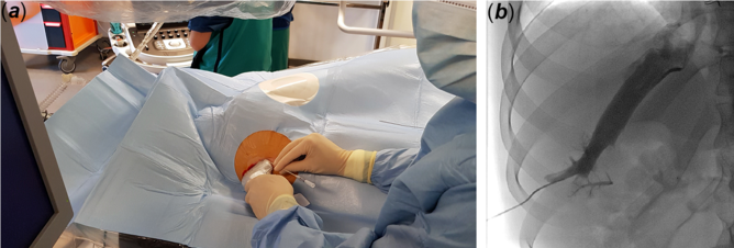
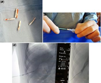
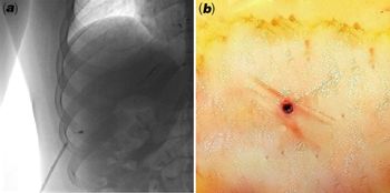
The procedure was performed under general anaesthesia, fluoroscopic control, and transoesophageal echocardiography guidance. The patient was kept therapeutic on aspirin and warfarin. At the start of the procedure, intravenous cefazolin was administrated, and the liver structure was sonographically assessed by an experienced interventional radiologist. The right hepatic vein was accessed with a 16 GA AngiocathTM (Becton Dickinson Infusion Therapy Systems, Inc., United States) (1.7 × 133 mm) under ultrasound guidance (Fig 1). The needle was removed and an angled 0.035 in × 180 cm Radifocus® Guidewire M Stiff Type (Terumo Corp., Japan) was advanced into the right atrium. At this point, the access was pre-dilated using an 11-cm-long, 6-F introducer sheath, and the patient received unfractionated heparin (5000 UI) to achieve an activated clotting time of 200 seconds. The defect was crossed smoothly with the 90° angle-approach despite right atrial dilatation and septum distortion. The sheath was upgraded to a 90-cm-long 8-F Flexor® Check-Flo® Introducer (Cook Medical, United States). The 6-mm-large defect with adequate margins was closed with a 10-mm AmplatzerTM septal Occluder (AGA Medical Corp., United States). The leftward and posterior orientation of the delivery sheath simplified the deployment sequence. Protamine sulphate was given to reverse heparinisation. Using the first angiography as an adjacent reference (Fig 1b), the sheath was gradually withdrawn from the lumen of the hepatic vein and positioned within the liver parenchymal sheath tract just outside the site where the hepatic vein was entered. A small volume of contrast was injected to ensure that the sheath tip was no longer within the vein. An absorbable haemostatic gelatin sponge, the SpongostanTM Standard (Ferrosan Medical Devices) was used to form multiple plugs that were rolled to fit into the delivery sheath (Fig 2a). Two plugs were successively pushed with to the end of the sheath (Fig 2b), which was then withdrawn while deploying the plugs between the hepatic vein and the liver capsule (Figs 2c, 3a). Successful haemostasis was confirmed by contrast injection through the sheath as it was completely withdrawn (Fig 3b). The overall procedure time was 30 minutes and required 12 minutes of fluoroscopy. Immediate liver ultrasound showed no complication. The patient was transferred to general wards for 48-hour observation with puncture site regularly checked. Before hospital discharge, there were no abnormalities detected in his liver function tests, chest X-ray, cardiac, and hepatic ultrasound control.
Ultrasound-guided puncture below the right costal margin, between the midclavicular and right anterior axillary line (at a 30° to 40° angle) directing the needle superiorly, posteriorly, and medially towards the patient’s left shoulder (a). Angiographic verification of the transhepatic access before sheath introduction (b).

Absorbable haemostatic gelatin sponge rolled into small plugs (a) and loaded into the delivery sheath to be pushed using the dilator into the desired position (b) under fluoroscopic control (c).

Angiographic view confirming proper plugs deployment (a). Successful entry site haemostasis (b).

Discussion
Obtaining the best access for a planned cardiac catheterisation is the primary key point for success. In the absence of femoral access, transjugular and transhepatic routes stand out as reasonable alternates for atrial septal defect closure. In adults, the internal jugular vein is a more classical entry point for catheterisation but addressing a centrally located atrial septal defect at a perpendicular angle may be encountered with some technical difficulties especially in patients with dilated right cavities. The puncture of the subclavian vein has been reported in complex catheterisations but can be easily complicated with pneumothorax and bleeding in a noncompressible site. The straight forward access to the atrial cavities via transhepatic access and the ease to introduce large delivery systems without serious risk of vascular injury motivated us to consider this route for defect closure in our patient with significant morbidities.Reference Shim, Lloyd and Beekman5,Reference Ebeid6 The occluder alignment with the plane of the septum avoided caudal traction on the delivery cable. This feature was also highlighted in closing the large atrial defects with deficient rims, avoiding complex manoeuvers, and prolapse of any part of the occluder.Reference Ebeid6
Some congenital interventionists have taken over the technique in some institutions and reported favourable experiences with variable success rates.Reference Shim, Lloyd, Cho, Moorehead and Beekman1,Reference Emmel, Sreeram, Pillekamp, Boehm and Brockmeier7,Reference McLeod, Houston, Richens and Wilson8 McLeod et al. even reported obtaining the hepatic access blind without the assistance of ultrasound and stated that as operator experience increases, the need for ultrasound guidance is reduced.Reference McLeod, Houston, Richens and Wilson8 However, we believe that the presence of an experienced interventional radiologist is unvoidable during first attempts. The knowledge and the skills to perform this approach should be acquired by the cardiac interventionalist, to be used when deemed necessary.Reference Ebeid6 Appropriate ultrasound evaluation of the liver anatomy and location is mandatory in grown-up congenital heart patients to identify the suitable hepatic vein for access. Ultrasound guidance is also needed during needle entry and sheath placement, in adjunction to fluoroscopy to optimise procedural safety and outcomes.
The transhepatic approach can be easily complicated with liver hematoma and intraperitoneal bleeding although the absolute risk is still not certain.Reference McLeod, Houston, Richens and Wilson8 The liver can store up to 10% of the total blood volume at any given time, and this volume can significantly increase in cases of elevated central venous pressure.Reference Guyton and Hall9 The antithrombotic state was carefully evaluated before the intervention. The medication was uninterrupted since our patient was estimated at a high risk of thromboembolism and heparin was added for atrial manipulations and bidirectional shunting. Access closure and heparin reversal before sheath withdrawal remain until now a matter for debate.Reference Ebeid6 Some authors reported achieving haemostasis in children only by manual external compression of skin at the puncture site with no significant complications.Reference Emmel, Sreeram, Pillekamp, Boehm and Brockmeier7,Reference McLeod, Houston, Richens and Wilson8 Others consider the closure of the tract only in patients who are on aspirin or in those with elevated right atrial pressure.Reference Ebeid6 Our patient had a basal right atrial pressure of 12 mmHg, required the use of a large sheath, and his procedure unexpectedly elapsed fast. Active clotting time had to be kept below 200 seconds at the end of the procedure before Gelfoam plugs were used to achieve fast and efficient hepatic vein haemostasis and allow early mobilisation. Although it does not cause artefacts in MRI, the gelfoam radiolucency was considered as an inconvenience for controlled and precise parenchymal access embolisation. Only detachable coils and vascular plugs were reported as haemostatic alternatives in similar clinical situations.Reference Alkhouli, Campsey, Higgins, Badhwar, Diab and Sengupta3,Reference Jolly, Dhar and Amin4,Reference Ebeid6 However, the risk of infectious complications has to be considered when using vascular plugs. Dollinger et al. reported a case of focal liver abscess adjacent to a vascular plug.Reference Dollinger, Goessmann, Mueller-Wille, Wohlgemuth, Stroszczynski and Heiss10 With this in mind, Uller et al. state that the gelfoam application does not increase the risk of local inflammation in comparison to permanent embolic agents offering a major advantage especially in patients with diminished immunity.Reference Uller, Müller-Wille and Grothues11
Conclusion
Transhepatic access can be efficiently established in adult patients with compromised femoral veins, and the atrial septal defect closure can be performed as planned. With many technical and intrinsic advantages, this approach may be favoured over the neck approach when accessing the interatrial septum. Procedure success and haemostasis control can be optimised with active sealing of the access site even in antithrombotic states, and the procedure being undertaken in collaboration with an experienced interventional radiologist.
Acknowledgements
We wish to thank Pr. Marc Gewillig for his useful technical suggestions.
Author contributions
RH collected clinical data and took the lead in writing the manuscript. All authors have read and approved the final version of the manuscript.
Financial support
This research received no specific grant from any funding agency, commercial or not-for-profit sectors.
Conflict of interest
None.
Ethical standards
The authors assert that all procedures contributing to this work comply with the ethical standards of the relevant national guidelines on human experimentation, and with the Helsinki Declaration of 1975, as revised in 2008. The patient’s written informed consent was obtained to perform the procedure and for the publication of this report.