Introduction
Species of Mawsonascaris Sprent, 1990 are rare ascaridoid nematodes, occurring in the digestive tract of elasmobranchs (Sprent, Reference Sprent1990; Ali et al., Reference Ali, Zhang, Al-Salim and Li2012; Li et al., Reference Li, Xu and Zhang2012). To date, only six species have been reported, namely M. australis (Johnston & Mawson, 1943), M. laymani (Mozgovoy, 1950), M. pastinacae (Rudolphi, 1819), M. zhoui Li, Xu & Zhang, 2012, M. myliobatum (Yin & Zhang, 1983) and M. parva Ali, Zhang, Al-Salim & Li, 2012, from the Atlantic Ocean, Arabian Gulf, and Australian and Chinese waters (Johnston & Mawson, Reference Johnston and Mawson1943; Mozgovoy, Reference Mozgovoy1950; Yin & Zhang, Reference Yin and Zhang1983; Sprent, Reference Sprent1990; Ali et al., Reference Ali, Zhang, Al-Salim and Li2012; Li et al., Reference Li, Xu and Zhang2012). During a helminthological survey of Chinese marine fishes, several nematode specimens, identified morphologically as M. australis, were collected from the brown guitarfish Rhinobatos schlegelii Müller & Henle (Rhinopristiformes: Rhinobatidae) in the Taiwan Strait. Their examination, using light microscopy, indicated the presence of a remarkable morphometric difference in the relative lengths of the right and left spicules of some individuals (left spicule approximately twice the length of the right spicule in most of the individuals; left spicule only about 1.2 times the length of the right spicule in one specimen). In order to determine whether the above-mentioned morphometric difference represents intraspecific or interspecific variability, specimens of M. australis were characterized using molecular approaches, by sequencing and analysing the ribosomal (internal transcribed spacer (ITS)) and mitochondrial (cytochrome c oxidase subunit 1 (cox1)) target regions.
Materials and methods
Light and scanning electron microscopy
A single specimen of R. schlegelii (body size about 42.0 cm), collected by a commercial trawler from the Taiwan Strait (off Xiamen (118°04′E; 24°26′N), Fujian Province, China), was examined for parasites. Nematodes isolated from the stomach were washed in physiological saline and then stored in 80% ethanol until studied. For study by light microscopy, specimens were cleared in lactophenol. Drawings were made with the aid of a Nikon microscope drawing attachment (Nikon, Tokyo, Japan). For scanning electron microscopy (SEM), selected specimens were fixed in 1% osmium tetroxide, dehydrated using an ethanol series and acetone, and then critical-point dried. The specimens were coated with gold and examined using a Hitachi S-4800 scanning electron microscope at an accelerating voltage of 20 kV (Hitachi, Tokyo, Japan). Measurements (the range, followed by the mean in parentheses) are given in micrometres unless otherwise stated. Voucher specimens are deposited in the College of Life Sciences, Hebei Normal University, Hebei Province, P.R. China.
DNA extraction and molecular analysis
Three selected male specimens of M. australis (two with the left spicule approximately twice as long as the right, one with the left spicule only about 1.2 times longer than the right) were subjected to molecular analysis of their ITS and cox1 sequences. Genomic DNA was extracted using a Column Genomic DNA Isolation Kit (Shanghai Sangon, China) according to the manufacturer's instructions. DNA was eluted in elution buffer and kept at −20°C until use. The ITS region was amplified by polymerase chain reaction (PCR) using the primers A (forward: 5′-GTC GAA TTC GTA GGT GAA CCT GCG GAA GGA TCA-3′) and B (reverse: 5′-GCC GGA TCC GAA TCC TGG TTA GTT TCT TTT CCT-3′) (D'Amelio et al., Reference D'Amelio, Mathiopoulos, Santos, Pugachev, Webb, Picanco and Paggi2000). The cox1 region was amplified by PCR using the primers CO1 F (forward: 5′-TTT TTT GGT CAT CCT GAG GTT TAT-3′) and CO1R (reverse: 5′-ACA TAA TGA AAA TGA CTA ACA AC-3′) (Lazarova et al., Reference Lazarova, Malloch, Oliveira, Hübschen and Neilson2006). All PCRs were performed in 50 μl of PCR reaction buffer with 10 mm Tris HCl at pH 8.4, 50 mm KCl, 3.0 mm MgCl2, 250 μm of each deoxyribonucleoside triphosphate (dNTP), 50 pmol of each primer and 1.5 U of Taq polymerase (Takara, Dalian, China) in a thermocycler (2720, Applied Biosystems, Foster City, California, USA) under the following conditions: 94°C for 5 min (initial denaturation); followed by 30 cycles of 94°C for 30 s (denaturation), 55°C for 30 s (annealing) and 72°C for 70 s (extension); and a final extension of 72°C for 7 min. PCR products were checked on GoldView-stained 1.5% agarose gel and purified using the Column PCR Product Purification Kit (Shanghai Sangon, China). Sequencing was carried out for both strands using a DyeDeoxyTerminator Cycle Sequencing Kit (v.2, Applied Biosystems) and an automated sequencer (ABI-PRISM 377). Sequences were aligned using ClustalW2 (Thompson et al., Reference Thompson, Higgins and Gibson1994) and adjusted manually. The target sequences (ITS and cox1) determined were compared (using the algorithm BLASTn) with those available in the National Center for Biotechnology Information (NCBI) database (http://www.ncbi.nlm.nih.gov).
Results
Mawsonascaris australis (Johnston & Mawson, 1943)
Redescription
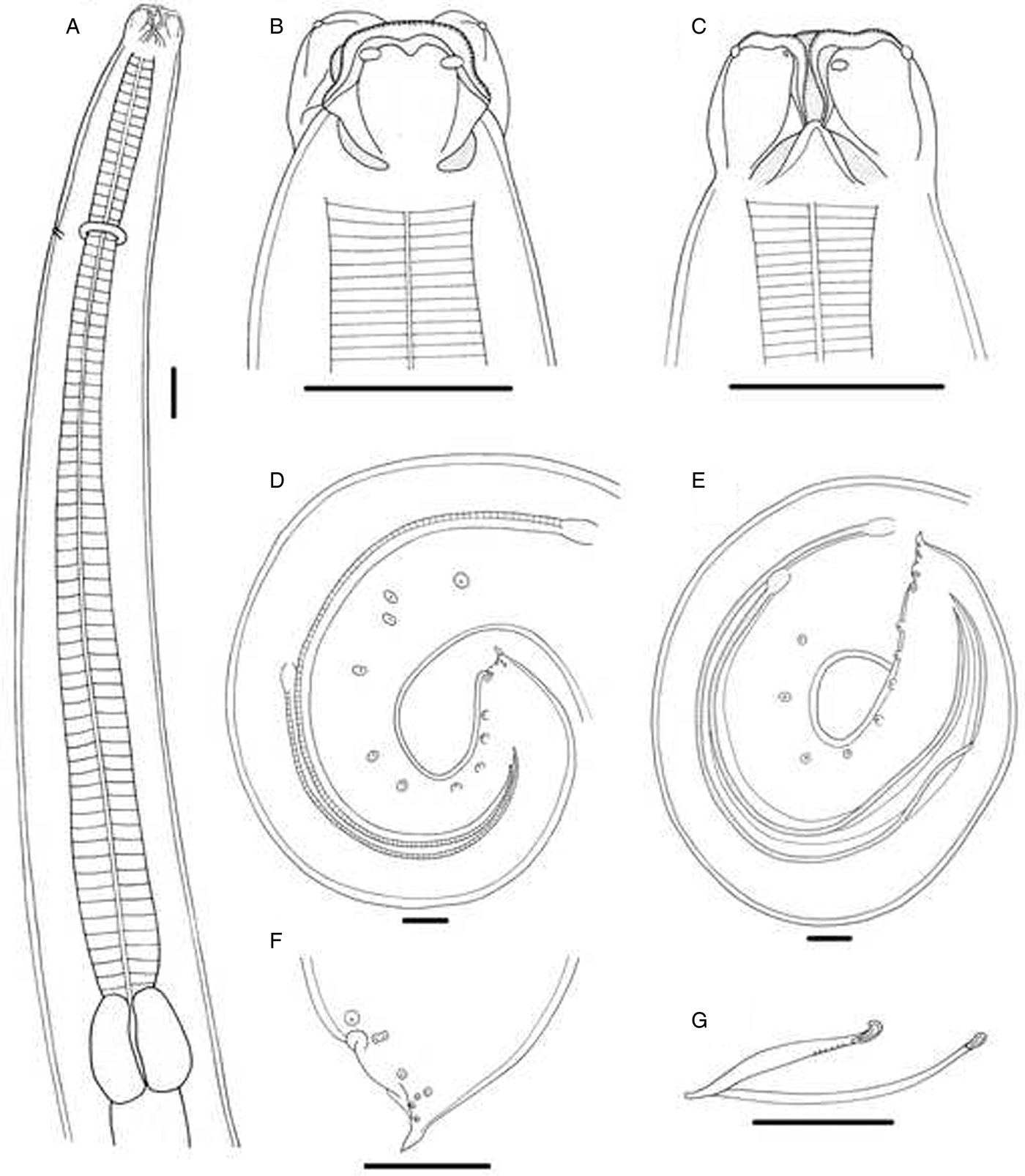
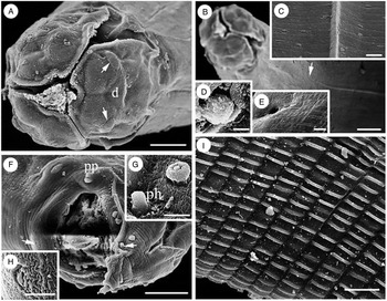
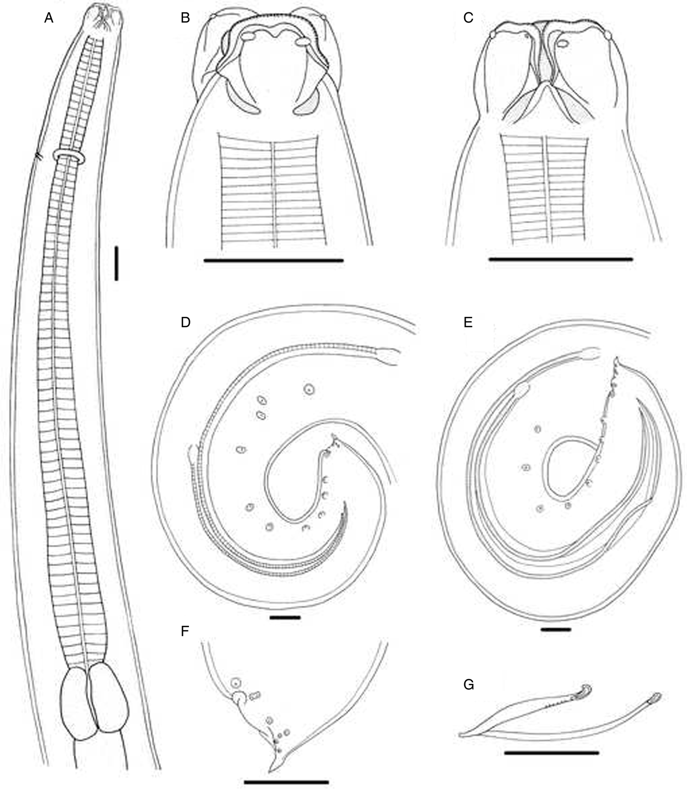
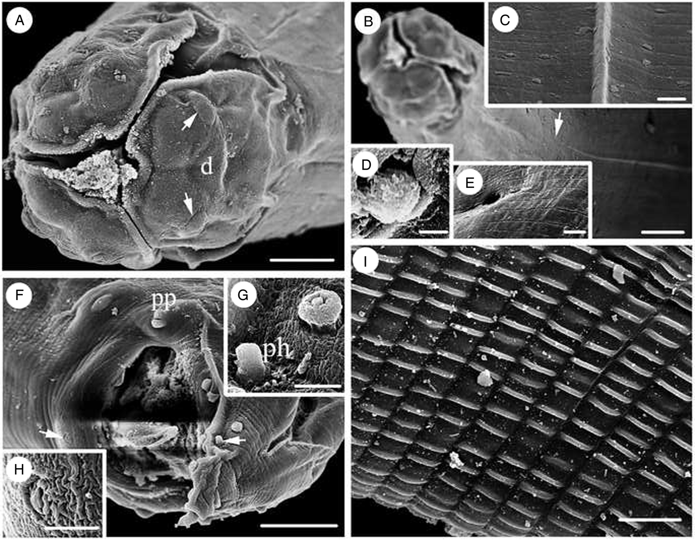
Male. Based on five mature specimens. Medium size, whitish nematodes. Body 20.1–24.2 (22.2) mm long, maximum width 588–637 (613). Cuticle with fine, transverse striations. Anterior end with three prominent lips, approximately equal in size, 80–104 (94) long, 92–101 (96) wide; lateral membranous flanges prominent (fig. 1A–C); labial pulp possesses two finger-like lobes extending into lateral membranous flanges (fig. 1B, C). Distal part of each lip with inconspicuous, obtusely triangular, medio-apical notch (fig. 2A). Small dentigerous ridges present along anterior margins of lips (fig. 1B, C). Dorsal lip with two lateral double papillae approximately in anterior third of its length (figs 1B, C, 2A); ventrolateral lips each with a single double papilla, small single papilla and amphid. Interlabia triangular, well developed, 29–35 (31) long (figs 1A–C, 2A). Lateral alae very narrow, extending from well posterior to base of ventrolateral lips to middle of body (fig. 2B, C). Oesophagus muscular, distinctly wider posteriorly (posterior part about twice width of anterior part), 1.69–2.01 (1.85) mm long, 128–157 (150) in maximum width, representing 8.31–8.41 (8.33)% of body length (fig. 1A). Nerve ring and excretory pore 431–554 (500) and 490–574 (528) from anterior extremity, respectively (figs 1A, 2E). Deirids small, rounded, approximately at same level as excretory pore (fig. 2D). Ventriculus nearly oval, clearly demarcated from muscular oesophagus and intestine, 181–226 (211) long by 196–216 (207) wide, slightly wider than posterior part of oesophagus (fig. 1A). Intestinal caecum and ventricular appendix absent. Posterior end of body distinctly curved ventrally. Precloacal ventral, longitudinal stockade-like ornamentation of cuticle well developed on posterior third of body (fig. 2I). Spicules slender, alate, distinctly unequal, ending in sharp point (fig. 1D, E); left spicule 1.40–2.71 (1.97) mm long, representing 7.0–11.2 (8.9)% of body length; right spicule 0.81–2.58 (2.04) mm long, representing 4.0–10.7 (9.2)% of body length. Gubernaculum present, V-shaped, 109–152 (128) long (fig. 1G). Caudal papillae 11–15 pairs in total, arranged as follows: 6–10 pairs of small precloacal papillae, 1 pair of double paracloacal papillae and 4 pairs of postcloacal papillae (3 subventral and 1 subdorsal) (figs 1D–F, 2F, G). Single small, median, precloacal papilla present on anterior margin of cloaca (fig. 2F, H). Tail conical, with sharply pointed tip, 98.0–121 (109) long (figs 1D–F, 2F). Lateral phasmids well developed, situated at about two-thirds of distance from cloaca (fig. 2F, G).
Female. Unknown.
Fig. 1.Mawsonascaris australis (Johnston & Mawson, 1943) collected from Rhinobatos schlegelii Müller & Henle (Rhinopristiformes: Rhinobatidae), male. (A) Anterior part of body, lateral view; (B) cephalic extremity, dorsal view; (C) cephalic extremity, lateral view; (D) posterior end of body (showing left spicule approximately twice length of right), lateral view; (E) posterior end of body (showing left spicule only about 1.2 times length of right), lateral view; (F) tail (phasmid arrowed), lateral view; (G) gubernaculum, lateral view. Scale bars: (A) 100 μm; (B–F) 120 μm; (G) 60 μm.
 Fig. 2.
Scanning electron micrographs of Mawsonascaris australis (Johnston & Mawson, 1943) collected from Rhinobatos schlegelii Müller & Henle (Rhinopristiformes: Rhinobatidae), male. (A) Cephalic extremity (double papillae arrowed), apical view; (B) anterior part of body (the starting position of lateral alae arrowed), lateral view; (C) magnified image of lateral ala and transverse cuticular striations; (D) magnified image of deirid; (E) magnified image of excretory pore; (F) tail (precloacal median papilla and phasmid indicated by white arrows; postcloacal papillae indicated by black arrows); ventrolateral view; (G) magnified image of phasmid and second postcloacal papilla; (H) magnified image of precloacal median papilla; (I) magnified image of longitudinal precloacal stockade-like ornamentation of cuticle. Abbreviations: d, dorsal lip; pp, paracloacal papillae; ph, phasmid. Scale bars: (A, F) 20 μm; (B) 30 μm; (C, D, E, G, H) 3 μm; (I) 10 μm.
Taxonomic summary
Type host and locality. Common stingaree Trygonoptera testacea Müller & Henle (Myliobatiformes: Urolophidae); off Sydney, Australia.
Host and locality of present specimens. Brown guitarfish Rhinobatos schlegelii Müller & Henle (Rhinopristiformes: Rhinobatidae); Taiwan Strait (off Xiamen (118°04′E, 24°26′N), Fujian Province, China).
Site of infection. Stomach.
Prevalence and intensity of infection. Single R. schlegelii infected with five nematodes.
Voucher specimens. Five males (HBNU–F20170010L).
Molecular characterization
Three ITS sequences of M. australis, 957 bp in length, were obtained, representing two different genotypes, which displayed 0.10% nucleotide divergence. No ITS sequences of Mawsonascaris species were available in GenBank. A comparison of the ITS sequences of M. australis with the only acanthocheilid species available in GenBank, Pseudanisakis rajae (Yamaguti, 1941) (JN392470), resulted in a 14.1% nucleotide divergence. Three cox1 sequences of M. australis, 384 bp in length, represented three different genotypes, which displayed a 1.04–2.08% nucleotide divergence. No cox1 sequences of Mawsonascaris species were available in GenBank. A comparison of the cox1 sequences of M. australis with the only acanthocheilid species available in GenBank, Pseudanisakis argentinensis Irigoitia, Braicovich, Farber & Timi, 2017 (KY765915–KY765923), exhibited a nucleotide divergence of more than 30.0%. The ITS (MF918573–MF918575) and cox1 sequences (MF918570–MF918572) of M. australis were deposited in the GenBank database (http://www.ncbi.nlm.nih.gov).
Discussion
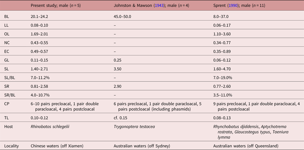
Johnston & Mawson (Reference Johnston and Mawson1943) described Paranisakis australis Johnston & Mawson, 1943 from the common stingaree Trygonoptera testacea Müller & Henle (Myliobatiformes: Urolophidae) in Australian waters (Johnston & Mawson, Reference Johnston and Mawson1943). Sprent (Reference Sprent1990) re-examined the type specimens of P. australis and considered that this species should be placed in a new genus, Mawsonascaris Sprent, 1990. He also redescribed the species based on the type specimens and some newly collected material from four additional fish hosts, including Aptychotrema rostrata (Shaw) (Rhinopristiformes: Rhinobatidae), Glaucostegus typus (Anonymous [Bennett]) (Rhinopristiformes: Rhinobatidae), Rhynchobatus djiddensis (Forsskål) (Rhinopristiformes: Rhinidae) and Taeniura lymma (Forsskål) (Myliobatiformes: Dasyatidae). The morphology and measurements of our material agree well with the description of M. australis by both Johnston & Mawson (Reference Johnston and Mawson1943) and Sprent (Reference Sprent1990), including body size, the morphology and size of the lips, the position of the nerve ring and excretory pore, the lengths of the oesophagus, gubernaculum and spicules, the number and arrangement of the caudal papillae, and the morphology and length of the tail (see table 1 for details). Moreover, the present nematodes were collected from the brown guitarfish R. schlegelii, which also occurs in Australian waters (Froese & Pauly, Reference Froese and Pauly2017), and some of the fish hosts of M. australis reported previously by Sprent (Reference Sprent1990) (i.e. A. rostrata and G. typus) also belong to the same family as R. schlegelii (the Rhinobatidae). Consequently, we considered our specimens to be conspecific with M. australis. Sprent (Reference Sprent1990) described the presence of one pair of single and one pair of double paracloacal papillae in his material. In fact, the pair of single paracloacal papillae (very close to the cloaca) should be regarded as precloacal papillae according to Fagerholm (Reference Fagerholm1991). Sprent (Reference Sprent1990) reported narrow lateral alae, starting from the anterior end and reaching to the middle of the body. However, in our material, under the SEM, we observed the lateral alae beginning well posterior to the base of ventrolateral lips. In addition, neither Johnston & Mawson (Reference Johnston and Mawson1943) nor Sprent (Reference Sprent1990) mentioned a single small, precloacal, median papilla, which our SEM observations showed to be present on the anterior margin of the cloaca. Furthermore, the present SEM observations show, for the first time, the detailed morphology of the lateral alae, deirids, excretory pore, precloacal median papilla and phasmids. Mawsonascaris australis is recorded in Chinese waters for the first time, and the brown guitarfish R. schlegelii represents a new host.
Morphometric comparisons of Mawsonascaris australis (Johnston & Mawson, 1943) between our present material and the previous studies (measurements in millimetres).
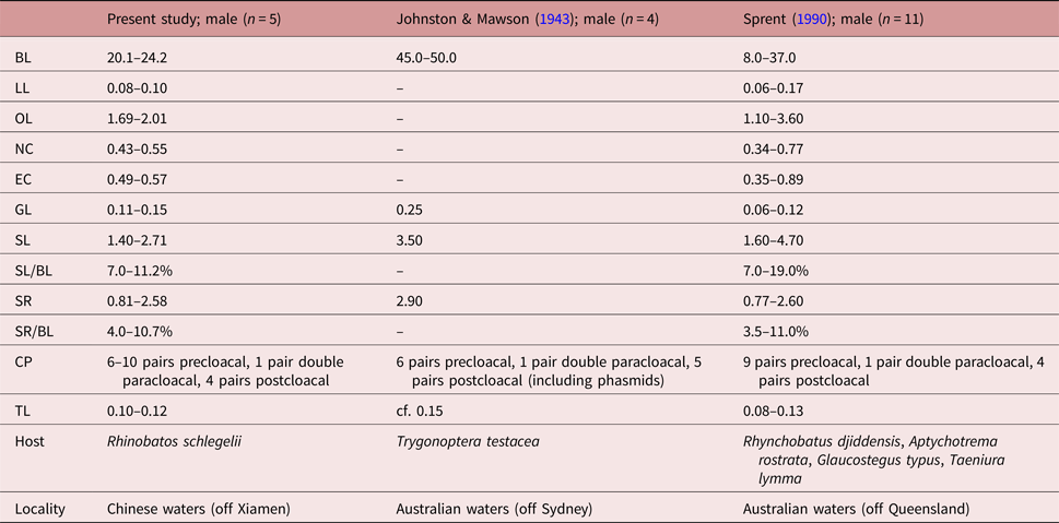
BL, Body length; LL, length of lip; OL, length of oesophagus; SL, length of left spicule; SR, length of right spicule; GL, length of gubernaculum; CP, numbers of caudal papillae; TL, length of tail; NC, distance of nerve ring to cephalic end; EC, distance of excretory pore to cephalic end.
According to Sprent (Reference Sprent1990), the left spicule of M. australis is about twice the length of the right. However, in the present study, in one individual, we found the left spicule to be only approximately 1.2 times longer than the right. The relative length of the left and right spicules is a diagnostic morphological character for distinguishing and identifying species of this genus (Sprent, Reference Sprent1990; Ali et al., Reference Ali, Zhang, Al-Salim and Li2012; Li et al., Reference Li, Xu and Zhang2012). The molecular analysis showed a low level of nucleotide variation in the ITS (only 0.10% nucleotide divergence) and cox1 sequences (only 1.04–2.08% nucleotide divergence) between the two different morphotypes, which is far lower than the interspecific nucleotide variations between M. australis and P. rajae/P. argentinensis recorded in GenBank (14.1% nucleotide differences in the ITS region; >30.0% nucleotide differences in the cox1 region). Consequently, the molecular evidence strongly supports the above-mentioned morphometric differences between the two different morphotypes as representing intraspecific variability, which is possibly a result of aberrant development or as yet unidentified factors. Mawsonascaris australis is the first species of this genus to have its ITS and cox1 genes sequenced for the purpose of species identification.
There are two previously reported species of Mawsonascaris from the Taiwan Strait, including M. myliobatum from the Japanese eagle ray Myliobatis tobijei Bleeker (Rajiformes: Myliobatidae) and M. zhoui from the granulated guitarfish Glaucostegus granulatus (Cuvier) (Rajiformes: Rhinobatidae) (Yin & Zhang, Reference Yin and Zhang1983; Li et al., Reference Li, Xu and Zhang2012). Mawsonascaris myliobatum differs from M. australis by having many more precloacal papillae (17–19 pairs in the former vs. 6–10 pairs in the latter). Mawsonascaris zhoui is very similar to M. australis in both its morphology and measurements, but it can be distinguished from M. australis by the absence of cervical alae, the presence of a particular volcaniform, precloacal, median papilla and slightly more precloacal papillae (10–11 pairs vs. 6–10 pairs in M. australis).
Acknowledgements
We are grateful to Dr David I. Gibson (Department of Life Sciences, Natural History Museum, London, UK) for improving the English of the manuscript.
Financial support
This study was supported by the National Natural Science Foundation of China (NSFC-31572231), Natural Science Foundation of Hebei Province (NSFH-C2016205088) and the Youth Top Talent Support Program of Hebei Province for L.L.
Conflict of interest
None.