Introduction
Members of the FMRFamide-like peptide (FLP) family of bioactive peptides have fundamental regulatory roles in nematodes (Li, Reference Li2005). They are especially important for neuromuscular control (Maule et al., Reference Maule, Mousley, Marks, Day, Thompson, Geary and Halton2002), behaviour (Kimber & Fleming, Reference Kimber and Fleming2005; Liu et al., Reference Liu, Kim, Li and Barr2007), feeding (Rogers et al., Reference Rogers, Reale, Kim, Chatwin, Li, Evans and deBono2003; Papaioannou et al., Reference Papaioannou, Marsden, Franks, Walker and Holden-Dye2005) and reproduction (Moffett et al., Reference Moffett, Beckett, Mousley, Geary, Marks, Halton, Thompson and Maule2003; Liu et al., Reference Liu, Kim, Li and Barr2007), and their genetic and biochemical complexities are well documented (Maule et al., Reference Maule, Mousley, Marks, Day, Thompson, Geary and Halton2002; Li, Reference Li2005; McVeigh et al., Reference McVeigh, Leech, Mair, Marks, Geary and Maule2005; Masler, Reference Masler and Kastin2006). A prominent feature of the FLP peptide family is a high level of sequence diversity. All nematodes surveyed have multiple flp genes (Li, Reference Li2005; McVeigh et al., Reference McVeigh, Leech, Mair, Marks, Geary and Maule2005) and a majority of these genes encode multiple FLP sequences. Such diversity may provide protection of critical physiological events through redundancy of bioactive peptide signals, or regulation of these events through maintenance of precise mixtures of such peptides (Masler, Reference Masler and Kastin2006). A key element in this regulation is the action of proteases that can metabolize bioactive peptides, attenuate signals and control cellular processes (Lopez-Otin & Overall, Reference Lopez-Otin and Overall2002; Husson et al., Reference Husson, Mertens, Janssen, Lindemans and Schoofs2007). The purpose of the present work was to explore the response of a variety of nematode FLPs to general proteolysis and determine how this response might vary among different bioactive sequences. The source of the proteases was the free-living nematode Panagrellus redivivus, and presented here are the first comparisons of FMRFamide-like peptide catabolism in this nematode.
Materials and methods
Nematode rearing and extraction
Panagrellus redivivus was reared in sterile liquid culture at 22°C (Chitwood et al., Reference Chitwood, Lusby, Thompson, Kochansky and Howarth1995). Worms were harvested after 1 week in culture as a mixed-age population and washed with autoclaved distilled and de-ionized (D/D) water to remove culture medium. The washed worms were collected in conical tubes on ice and allowed to settle as ~1 ml aliquots. Water was removed and the worms were stored at − 20°C. For extraction, worms were thawed in ice-cold D/D water (12 volumes of water/worm volume) and partially disrupted with a Polytron homogenizer (Brinkman Instruments, Westbury, New York, USA). After centrifugation (48,000 × g, 30 min, 10°C), the supernatant (S1) was collected, and the pellet processed further using a Bead Beater (BioSpec Products, Bartlesville, Oklahoma, USA). The pellet was suspended in 10 volumes of D/D water and processed with the Bead Beater using 0.5 mm zirconia/silica beads until disruption was complete. This was assessed by examining aliquots of the disruption solution at 40 × for the presence of intact or partially disrupted worms. The preparation was centrifuged as above and the supernatant (S2) was combined with S1. Total protein was determined in the pooled supernatant using the microBCA assay (Pierce Chemical Co., Rockford, Illinois, USA); aliquots were dried by vacuum centrifugation, and stored at − 20°C.
Neuropeptides and protease inhibitors
FMRFamide (FMRFa) was from Peptide Institute, Osaka, Japan or Sigma Chemical (St. Louis, Missouri, USA). FLRFa, dFMRFa, FdMRFa, FMdRFa, and FMRdFa were from Sigma. The FLP sequences examined are identified in the text according to the Caenorhabditis elegans flp gene numbering convention (Li, Reference Li2005) and, where more than one unique sequence is encoded by a flp gene, a letter is used to indicate the order in which the specific FLP sequence occurs in the transcript (WormBase, available at http://www.wormbase.org). KPNFLRFa (FLP-1-H), APKPKFIRFa (FLP-5-A), KNEFIRFa (FLP-8), RNKFEFIRFa (FLP-12), and KHEYLRFa (FLP-14) were gifts from D.P. Thompson, Pfizer Animal Health, Kalamazoo, Michigan, USA and KPSFVRFa (FLP-9) was a gift from R.E. Isaac, University of Leeds, Leeds, UK. The aminopeptidase inhibitor amastatin was from Sigma, and the serine protease inhibitor AEBSF was from Calbiochem (San Diego, California, USA).
Assay system
All components of the digestion reactions were prepared in assay buffer (100 mm TRIS, pH 7.86) and combined to a total volume of 5 μl/reaction. Reactions contained 0.4 μg μl− 1 extract protein and 200 pmol μl− 1 peptide substrate. Inhibitors were used at 2 mm each. Control reactions (T0) contained extract and substrate but were stopped immediately by the addition of 50 μl acetonitrile (CH3CN)/0.1% trifluoroacetic acid (TFA). Digestion reactions (T1) were started by the addition of peptide substrate, incubated for 1 h at 27°C, and then stopped as above. Control reactions (T1), containing extract only, were used to account for non-substrate derived peaks. In inhibitor tests, extract and inhibitor were incubated for 30 min prior to the addition of peptide substrate, then incubated for an additional hour and stopped as above. Parallel controls were incubated without inhibitor. All stopped samples were dried under vacuum, residues were dissolved in 55 μl 0.1%TFA, then 50 μl were injected for high performance liquid chromatography (HPLC) fractionation. The chromatographic system comprised a reversed-phase C8 column (Hewlett–Packard 5 mm id × 150 mm, 5 μm particle size; Hewlett–Packard, Avondale, Pennsylvania, USA), a flow rate of 0.5 ml min− 1, and a linear gradient of 5–45% CH3CN in 0.1% TFA over 20 min (2% CH3CN/min). Absorbance was monitored at 210 nm with retention times and peak dimensions determined by on-board integration software (ChemStation Rev. B.03.01, Hewlett–Packard).
Data analysis
Overall digestion of substrate peptides is expressed as the mean percentage of substrate remaining after 1 h (TS1) and calculated by comparing the height (mAU, 210 nm) of the integrated substrate peaks detected in 1 h (TS1) and time zero (TS0) reactions using the formula: [(mAU 210 nm TS1/mAU 210 nm TS0)] × 100. Product peak (PP) levels were compared using the formula: [(mAU 210 nm TPPx/mAU 210 nm TPPy)] × 100, where PPx and PPy represent heights of the same product peak resulting from different (x versus y) treatments. A minimum of three independent reactions and HPLC fractionations were done to provide mean percentage peak heights for all peaks examined, and all mean comparisons were done using ANOVA (KaleidaGraph, Synergy Software, Reading, Pennsylvania, USA).
Results
Neuropeptide digestion
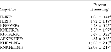
All neuropeptides examined were subject to digestion by P. redivivus extract (table 1), with substrate peak heights of six of the eight tested unmodified peptides reduced by more than 90% after 1 h. Two nematode peptides, KHEYLRFa (FLP-14) and RNKFEFIRFa (FLP-12) were significantly less susceptible to digestion than the others. RNKFEFIRFa retained 29% of the control level after 1 h incubation, and was significantly less susceptible (P < 0.0001) to digestion than all other tested peptides. KHEYLRFa retained over 16% of the control level after 1 h, and was significantly less susceptible than FMRFa, FLRFa, KPSFVRFa (FLP-9) and KNEFIRFa (FLP-8) (P < 0.0001), APKPKFIRFa (FLP-5-A) (P = 0.0001), and KPNFLRFa (FLP-1-H) (P = 0.0004). There was no apparent correlation between either sequence length or sequence identity and the extent of digestion (loss of substrate).
Comparison of the susceptibility of selected invertebrate neuropeptides to proteolytic digestion by the soluble fraction of homogenates of Panagrellus redivivus.
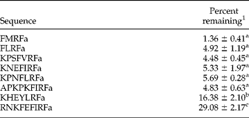
1 The mean percentage of peptide remaining was calculated as the ratio of peak height (mAU 210 nm) after 1 h digestion at 27°C relative to peak height of non-incubated controls. Each mean is the result of 3–5 separate digestions. Means were compared by ANOVA and those followed by different letters are significantly different (P < 0.0001; except KHEYLRFa versus APKPKFIRFa, P = 0.0001; and KHEYLRFa versus KPNFLRFa, P = 0.0004).
Inhibitors
RNKFEFIRFa, the least susceptible sequence, APKPKFIRFa, a nonapeptide significantly more susceptible to digestion than RNKFEFIRFa, and FMRFa, one of the two shortest peptides, were used as substrates to examine the effects of protease inhibitors on substrate loss (table 2) and digest patterns (figs 1–3). Amastatin significantly (P < 0.0005) reduced the loss of FMRFa from over 97% to less than 61%, and marginally protected RNKFEFIRFa (P < 0.03), decreasing loss by 18% over controls (table 2). In contrast, amastatin had no protective effect on APKPKFIRFa (table 2). AEBSF had no effect on FMRFa digestion, but significantly reduced the digestion of both APKPKFIRFa (P < 0.0001) and RNKFEFIRFa (P < 0.03) compared with controls (table 2). The combination of both amastatin and AEBSF increased the protection of FMRFa above that provided by either inhibitor alone (P < 0.0005), but afforded no additional protection for either APKPKFIRFa or RNKFEFIRFa (table 2).
Effect of inhibitors on digestion of selected invertebrate neuropeptides by the soluble fraction of homogenates of Panagrellus redivivus.

1 1 nmol peptide was incubated for 1 h at 27°C with 2 μg extract proteins with or without inhibitors (2 mm each). Identical reactions prepared in parallel but immediately stopped were used as controls.
2 The mean percentage of peptide remaining is the ratio of peak height (mAU 210 nm) after 1 h digestion at 27°C relative to peak height of non-incubated controls. Means for each peptide followed by different letters are significantly different (FMRFa, P < 0.0005; APKPKFIRFa, P < 0.0001 except AEBSF treatment versus both inhibitors, P = 0.0008; RNKFEFIRFa, P < 0.03).
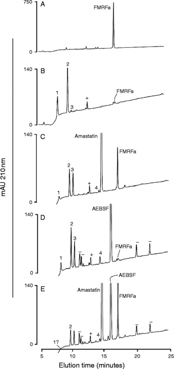
RP-HPLC fractionation of FMRFa and FMRFa metabolites following incubation with Panagrellus redivivus homogenate soluble fraction. Each incubation contained 1 nmol peptide and 2 μg soluble P. redivivus protein. (A) Peptide plus P. redivivus protein non-incubated control; (B) peptide plus P. redivivus protein incubated 1 h at 27° C; (C) as in (B), plus 2 mm amastatin; (D) as in (B) plus 2 mm AEBSF; (E) as in (B) plus 2 mm each inhibitor. Product peaks are numbered. Peaks labelled as ‘+’ are from P. redivivus protein. Peaks labelled as ‘ − ’ are artefacts from the inhibitor. UV detection at 210 nm. Note different scales for absorbance (mAU, y-axes).

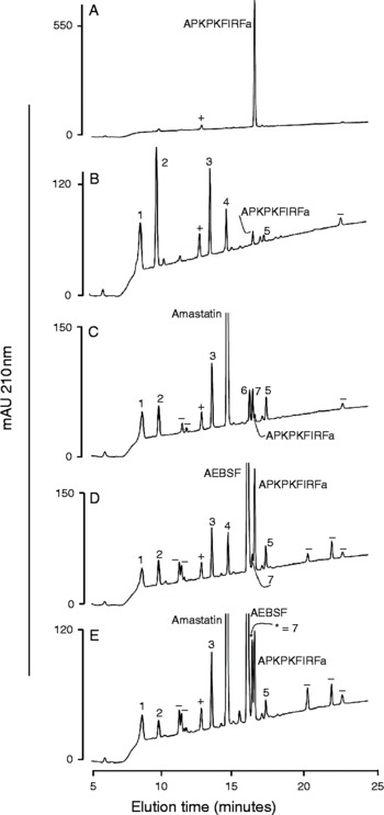
RP-HPLC fractionation of APKPKFIRFa (FLP-5-A) and APKPKFIRFa metabolites following incubation with Panagrellus redivivus homogenate soluble fraction. Each incubation contained 1 nmol peptide and 2 μg soluble P. redivivus protein. (A) Peptide plus P. redivivus protein non-incubated control; (B) peptide plus P. redivivus protein incubated 1 h at 27° C; (C) as in (B), plus 2 mm amastatin; (D) as in (B) plus 2 mm AEBSF; (E) as in (B) plus 2 mm each inhibitor. Product peaks are numbered. Peaks labelled as ‘+’ are from extract protein. Peaks labelled as ‘ − ’ are artefacts from the inhibitor. UV detection at 210 nm. Note different scales for absorbance (mAU, y-axes).

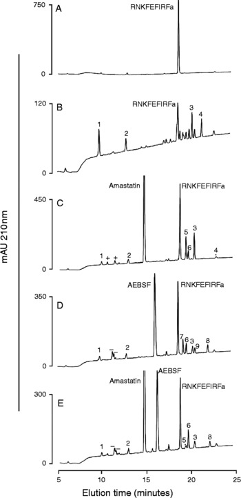
RP-HPLC fractionation of RNKFEFIRFa (FLP-12) and RNKFEFIRFa metabolites following incubation with P. redivivus homogenate soluble fraction. Each incubation contained 1 nmol peptide and 2 μg soluble P. redivivus protein. (A) Peptide plus P. redivivus protein non-incubated control; (B) peptide plus P. redivivus protein incubated 1 h at 27° C; (C) as in (B), plus 2 mm amastatin; (D) as in (B) plus 2 mm AEBSF; (E) as in (B) plus 2 mm each inhibitor. Product peaks are numbered. Peaks labelled as ‘+’ are from extract protein. Peaks labelled as ‘ − ’ are artefacts from the inhibitor. UV detection at 210 nm. Note different scales for absorbance (mAU, y-axes).

Digest patterns
FMRFa eluted at 17.2 min (fig. 1A) and its digestion resulted in at least three product peaks (fig. 1B) which eluted at 7.9, 9.6 and 10.2 min (peaks 1, 2 and 3, respectively). In the presence of amastatin and/or AEBSF (fig. 1C–E), an additional product peak (peak 4, 14.3 min) was detected. Peak 1 was reduced in size with all inhibitor applications, whereas peak 2 was significantly reduced only in the presence of amastatin (fig. 1B versus 1C, P = 0.07; fig. 1B versus 1E, P = 0.014). Peak 3 was increased by either amastatin or AEBSF (P < 0.04), even though amastatin was required for the overall reduction in FMRFa digestion (fig. 1B–E; table 2). All product peaks were reduced in size with the amastatin–AEBSF combination as compared with either amastatin or AEBSF alone (fig. 1E versus fig. 1C, D).
APKPKFIRFa eluted at 16.8 min (fig. 2A) and had a digest pattern (fig. 2B) slightly more complex than FMRFa, with product peaks 1 through 5 detected at 8.7, 9.9, 13.7, 14.9 and 17.6 min, respectively. Peaks 1 and 3 were little changed regardless of treatment, whereas peak 2 was significantly reduced (P < 0.0001) with all inhibitor combinations (fig. 2B–E). Both amastatin and AEBSF treatments increased peak 5 (P < 0.01), but the effects were not additive (fig. 2B–E). Peaks not present in control incubations appeared in the presence of amastatin (fig. 2C; peaks 6, 7), and peak 7 was also present with AEBSF (fig. 2D). Peak 6, however, may have been obscured by the AEBSF peak (fig. 2D). Peak 5 was the only product peak less polar than the substrate peak (fig. 2B–E).
RNKFEFIRFa, eluting at 18.9 min (fig. 3A), presented a more complex initial digestion pattern than either FMRFa or APKPKFIRFa (fig. 3B versus figs 1B and 2B), with four quantitatively major peaks observed at 9.9, 12.9, 20.6 and 21.7 min (fig. 3B; peaks 1–4, respectively), and a series of small product peaks eluting between the substrate peak and peak 3. None of these small peaks was present in any of the inhibitor treatments (fig. 3C–E). A notable feature of the RNKFEFIRFa digests is that most of the product peaks were less polar than the substrate peak (fig. 3B). This is in sharp contrast to results with FMRFa and APKPKFIRFa, where all of the product peaks, except for APKPKFIRFa product peak 5, were more polar than the substrate peaks (figs 1B and 2B). In response to all inhibitor treatments, RNKFEFIRFa peak 1 was significantly reduced (P < 0.0001), peak 2 was marginally reduced, and peak 4 was essentially eliminated. Peak 3 was significantly (P < 0.0001) increased with amastatin (fig. 3C) but was reduced in size in the presence of AEBSF (fig. 3D; P < 0.08). Finally, all additional peaks appearing in the presence of inhibitors (fig. 3C–E; peaks 5–9; 19.5, 19.6, 19.9, 20.9 and 22.4 min, respectively) were less polar than that for the RNKFEFIRFa substrate sequence.
Effect of FMRFa modification
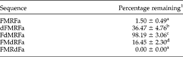
The substitution of d-amino acid isomers in FMRFa had a significant and position-dependent effect on the stability of the peptide in P. redivivus extract (table 3). Substitution of d-Phe at position 1 resulted in a highly significant (P < 0.0001) 24-fold increase in stability over the native peptide. The substitution of d-Met at position 2 resulted in nearly complete protection against proteolytic digestion, and substitution of d-Arg at position 3 resulted in a significant (P = 0.018) 11-fold increase in stability. However, substitution of d-Phe at position 4 had no protective effect. In fact, FMRdFa was the only peptide of the 12 peptides tested that was completely digested.
Position effect of d-amino acid analogues on the susceptibility of FMRFa to digestion by Panagrellus redivivus extract.
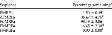
1 The mean percentage of peptide remaining is the ratio of peak height (210 nm) after 1 h digestion at 27° C relative to peak height (210 nm) in non-incubated controls. Each mean is the result of 3–5 separate digestions. Means followed by different letters are significantly different with P < 0.0001 except FMdRFa versus dFMRFa (P = 0.0033), FMdRFa versus FMRdFa (P = 0.0145), and FMdRFa versus FMRFa (P = 0.0176).
Discussion
The soluble fraction from P. redivivus aqueous extracts contains proteases capable of digesting a number of FMRFamide and FMRFamide-like sequences, including those that are associated with physiological events in nematodes (Li, Reference Li2005). As with any crude homogenate, substrates were exposed to combinations of enzymes that may not normally act simultaneously in vivo. Given this proteolytically aggressive environment, it is striking that while peptide substrates of varying lengths and sequence identities were extensively digested, only one peptide substrate (FMRdFa) was completely digested. In addition, one-third of the six FLP sequences exhibited significantly lower levels of digestion. Extrapolating this 33% to the large number of unique FLP sequences confirmed or predicted in nematodes (Li, Reference Li2005; McVeigh et al., Reference McVeigh, Leech, Mair, Marks, Geary and Maule2005) underscores the biochemical complexity resulting from FLP sequence diversity and illustrates that protease–FLP substrate interactions in P. redivivus can provide a regulatory mechanism for precise physiological control. In fact, diversity and complexity are hallmarks of the patterns of protease–substrate interactions that are emerging as the degradomes in which these interactions occur are elucidated (Lopez-Otin & Overall, Reference Lopez-Otin and Overall2002; Craig et al., Reference Craig, Isaac and Brooks2007).
Variation in proteolysis relative to peptide identity was even more apparent in examining digestion product patterns and evaluating the patterns relative to specific protease inhibitors. The significant protection of FMRFa afforded by amastatin clearly indicated aminopeptidase involvement in FMRFa metabolism. However, since the protective effect of amastatin was only partial, other classes of proteases, or aminopeptidases not sensitive to amastatin, must also be involved in initial cleavage. One of these may be deamidase, which is involved in invertebrate neuropeptide metabolism (Sajid et al., Reference Sajid, Keating, Holden-Dye, Harrow and Isaac1996; Morishita et al., Reference Morishita, Matsushima, Furukawa and Minakata2003). Deamidase is a serine protease present in both membrane and supernatant fractions. The membrane form (Sajid et al., Reference Sajid, Keating, Holden-Dye, Harrow and Isaac1996; Morishita et al., Reference Morishita, Matsushima, Furukawa and Minakata2003), but not the supernatant form (Morishita et al., Reference Morishita, Matsushima, Furukawa and Minakata2003), is sensitive to the serine protease inhibitor PMSF. If this behaviour applies to AEBSF treatment, then a deamidase in P. redivivus may partially account for the metabolism of peptide substrates in the presence of this serine protease inhibitor.
Amastatin and AEBSF combined to prevent a large majority of FMRFa degradation coupled with a reduction in product peak size. This indicates that aminopeptidase and serine protease cleavage account for most of FMRFa metabolism. In this context, the effects of d-amino acid substitutions are informative. The presence of a d-amino acid at the second position renders aminopeptidases incapable of removing the N-terminal residue (Morishita et al., Reference Morishita, Matsushima, Furukawa and Minakata2003). The nearly complete resistance of FdMRFa to proteolysis in P. redivivus supernatant is unexpected since endopeptidases including serine endopeptidases, and deamidase, should continue to function. However, the effect of d-amino acid substitution on peptide half-life is positional (Hong et al., Reference Hong, Oh and Lee1999), and appears to be the case with the FMRFa isomers. It may be possible that the structural change in FdMRFa requires an extended exposure to P. redivivus proteases to effect metabolism. A comparison of half-lives of modified FMRFa sequences among other nematode species may be quite revealing.
In the context of the present survey of FMRFa and FLP digests, the behaviour of RNKFEFIRFa (FLP-12) is notable. It was the most resistant to proteolysis of all sequences and was equally protected by amastatin and AEBSF, suggesting that aminopeptidase and serine proteases contribute equally to its degradation. This contribution, however, represents only a minority portion since the difference in RNKFEFIRFa degradation between the control treatment and the separate amastatin plus AEBSF treatments was only 32%. Also, differences between the product peak arrays in amastatin-treated and AEBSF-treated samples were real but rather minor, and the effect of combining the inhibitors was non-additive. These observations indicate that other classes of proteases must have important roles in the metabolism of RNKFEFIRFa, and the metabolism of this sequence may be more complex than the others tested. In this context it is interesting to note that the large majority of flp genes identified in Caenorhabditis elegans and other nematodes (Li, Reference Li2005; McVeigh et al., Reference McVeigh, Leech, Mair, Marks, Geary and Maule2005) encode multiple FLPs. The small minority of those genes that encode a single copy of a single sequence includes flp-12, and the FLP-12 sequence is highly conserved. All nematode species have the xNKFEFIRFa core with only single amino acid variants (K, N, R) at the N-terminus. Whether low copy number and high conservation are related to the fate of RNKFEFIRFa within the nematode degradome is an intriguing question, and should be addressed through a comprehensive analysis of the metabolism of additional singly encoded FLPs. This may help to determine if the catabolic behaviour of RNKFEFIRFa is typical of such FLPs.
The contrast between the complex pattern of RNKFEFIRFa digestion and the relatively simple pattern for APKPKFIRFa is striking, especially since digestion of APKPKFIRFa was more complete. One explanation is that the products of the initial cleavage of APKPKFIRFa were higher affinity protease substrates than those from RNKFEFIRFa and were degraded to component amino acids before the reaction was terminated. Also, the presence of two internal lysines in APKPKFIRFa as opposed to a single lysine residue in RNKFEFIRFa may contribute to the more rapid digestion of APKPKFIRFa. The failure of amastatin to reduce the level of digestion of APKPKFIRFa clearly demonstrates that initial cleavages of this FLP do not involve aminopeptidase, and may be attributed to the presence of proline (Mentlein, Reference Mentlein1988). In fact, the stability of FLPs is enhanced by the presence of proline at the second position, as was demonstrated with KPNFIRFa (Kubiak et al., Reference Kubiak, Maule, Marks, Martin and Wiest1996). Digestion of APKPKFIRFa likely involves the action of a soluble aminopeptidase P, similar to that described in C. elegans (Laurent et al., Reference Laurent, Brooks, Coates and Isaac2001), which specifically removes lysine, arginine or alanine from peptides containing Pro2. APKPKFIRFa digestion may also require the action of prolyl endopeptidases (Mentlein, Reference Mentlein1988). Serine proteases account for approximately 17% of proteases predicted from the C. elegans genome (Coates et al., Reference Coates, Siviter and Isaac2000; MEROPS database, Rawlings et al., Reference Rawlings, O'Brien and Barrett2002). Thus, the limited effect of AEBSF on the metabolism of APKPKFIRFa, as well as RNKFEFIRFa, might be expected.
A comprehensive set of proteases, protein and peptide substrates, and endogenous inhibitors, the degradome (Lopez-Otin & Overall, Reference Lopez-Otin and Overall2002), exists in nematodes as in other animals. Regulation of behaviour, development and metabolism is dependent upon the interactions of members of this set. These interactions vary by tissue location and developmental time, making this system both dynamic and complex, and it is now being explored in nematodes (Craig et al., Reference Craig, Isaac and Brooks2007). A core component of the nematode system involves the association of proteases with neuropeptides (Coates et al., Reference Coates, Siviter and Isaac2000; Husson et al., Reference Husson, Mertens, Janssen, Lindemans and Schoofs2007). Their ubiquity and physiological importance to nematodes make FMRFamide-like peptides an important group to examine. The variety of responses of these peptides to digestion by nematode proteases, and the varied effects of inhibitors of only two classes of proteases, are striking. An important caveat with the current study is that neuropeptides were exposed to proteases in combinations that they might not encounter in vivo. Exposure of FLPs to selected subcellular fractions (e.g. neural tissue preparations) can be used to generate more physiologically authentic metabolic patterns. Nevertheless, this initial survey clearly indicates that the continued exploration of FLP metabolism, utilizing additional FLP sequences, inhibitors of additional protease classes and identification of metabolic products, will provide important insight into neuropeptide control of nematode physiology.