INTRODUCTION
Worldwide Cryptosporidium is associated with diarrhoea in young calves (de Graaf et al. Reference de Graaf, Vanopdenbosch, Ortega-Mora, Abbassi and Peeters1999). Parasite prevalence varies from 1% (Kvac et al. Reference Kvac, Kouba and Vitovec2006) to 59% (Olson et al. Reference Olson, Guselle, O'Handley, Swift, McAllister, Jelinski and Morck1997). Next to differences in breed (Geurden et al. Reference Geurden, Goma, Siwila, Phiri, Mwanza, Gabriel, Claerebout and Vercruysse2006b; Kvac et al. Reference Kvac, Kouba and Vitovec2006), the variation in prevalence is partially due to the choice of diagnostic technique, and several diagnostic assays have therefore been evaluated for use in epidemiological studies (Geurden et al. Reference Geurden, Berkvens, Geldhof, Vercruysse and Claerebout2006a).
Since not all Cryptosporidium species in cattle have zoonotic potential, epidemiological research should include molecular identification to elucidate the relevance of cattle in the epidemiology of human cryptosporidiosis (Traub et al. Reference Traub, Monis and Robertson2005). Apart from the ubiquitous and zoonotic C. parvum, the ruminant-specific species C. bovis and C. andersoni, and the Cryptosporidium cervine genotype have been frequently reported in cattle (Caccio et al. Reference Caccio, Thompson, McLauchlin and Smith2005). Occasionally C. suis (Fayer et al. Reference Fayer, Santin, Trout and Greiner2006), C. felis (Bornay-Llinares et al. Reference Bornay-Llinares, da Silva, Moura, Myjak, Pietkiewicz, Kruminis-Lozowska, Graczyk and Pieniazek1999), C. canis (Fayer et al. Reference Fayer, Trout, Xiao, Morgan, Lai and Dubey2001) and C. hominis (Smith et al. Reference Smith, Nichols, Mallon, Macleod, Tait, Reilly, Browning, Gray, Reid and Wastling2005) are reported in cattle. Human cryptosporidiosis can be due to infections with either the human-specific C. hominis or the zoonotic C. parvum, and in a more limited number of cases with C. meleagridis, which is also zoonotic (Pedraza-Diaz et al. Reference Pedraza-Diaz, Amar, McLauchlin, Nichols, Cotton, Godwin, Iversen, Milne, Mulla, Nye, Panigrahl, Venn, Wiggins, Williams and Youngs2001). In Europe C. parvum is the main cause of human cryptosporidiosis, while worldwide C. hominis is most prevalent (Caccio et al. Reference Caccio, Thompson, McLauchlin and Smith2005). At least part of the human C. parvum infections can be ascribed to contact with livestock (Hunter and Thompson, Reference Hunter and Thompson2005), although the identification of C. parvum in stool samples does not conclusively implicate farm animals as the source of infection. Subtype analysis at the 60 kDa glycoprotein locus (Peng et al. Reference Peng, Matos, Gatei, Das, Stantic-Pavlinic, Bern, Sulaiman, Glaberman, Lal and Xiao2001; Alves et al. Reference Alves, Xiao, Antunes and Matos2006; Trotz-Williams et al. Reference Trotz-Williams, Martin, Gatei, Cama, Peregrine, Martin, Nydam, Jamieson and Xiao2006), revealed both human specific and zoonotic subtypes within C. parvum. In most waterborne outbreaks, human sources of infection were identified (Peng et al. Reference Peng, Xiao, Freeman, Arrowood, Escalante, Weltman, Ong, Mac Kenzie, Lal and Beard1997). Since the majority of human cryptosporidial infections are, however, not outbreak related (Feltus et al. Reference Feltus, Giddings, Schneck, Monson, Warshauer and McEvoy2006), the importance of zoonotic transmission might be underestimated.
The objective of the present study was to obtain a reliable estimate of the parasite prevalence in dairy and beef calves using a previously evaluated diagnostic technique, and to identify the Cryptosporidium species in dairy, beef and clinically affected calves. By subgenotyping of the C. parvum-positive samples, the potential of calves as zoonotic reservoir for human infection was studied.
MATERIALS AND METHODS
Study design
A cross-sectional epidemiological study was conducted. In total, 100 dairy farms and 50 beef farms in the province of East-Flanders (Belgium) were randomly selected and visited on a single occasion between September 2001 and August 2005. All dairy calves were Holstein or local breed crossed with Holstein. All beef calves were Belgian White-Blue (BWB) or local breed crossed with BWB. Faecal specimens were collected from all calves below 10 weeks of age, present on the farm at the time of the visit. Faeces were scored either as normal, pasty or watery. The faeces were examined in the laboratory within 48 h after collection using a quantitative immunofluorescence assay (IFA; Merifluor Cryptosporidium/Giardia kit; Meridian Diagnostics Inc., Cincinnati, Ohio), using the same procedure as described by Geurden et al. (Reference Geurden, Berkvens, Geldhof, Vercruysse and Claerebout2006a). In short, 1 g of faeces was suspended in distilled water and strained through a layer of surgical gauze to withold large debris. After sedimentation for at least 1 h and centrifugation at 3000 g for 5 min, the sediment was resuspended in 1 ml of distilled water. After thorough vortexing, an aliquot of 10 μl was pipetted onto the IFA-slide. After staining, the entire slide was examined at a 400× magnification under a fluorescence microscope. A sample was considered positive if at least 1, clearly recognizable Cryptosporidium oocyst was identified. The number of Cryptosporidium oocysts per gram faeces was obtained by multiplying the total number of oocysts on the slide by 100. A subset of the dairy samples was used in a previous Bayesian analysis, in order to estimate both the sensitivity and the specificity of different diagnostic assays, including the IFA, in a bovine population (Geurden et al. Reference Geurden, Berkvens, Geldhof, Vercruysse and Claerebout2006a).
Statistical analysis
The results of the IFA were used to obtain the true calf prevalence, the farm prevalence, the intensity of cyst excretion and the prevalence in each age category. The true calf prevalence was estimated based on the number of negative and positive samples found in the present study, and on the 95% probability intervals of the sensitivity (19–34%) and specificity (88–99%) of the IFA (Geurden et al. Reference Geurden, Berkvens, Geldhof, Vercruysse and Claerebout2006a). The farm prevalence was calculated as the number of farms with at least 1 positive calf, compared to the total number of farms in the study. The difference in age between dairy and beef calves, and between C. parvum and the C. bovis-positive calves was compared with a two-way Mann-Whitney U test, as was the difference in faecal consistency between infected and non-infected calves. Probability (P) values <0·05 were considered to indicate a significant difference.
Molecular characterization
In order to have a representative view of the different Cryptosporidium species and subtypes, Cryptosporidium positive isolates from both dairy and beef calves from the epidemiological study were selected for genotyping. Next to the samples from the epidemiological study, samples from calves younger than 6 weeks with clinical cryptosporidiosis, submitted to the diagnostic laboratory by practicing veterinarians, were selected.
DNA was extracted using the QIAamp® Stool Mini Kit (Qiagen) according to the manufacturer's instructions, incorporating an initial step of 3 freeze-thaw cycles (freezing in liquid nitrogen for 5 min and heating at 95°C for 5 min) in the protocol to maximize oocyst lesions. The eluted DNA was dissolved in 15 μl of ultra-pure water.
For the amplification of the Cryptosporidium 70 kDa heat shock protein (HSP-70) and the 18S ribosomal DNA gene (18S rDNA) gene, previously described PCR protocols were used (Morgan et al. Reference Morgan, Monis, Xiao, Limor, Sulaiman, Raidal, O'Donoghue, Gasser, Murray, Fayer, Blagburn, Lal and Thompson2001; Ryan et al. Reference Ryan, Xiao, Read, Zhou, Lal and Pavlasek2003). For the subgenotyping of C. parvum positive samples, the 60 kDa glycoprotein (gp60) was targeted (Peng et al. Reference Peng, Matos, Gatei, Das, Stantic-Pavlinic, Bern, Sulaiman, Glaberman, Lal and Xiao2001). In all the above-mentioned PCR reactions bovine serum albumin (BSA) was added to a final concentration of 0·1 μg BSA/μl reaction mixture. Amplification products were visualized on 1·5% agarose gels with ethidium bromide. A positive (Plasmid DNA) and negative (PCR water) control sample were included in each PCR reaction. PCR products were purified using the Qiaquick® purification kit (Qiagen) and fully sequenced using the Big Dye Terminator V3.1 Cycle sequencing Kit (Applied Biosystems). Sequencing reactions were analysed on a 3100 Genetic Analyzer (Applied Biosystems) and assembled with Seqman II (DNASTAR, Madison WI, USA). Sequences were compared with known sequences by BLAST-analysis (Altschul et al. Reference Altschul, Madden, Schaffer, Zhang, Zhang, Miller and Lipman1997) against the NCBI database. Furthermore, phylogenetic analysis was performed on more conserved positions of the nucleotide alignment created with MUSCLE (Edgar, Reference Edgar2004). The sequence alignments were manually improved using BioEdit (Hall, Reference Hall1999). Distance matrices were calculated based on Poisson correction and trees were constructed with the neighbour-joining algorithm using the software package TREECON (Van de Peer and De Wachter, Reference Van de Peer and De Wachter1994). Bootstrap analysis with 500 replicates was performed to test the significance of the nodes.
RESULTS
Epidemiological study
A total of 499 dairy and 333 beef calves were examined with IFA for the presence of Cryptosporidium oocysts. There was an average number of 5 calves on these dairy farms, ranging from 1 to 19. The mean age of the dairy calves was 26·43±0·93 (mean±standard error) days ranging from 1 to 70 days, with 59% younger than 4 weeks and 40% younger than 2 weeks of age. Seventy-four percent of the dairy calves were female and 26% were male. On the beef farms there was an average number of 7 calves, ranging from 1 to 17. The mean age of the beef calves was 32·7±1·1 days ranging from 1 to 70 days, with 47% younger than 4 weeks and only 22% younger than 2 weeks. The mean age of the calves on beef farms was significantly (P<0·001) higher than on dairy farms. Fifty-two percent of the beef calves were female and 48% were male.
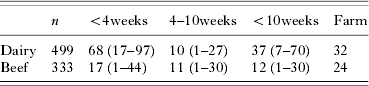
The true prevalence of Cryptosporidium is presented in Table 1. In the dairy calves a peak in prevalence was observed in calves between the ages of 2 and 4 weeks, whereas in beef calves no peak prevalence was observed. The number of excreted oocysts per gram of faeces (OPG) was determined using IFA. The geometric mean oocyst excretion in the dairy calves was 29 425 OPG, ranging from 100 to 7×10⁷ OPG. The highest individual OPG was observed in a 15-day-old calf. On the beef farms the geometric mean oocyst excretion in beef calves was 780 OPG, ranging from 50 to 3·2×104 OPG. The highest individual OPG was observed in a 20-day-old calf. In some of the Cryptosporidium positive calves diarrhoea was observed, but both in the dairy and beef calves there was no correlation between faecal consistency and the presence of Cryptosporidium oocysts in the faeces (P>0·05).
The number (n) of animals included in the study, the true Cryptosporidium prevalence (in percentage), with the 95% Probability Interval, for different age categories in dairy and beef farms, and the farm prevalence

Molecular characterization
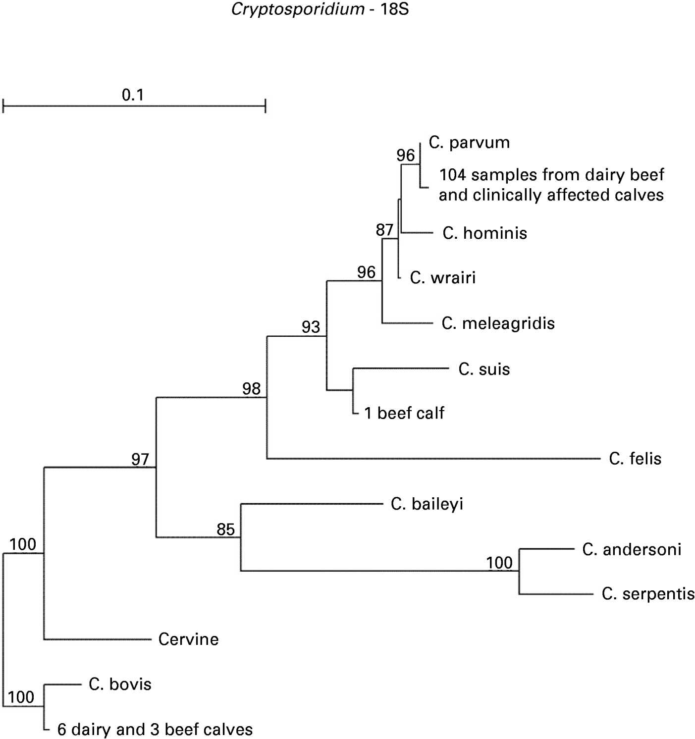
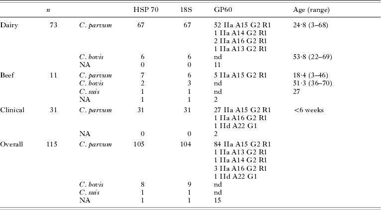
The number of samples and the results of the molecular characterization are presented in Table 2. At least 1 isolate from each of the 32 Cryptosporidium-positive dairy farms was included. Isolates from 9 of the 12 positive beef farms were included, since no amplification product could be obtained from isolates of the 3 remaining farms. Both in beef and in dairy farms C. parvum was predominantly identified, next to the cattle-specific C. bovis. All but 1 of the calves infected with C. bovis were older than 4 weeks and the average age of the C. bovis-positive calves was higher (P<0·001) than the age of the C. parvum-positive calves. The isolate of 1 beef calf was found to be closely related to the porcine-specific C. suis. In the samples from the calves with clinical cryptosporidiosis only C. parvum was identified. The identification on the 18S rDNA concurred with the identification on the HSP-70 gene in all but 2 samples, in which 1 of the 2 genes did not amplify. The phylogenetic analysis on the 18S rDNA is presented in Fig. 1. Four different subtypes of the C. parvum allele IIa were identified (IIaA13G2R1 (n=1), IIaA14G2R1 (n=1), IIaA15G2R1 (n=84), IIaA16G2R1 (n=3)). In 1 sample from a clinically affected calf, the subtype IIdA22G1 was identified. The presence of any specific Cryptosporidium species or C. parvum subtype was not correlated with clinical symptoms.
Phylogenetic relationship of Cryptosporidium isolates (18S rDNA). Only bootstrap-values >70 are reported.

Molecular characterization of Cryptosporidium in the dairy, beef and clinically affected calves, with the mean age and age range for each species
(n, Total number of samples; HSP-70, 70 kDa Heat Shock Protein; 18S, 18S ribosomal DNA; gp60, 60 kDa glycoprotein; NA, No Amplification; nd, not done.)

DISCUSSION
The true Cryptosporidium prevalence was estimated taking the IFA test characteristics into account, which were previously estimated for use in epidemiological studies. The low sensitivity of IFA for the diagnosis of infection in calves older than 1 month is hereby taken into account, providing a more reliable estimate of the true prevalence (Geurden et al. Reference Geurden, Berkvens, Geldhof, Vercruysse and Claerebout2006a). The peak prevalence of Cryptosporidium in dairy calves was observed in calves younger than 1 month, confirming previous studies (O'Handley et al. Reference O'Handley, Cockwill, McAllister, Jelinski, Morck and Olson1999; Nydam et al. Reference Nydam, Wade, Schaaf and Mohammed2001; Maddox-Hyttel et al. Reference Maddox-Hyttel, Langkjaer, Enemark and Vigre2006). In the beef calves there was no obvious age-related infection pattern, possibly due to the overall low Cryptosporidium prevalence in beef calves. The low prevalence in beef calves can largely be attributed to the significantly higher age of the beef calves compared to dairy calves. Calf age is an important risk factor for infection with Cryptosporidium (O'Handley et al. Reference O'Handley, Cockwill, McAllister, Jelinski, Morck and Olson1999), with a maximum risk for infection between 9 and 12 days of age (Maldonado-Camargo et al. Reference Maldonado-Camargo, Atwill, Saltijeral-Oaxaca and Herrera-Alonso1998; Castro-Hermida et al. Reference Castro-Hermida, Gonzalez-Losada and Ares-Mazas2002a, Reference Castro-Hermida, Gonzalez-Losada, Mezo-Menendez and Ares-Mazasb). In the present study only 22% of the beef calves was younger than 2 weeks, compared to 40% of the dairy calves. In previous studies, housing and differences in management have also been advocated as a possible risk factor for infection (Kvac et al. Reference Kvac, Kouba and Vitovec2006). Often, beef calves are raised in cow-calf herds, with less risk for cryptosporidial infection (Gow and Waldner, Reference Gow and Waldner2006) due to the more efficient transfer of colostrum and lower calf stocking densities. Furthermore, cow-calf herds tend to stay on pasture, where the Cryptosporidium oocysts are more exposed to dessication and direct sunlight (Atwill et al. Reference Atwill, Johnson, Klingborg, Veserat, Markegard, Jensen, Pratt, Delmas, George, Forero, Philips, Barry, McDougald, Gildersleeve and Frost1999; Geurden et al. Reference Geurden, Goma, Siwila, Phiri, Mwanza, Gabriel, Claerebout and Vercruysse2006b). However, in the present study, both housing and management were similar on dairy and beef farms, with immediate separation of calves from the dam and rearing in either individual or group housing.
Since at least part of the human C. parvum infections can be ascribed to contact with livestock (Hunter and Thompson, Reference Hunter and Thompson2005), it has been advocated to include tools for molecular identification in epidemiological studies (Traub et al. Reference Traub, Monis and Robertson2005). However, only a limited number of studies did so in the United States (Santin et al. Reference Santin, Trout, Xiao, Zhou, Greiner and Fayer2004; Fayer et al. Reference Fayer, Santin, Trout and Greiner2006) and in Australia (Becher et al. Reference Becher, Robertson, Fraser, Palmer and Thompson2004). In Europe, a similar approach was used in Denmark and in Serbia-Montenegro (Langkjaer et al. Reference Langkjaer, Vigre, Enemark and Maddox-Hyttel2007; Misic and Abe, Reference Misic and Abe2007). In the present study, the guidelines for molecular identification of Cryptosporidium (Caccio et al. Reference Caccio, Thompson, McLauchlin and Smith2005) were taken into account, emphasizing that the genetic characterization of Cryptosporidium isolates should be based on 2 genetic loci, of which at least one is the conserved 18S rDNA gene. The genotyping results of the HSP-70 and 18S rDNA gene concurred in all but 2 samples, in which 1 of the 2 genes did not amplify. The majority of the Cryptosporidium-positive samples from the dairy and beef calves was identified as C. parvum. Since the calves included in the epidemiological study were all younger than 70 days, the current results are in accordance with previous findings that C. parvum is the most prevalent species in pre-weaned calves (Santin et al. Reference Santin, Trout, Xiao, Zhou, Greiner and Fayer2004). The 70-days age limit might also account for the limited number of C. bovis infections, and for the absence of C. andersoni or the Cryptosporidium cervine genotype in the present study. All but 1 of the C. bovis infections were identified in calves older than 1 month, confirming that C. bovis is more prevalent in older calves compared to neonatal calves (Santin et al. Reference Santin, Trout, Xiao, Zhou, Greiner and Fayer2004). In 1 beef calf C. suis was found, indicating a rather erratic parasitism of this Cryptosporidium species in cattle (Fayer et al. Reference Fayer, Santin, Trout and Greiner2006). In the clinically affected calves solely C. parvum was identified, confirming the relevance of C. parvum in the aetiology of neonatal diarrhoea (de Graaf et al. Reference de Graaf, Vanopdenbosch, Ortega-Mora, Abbassi and Peeters1999).
Of the Cryptosporidium species commonly found in calves, C. parvum is known to infect human patients. The molecular analysis on the gp60 gene identified all but 1 of the C. parvum positive calf isolates as the zoonotic IIa allele. All C. parvum IIa subtypes identified in the present study, except the IIaA14G2R1 subtype, have previously been described in calves. The IIaA15G2R1 subtype was predominantly identified, as in previous studies in Portugal (Alves et al. Reference Alves, Xiao, Antunes and Matos2006), in the UK (Chalmers et al. Reference Chalmers, Ferguson, Caccio, Gasser, Abs, Heijnen, Xiao, Elwin, Hadfield, Sinclair and Stevens2005), and in Northern America (Peng et al. Reference Peng, Wilson, Holland, Meshnick, Lal and Xiao2003; Trotz-Williams et al. Reference Trotz-Williams, Martin, Gatei, Cama, Peregrine, Martin, Nydam, Jamieson and Xiao2006; Xiao et al. Reference Xiao, Zhou, Santin, Yang and Fayer2007). Although there are no data on the distribution of C. parvum subtypes in human patients in Belgium, in other countries the IIaA15G2R1 subtype is known to frequently infect human patients (Chalmers et al. Reference Chalmers, Ferguson, Caccio, Gasser, Abs, Heijnen, Xiao, Elwin, Hadfield, Sinclair and Stevens2005; Feltus et al. Reference Feltus, Giddings, Schneck, Monson, Warshauer and McEvoy2006). The occurrence of subtype IId in calves is rare, but has previously been reported (Alves et al. 2003, Reference Alves, Xiao, Antunes and Matos2006). Contrary to a previous report (Xiao et al. Reference Xiao, Zhou, Santin, Yang and Fayer2007), the distribution of the C. parvum subtypes did not differ geographically in the present study, and the occurence of a subtype other than the predominant IIaA15G2R1 was not linked to a particular region. As expected, the anthroponotic C. parvum allele IIc was not found in any of the calves.
The present study demonstrated a high prevalence of C. parvum infections in Belgium, especially in dairy calves and to a lesser extent in beef calves. The results of the subtyping confirm that young calves should be considered as potential zoonotic reservoirs for human infections, although further studies are needed to elucidate the distribution of C. parvum subtypes in human patients in Belgium.