Introduction
Infertility in couples is defined as 12 months of unprotected intercourse without a resulting successful pregnancy. This worldwide reproductive health problem involves approximately 15% of couples. Male factors are the cause of half the cases of infertility (Okada et al., Reference Okada, Tajima, Shichiri, Tanaka, Tanaka and Inoue2008). Reduced fertility in men has several causes including varicocele, immunological and genetic factors, endocrine disturbances and urogenital aberrations. For 60–75% of men, the clear cause of unexplained infertility could not be detected, resulting in the term ‘idiopathic male infertility’. Results of physical examinations are usually normal without any previous history of fertility problems (Quaas and Dokras, Reference Quaas and Dokras2008; Guzick et al., Reference Guzick, Overstreet, Factor-Litvak, Brazil, Nakajima, Coutifaris, Carson, Cisneros, Steinkampf, Hill, Xu and Vogel2001). Genetic anomalies are involved in some men with idiopathic infertility, which is divided into two major categories of quantitative and qualitative abnormalities that include azoospermia, cryptozoospermia, oligoasthenozoospermia and asthenospermia, teratozoospermia and necrospermia, respectively (Hargreave, Reference Hargreave2000).
Invasive and non-invasive diagnostic methods for male infertility can be applied. Identification of subsets of patients is based on testicular and prostate biopsy, but these invasive diagnostic approaches may cause some impairment. Similarly, a slightly less invasive procedure, fine-needle aspiration, is often used in gene expression studies to evaluate epigenetics, cytological and histologic diagnoses, provided that inadequate amounts of sample are collected. The use of non-invasive diagnostic methods, therefore, can play a major role in addressing the issue of male infertility (Hamada et al., Reference Hamada, Seguin, Bujan, Barthelemy, Mieusset and Lansac1998; Kline et al., Reference Kline, Treat, Averna, Davis, Smith and Sillerud2006; Huang et al., Reference Huang, Li, Ding and Xiong2009). The assessment of causes of male infertility requires a total history alongside semen examination, however this evaluation alone is insufficient to recognize men with unexplained infertility. Therefore, non-invasive molecular investigation is needed to gain valuable information when assessing and diagnosing these men (Khazaie and Nasr Esfahani, Reference Khazaie and Nasr Esfahani2014). The study of miRNA is a major area of interest within the field of non-invasive diagnostic biomarkers of diseases (Baldassarre et al., Reference Baldassarre, Felli, Prantera and Masotti2017). In patients with various types of male infertility, a quantitative miRNA expression profile of testicular tissue, sperm, and original plasma can prove to be a helpful molecular assessment, complementing histopathologic diagnostics and governing the level of spermatogenic weakening (Wang et al., Reference Wang, Yang, Chen, Yao, Yang, Zhu, Li, Wang, Li, Shao, Liu, Ji, Zhang, Zen, Zhang and Zhang2011; Barceló et al., Reference Barceló, Lluís Bassas and Exosomal2018).
miRNAs are short (20–23 nucleotides), single-stranded non-coding RNA molecules that manage post-transcriptional gene silencing, and initiate translational hindrance or restraint (He and Hannon, Reference He and Hannon2004). miRNAs play a crucial role in different biological procedures including differentiation, growth, cell propagation and apoptosis (Bartel, Reference Bartel2004; He and Hannon, Reference He and Hannon2004; Pasquinelli et al., Reference Pasquinelli, Hunter and Bracht2005). Recent research has demonstrated that miRNAs are produced in serum, plasma, semen, and other body fluids (Poon et al., Reference Poon, Leung, Lau and Lo2000; Li et al., Reference Li, Zhou, St John and Wong2004). Therefore, changes in miRNA expression and in their recognized locations are likely to contribute to human illness, including sperm defects. There is some evidence to suggest that miRNAs concentrations in seminal plasma, sperm, and testicular biopsy may offer valuable data on gene expression in the male generative system. These data could also provide a new way to develop innovative non-invasive biomarkers of male infertility (Wang et al., Reference Wang, Yang, Chen, Yao, Yang, Zhu, Li, Wang, Li, Shao, Liu, Ji, Zhang, Zen, Zhang and Zhang2011).
As miRNAs have a pivotal role in spermatogenesis and also fertilization of the oocyte, offspring phenotype may also be affected (Kotaja, Reference Kotaja2014; Reilly et al., Reference Reilly, McLaughlin, Stanger, Anderson, Hutcheon, Church, Mihalas, Tyagi, Holt, Eamens and Nixon2016). The issue of altered miRNAs has received considerable critical attention in the field of male infertility in recent years. Aberrant expression of certain miRNAs is linked to certain male infertility dysfunctions. For this reason, miRNA expression might act as a suitable molecular biomarker for diagnosis of male infertility disorders and provide non-invasive biomarkers to diagnose patients more effectively (Lian et al., Reference Lian, Zhang, Tian, Liang, Wang, Liang, Li and Sun2009; Wang et al., Reference Wang, Yang, Chen, Yao, Yang, Zhu, Li, Wang, Li, Shao, Liu, Ji, Zhang, Zen, Zhang and Zhang2011; Salas-Huetos et al., Reference Salas-Huetos, Blanco, Vidal, Godo, Grossmann, Pons, F-Fernández, Garrido and Anton2015). This study was undertaken to review recent research on altered expression of microRNA in semen, sperm and testicular biopsy samples from males with different types of unexplained infertility.
Mechanisms of the function of microRNA
MicroRNAs are usually synthesized in the nucleus using RNA polymerase II to produce a primary miRNA transcript (pri-miRNA). Drosha, an RNase III endonuclease, together with DGCR8, form part of the Microprocessor complex and recognize the double-stranded RNA (dsRNA) hairpin structure in pri-miRNA (Han et al., Reference Han, Lee, Yeom, Kim, Jin and Kim2004; Landthaler et al., Reference Landthaler, Yalcin and Tuschl2004). Two strands of pri-miRNA are cut and, accordingly, produce a precursor miRNA (pre-miRNA) (Lee et al, Reference Lee, Ahn, Han, Choi, Kim, Yim, Lee, Provost, Rådmark, Kim and Kim2003). Pre-miRNAs are exported into the cytoplasm and the RNase III enzyme Dicer cleaves the hairpin structure of pre-miRNA. This endoribonuclease interacts with 5′ and 3′ modifications of the hairpin and removes the loop joining the 3′ and 5′ arms. Therefore, an incomplete miRNA of around 22 nucleotides in length is created. The subsequent miRNA duplex is then untwisted by the action of a helicase and, although the miRNA strand (the developed strand) is cleaved, it is incorporated into the RNA-induced silencing complex (RISC) (Yi et al., Reference Yi, Qin, Macara and Cullen2003; Lund et al., Reference Lund, Guttinger, Calado, Dahlberg and Kutay2004; Wilson and Doudna, Reference Wilson and Doudna2013). RISC incorporates the mature miRNA strand to form a complex, which acts to cleave an mRNA target. Resulting miRNA/mRNA complementarity success leads to both translational suppression and mRNA neutralization. MicroRNAs can act by diminishing target mRNA levels or by hindering their translation (Wahid et al., Reference Wahid, Shehzad, Khan and Kim2010; Kotaja, Reference Kotaja2014).
MicroRNAs in spermatogenesis
Self-reestablishment and propagation of spermatogonia, the meiotic division of spermatocytes, and postmeiotic separation of spermatids into spermatozoa are three important phases in the complex differentiation process of spermatogenesis. This procedure, at both the transcriptional and the post-transcriptional levels, is a firmly regulated process that allows continuous production of a gamete throughout adulthood (Bouhallier et al., Reference Bouhallier, Allioli, Lavial, Chalmel, Perrard, Durand, Samarut, Pain and Rouault2010; Ni et al., Reference Ni, Hu, Liu, Liu, Lu, Zhang, Zhang and Zhang2011). An important regulator of spermatogenesis is the innovative application of post-transcriptional control that is controlled by microRNAs. Some examinations have evaluated the key role of non-coding RNAs as post-transcriptional controllers of spermatogenesis. Long non-coding RNAs basically are used as transcriptional repressors and chromatin alteration factors, piwi-interacting RNAs, which are known to control transposon silencing of microRNAs, and managing the expression of specific target messenger RNAs (McLachlan, Reference McLachlan2013; Kotaja, Reference Kotaja2014).
miRNAs are an important component of spermatogenesis and play vital roles in the progress of spermatozoa, principally in germ cells and somatic cells. However, it is widely accepted that any deregulation in miRNA expression can have significant effects on spermatogenesis pathways and lead to reproduction aberrations.
Alteration in microRNA expression in different varieties of male infertility
Variation in miRNA expression patterns has been allied to dissimilar human testicular histopathologic patterns (Sertoli cell only, mixed atrophy, and germ-cell arrest), and spermatogenic impairments (asthenozoospermia, oligozoospermia, oligoasthenozoospermia). Evaluation of the seminal plasma of patients with azoospermia or with sperm with abnormal morphology has indicated that miRNAs could be used as special biomarkers to appraise human male fertility (Abu-Halima et al., Reference Abu-Halima, Hammadeh, Schmitt, Leidinger, Keller, Meese and Backes2013; Kotaja, Reference Kotaja2014).
The following section describes changes in the regulation of miRNA in some types of male infertility assessed by microarray; miRNA levels were confirmed by real-time qRT-PCR.
Azoospermia
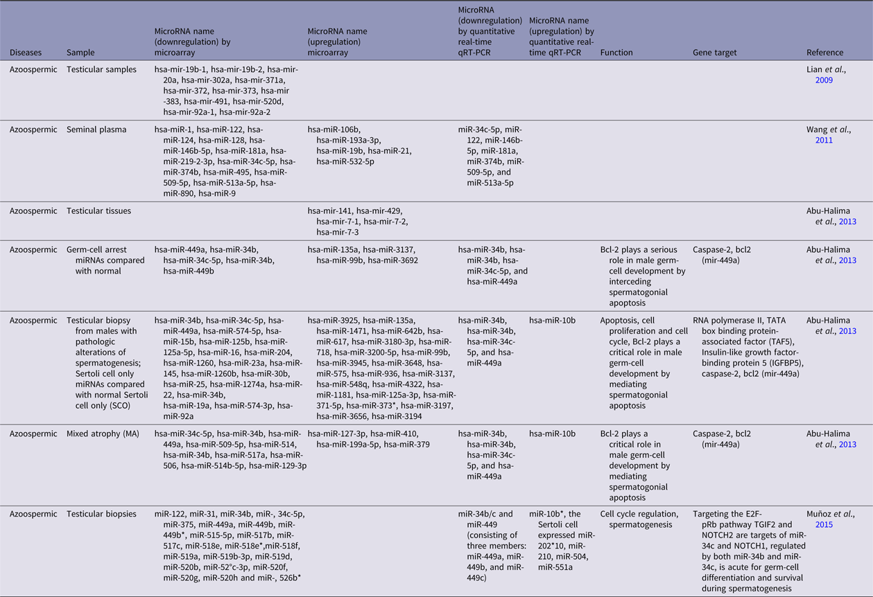
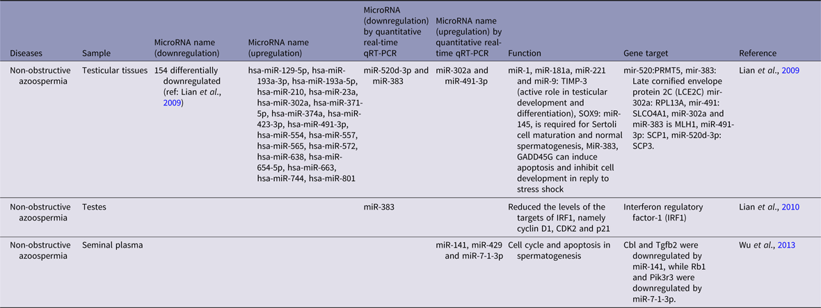
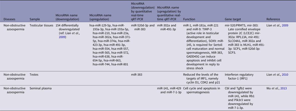
A large percentage of male infertility is due to azoospermia (the complete lack of spermatozoa upon semen assessment) instigated by blockage of the excurrent ductal system (obstructive azoospermia, OA) or by abnormal/absent testes (non-obstructive azoospermia, NOA), with the latter found in 1% of all mature men (Lian et al., Reference Lian, Tian, Liu, Zhang, Li, Deng, Yao, Yin and Sun2010; Wosnitzer et al., Reference Wosnitzer, Goldstein and Hardy2014). Tables 1 and 2 show a brief description of changes in miRNA regulation in azoospermia and non-obstructive azoospermia patients.
Changes in the regulation of miRNAs in the human testicular biopsy, semen, and sperm samples from azoospermia patients compared with the normal controls determined by microarray and quantitative real-time qRT-PCR

Changes in the regulation of miRNA in the human testicular biopsy, semen, and sperm samples from non-obstructive azoospermia patients compared with the normal controls determined by microarray and quantitative real-time qRT-PCR
Asthenozoospermia
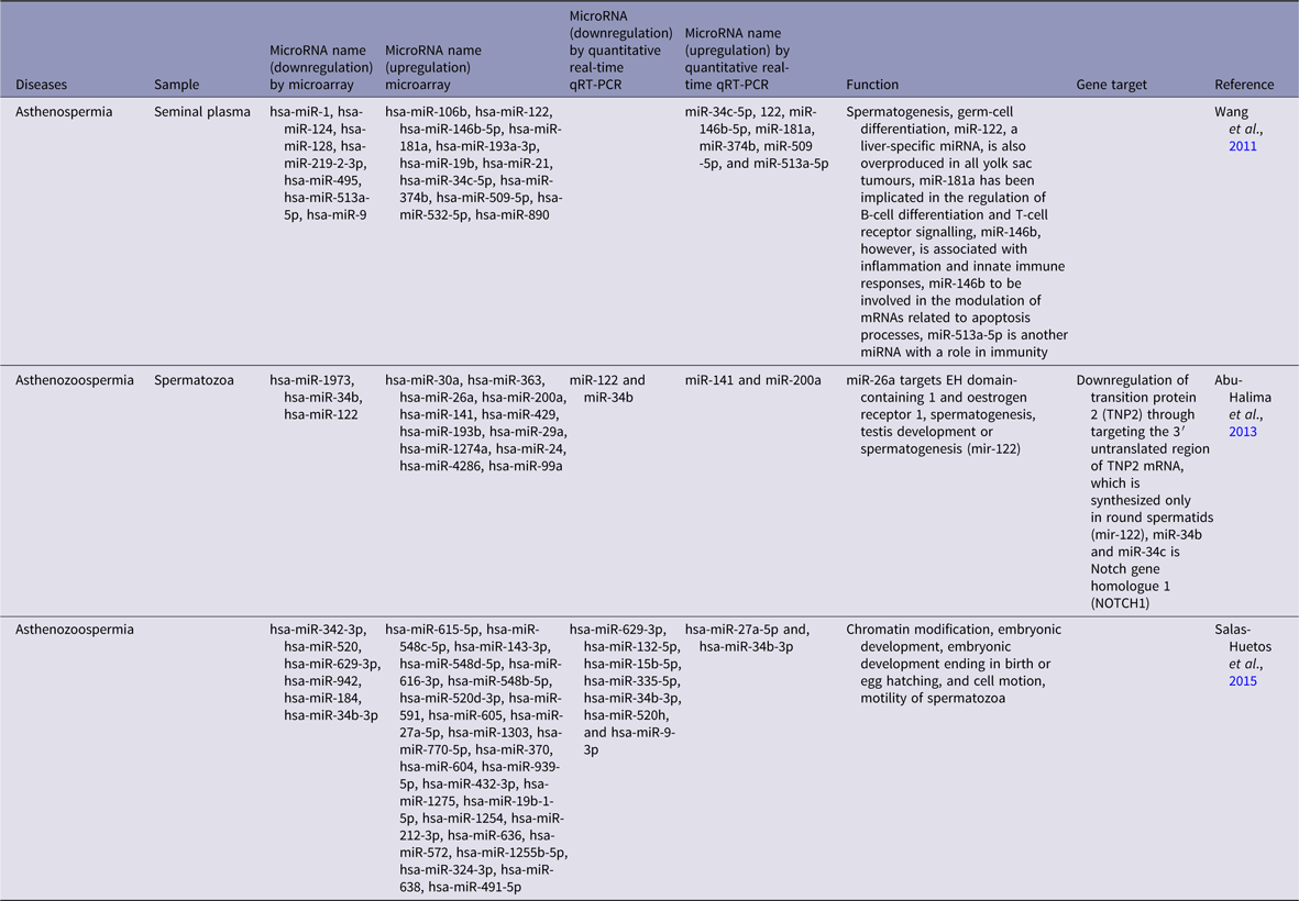
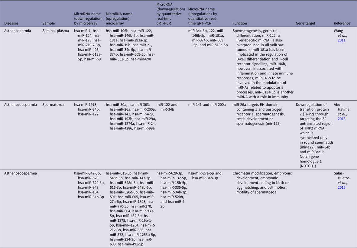
The term asthenozoospermia or asthenospermia refers to a biological event characterized by decline in sperm motility. It is diagnosed when the percentage of motile sperm is less than 50% or when the percentage of motile sperm moving in a straight line and with a speed of 25 µm/s is less than 25% (Youn et al., Reference Youn, Cha, Park, Yang, Kim, Koong, Kang, Song and Han2011). Table 3 presents the results from assessment of changes in miRNA expression in asthenozoospermia patients.
Changes in the regulation of miRNAs in human testicular biopsy, semen, and sperm samples from asthenozoospermia patients compared with the normal controls determined by microarray and quantitative real-time qRT-PCR
Oligoasthenoteratozoospermia
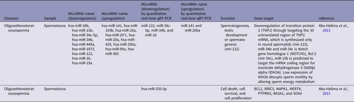
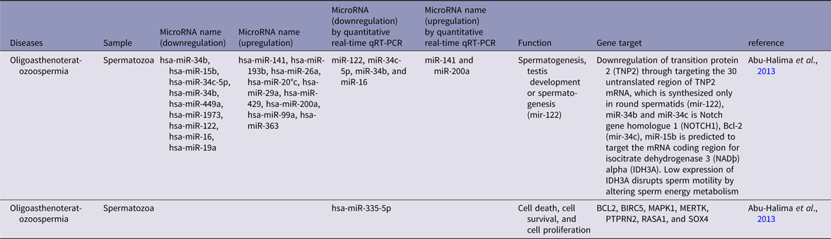
Previous studies have mostly defined oligospermia and oligozoospermia as male infertility due to low concentrations of sperm in semen. Often semen with a reduced sperm concentration may also show significant abnormalities in sperm morphology and motility (McLachlan, Reference McLachlan2013). Microarray and quantitative real-time qRT-PCR results for oligoasthenoteratozoospermia patients related to changes in miRNA regulation are summarized in Table 4.
Changes in the regulation of miRNAs in human testicular biopsy, semen, and sperm samples from oligoasthenoteratozoospermia patients compared with the normal controls determined by microarray and quantitative real-time qRT-PCR
Teratozoospermia
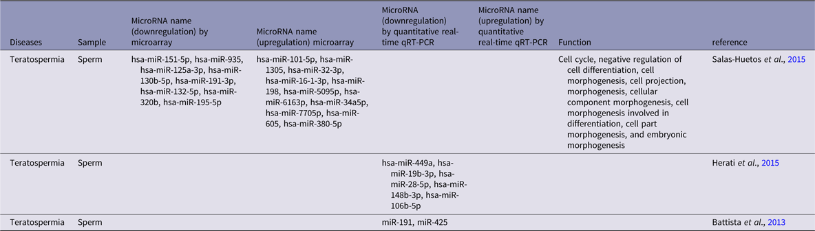
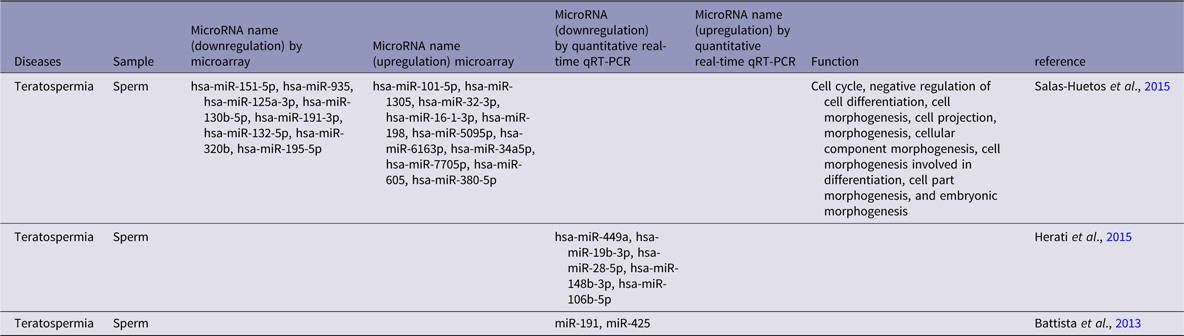
The broad use of the term teratospermia or teratozoospermia is characterized by the presence of sperm with irregular morphology that affect male fertility. According to the criteria in a World Health Organization manual (5th edition) (WHO, 2010) teratozoospermia is defined when the percentage of normal spermatozoa in the ejaculate is less than 4% (Herati et al., Reference Herati, Mielnik, Schlegel and Paduch2015). As seen from the data in Table 5, teratospermia patients exhibit changes in miRNAs regulation.
Changes in the regulation of miRNA in human testicular biopsy, semen, and sperm samples from teratospermia patients compared with the normal controls determined by microarray and quantitative real-time qRT-PCR
Oligozoospermia
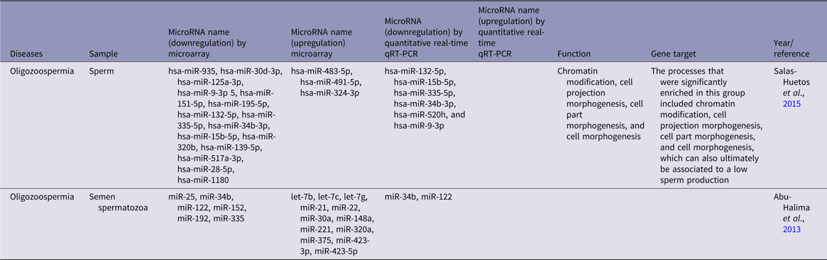
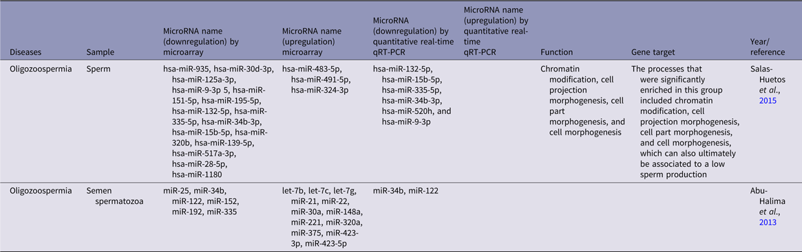
Another problem in male infertility is oligospermia. This is characterized by a low sperm count. The WHO classifies sperm counts at or above 15 × 106/ml sperm in semen as average. Anything below this number is considered low and is diagnosed as oligospermia (Kumar and Singh, Reference Kumar and Singh2015). From Table 6, we can see changes in the regulation of miRNAs in patients with oligospermia.
Changes in the regulation of miRNA in human testicular biopsy, semen, and sperm samples from oligospermia patients compared with the normal controls determined by microarray and quantitative real-time qRT-PCR
Discussion
miRNA analysis is a suitable option for diagnosing infertility
Grading of diagnostic markers is based on ease of measurement, harmlessness, rate efficiency, variability with use, and consistency across gender and ethnic groups. Currently, low specificity, affectability, and false-positive outcomes are the major limitations of available biomarkers. There are several reasons why miRNAs have become a suitable option for the diagnosis of infertility. These markers are evolutionarily conserved small non-coding RNAs that are also acknowledged as micro-coordinators of gene expression. They are effective tools as potential new diagnostic and predictive biomarkers with great precision and sensitivity. The role of miRNAs in the regulation of progressive and physiological processes is critical and their lack has been associated with several diseases. In addition, miRNAs are stable and can be easily isolated and quantified from body fluids and tissues (Faruq and Vecchione, Reference Faruq and Vecchione2015). Recently, the practicalities of using miRNAs or miRNA boards as biomarkers have been under consideration in the diagnosis of cancer. The expression patterns of these biomarkers in oncology have initiated new and widespread discussion. Recent experiments have shown that miRNAs are impaired in cancer and are assumed to have an indispensable role in the development and advancement of this disease. Accordingly, the exploration of such molecules is a goal for the cancer diagnosis, treatment and prediction, offering promising methodology (Hamam et al., Reference Hamam, Hamam, Alsaleh, Kassem, Zaher, Alfayez, Aldahmash and Alajez2017).
There is no reliable molecular procedure to identify male infertility as a cohort diagnosis from semen analysis or to corroborate conservative histology of testes biopsies. Variations in miRNA expression profiles in patients with dissimilar spermatogenic and histopathologic deficiencies could potentially to be used as new biomarkers for diagnostic purposes. This literature review assesses the importance of miRNA applications in different types of male infertility as a non-invasive indicator of diagnosis. One or more miRNAs could be considered as indicators for diagnosis of some causes of male infertility.
Changes in miRNA profiles in patients with various spermatogenic and histopathologic impedances can possibly be used as new biomarkers related to symptoms. In total, 30% of protein-coding events are orchestrated by miRNAs either as a clear mRNA target or preceding it.
Benefits and disadvantages of miRNA as a diagnostic marker
The many benefits of miRNAs as biomarkers have drawn attention. These benefits include:
1. In the human 3′ untranslated region (UTR), over 45,000 miRNAs target destinations are available and over 60% of human protein-coding genes are presumably organized by multiple miRNAs. It is predicted that more than 30% of protein-coding genes are arranged by miRNAs, either by linking target mRNA or by linkage to mutated regions in the 3′-UTR, resulting in regulation of gene expression at the translational level.
2. MicroRNAs are endogenous, naturally plentiful, relatively stable, evolutionarily conserved, small non-coding RNAs that operate in post-transcriptional gene regulation in important biological and pathological processes.
3. Stable miRNAs have been found in several biological fluids such as plasma, serum, saliva and milk, and the expression motifs of serum miRNAs are known to be related to several illnesses.
4. MicroRNAs have been identified in blood plasma, even under unfavourable conditions (long-term deposit at room temperature, high or low pH, or bubbling due to freeze–thaw cycles).
5. Using microarray, hybridization, deep-sequencing and qRT-PCR analysis, miRNA quantitation it is easy to perform.
6. MicroRNAs play the main role in an extensive range of developmental processes including cell proliferation, stem cell separation, muscle differentiation, metabolism, apoptosis, and brain morphogenesis (Friedman et al., Reference Friedman, Farh, Burge and Bartel2009; Felekkis et al., Reference Felekkis, Touvana, Ch and Deltas2010; Yi and Fuchs, Reference Yi and Fuchs2011; Faruq and Vecchione, Reference Faruq and Vecchione2015).
As with all diagnostic methods, this approach has some advantages and disadvantages, In reviewing the literature regarding the challenges facing miRNA use as diagnostic biomarkers, we concluded that all miRNA analysis need confirmatory steps, appropriate standardization of analytical procedures, and control of potential technical biases before being considered for use in clinical practices (Moldovan et al., Reference Moldovan, Batte, Trgovcich, Wisler, Marsh and Piper2014). The capacity to take an extracellular miRNA signature from body fluids by non-invasive means exists, but ways to separate extracellular RNAs from plasma or serum must be extrapolated to other body liquids such as spittle or urine. Standardized and reliable methods need to be applied at many levels from the whole blood to plasma/serum preparation; handling and methods of RNA extraction and miRNA evaluation. To improve the efficiency of miRNA results, there should be minimal differences in procedures between laboratories and ways to limit inconsistencies among different operators. If methodological parameters are not considered in the design of these study, some individual and environmental factors in miRNA analysis could not be properly evaluated, and could plausibly influence miRNA application in clinical practices (Tiberio et al., Reference Tiberio, Callari, Angeloni, Daidone and Appierto2015).
Biological role of miRNA as a means for the diagnosis of infertility
miRNAs as biomarkers have increasingly gained a role in diagnosing abnormalities in spermatogenesis. The studies presented in Table 1 showed mir-499a in relation to caspase-2, bcl-2, RNA polymerase II, TATA box binding protein-associated factor (TAF5), and insulin-like growth factor-binding protein 5 (IGFBP5) gene expression, plus mir-449a playing an imperative role in apoptosis, cell proliferation and the cell cycle (Abu-Halima et al., Reference Abu-Halima, Hammadeh, Schmitt, Leidinger, Keller, Meese and Backes2013). Bcl-2 plays a pivotal role in male germ-cell improvement by interceding in spermatogonial apoptosis. In azoospermia, miR-34b and miR-34c downregulate E2F-pRb, TGIF2 and NOTCH2, targets of miR-34c and miR-34b that are necessary for germ-cell differentiation and subsistence during spermatogenesis. In non-obstructive azoospermia (Table 2) miR-1, miR-181a, miR-221 and miR-9 affected the TIMP-3 gene that has an intrinsic role in testicular progress and differentiation. miR-145 affects SOX9, which is essential for Sertoli cell maturation and normal spermatogenesis; and miR-383 regulates GADD45G, which can induce apoptosis and obstruct cell growth in answer to stress shocks. Cbl and Tgfb2 were downregulated by miR-141, and Rb1 and Pik3r3 were downregulated by miR-7-1-3p, which affects the cell cycle and apoptosis in spermatogenesis (Lian et al., Reference Lian, Zhang, Tian, Liang, Wang, Liang, Li and Sun2009, Reference Lian, Tian, Liu, Zhang, Li, Deng, Yao, Yin and Sun2010; Wu et al., Reference Wu, Qin, Li, Dong, Dai, Lu, Guo, Zhao, Zhu, Zhang, Hang, Sha, Shen, Xia, Hu and Wang2013). In men with asthenospermia, miR-122 is important for spermatogenesis and germ-cell differentiation, miR-181a which is a liver-specific miRNA, and miR-146b which is related to the inflammation and the innate immune response, are involved in modulation of mRNAs linked to apoptosis. miR-513a-5p is another miRNA with a role in immunity that showed increase in expression (Wang et al., Reference Wang, Yang, Chen, Yao, Yang, Zhu, Li, Wang, Li, Shao, Liu, Ji, Zhang, Zen, Zhang and Zhang2011; Salas-Huetos et al., Reference Salas-Huetos, Blanco, Vidal, Godo, Grossmann, Pons, F-Fernández, Garrido and Anton2015). Abu-Halima et al. (Reference Abu-Halima, Hammadeh, Schmitt, Leidinger, Keller, Meese and Backes2013) showed that mir-122 was downregulated in infertile men, leading to the downregulation of transition protein 2 (TNP2), targeting the untranslated area of TNP2 mRNA, which is synthesized uniquely in round spermatids. This miRNA affects spermatogenesis, testis development and spermatogenesis. In teratospermia, miRNAs play a central role in cell cycle, negative control of cell differentiation, cell morphogenesis, cellular component morphogenesis and cell morphogenesis and were involved in differentiation, and embryonic morphogenesis. Salas-Huetos et al. (Reference Salas-Huetos, Blanco, Vidal, Godo, Grossmann, Pons, F-Fernández, Garrido and Anton2015) showed that changes in miRNA expression in teratospermia coincided with a reduction in normal sperm, so changes in some miRNAs could be used as new biomarkers in diagnosing men with teratospermia.
A strong relationship has been reported between changes in miRNA expression in male infertility and gene expression. Based on evidence of miRNA changes in infertility, miRNA can be used as a diagnostic indicator for individual patients. Researchers have stated that they would focus on one miRNA for any infertile disease, so results could be used to provide a dedicated kit.
The following conclusions can be drawn from the present review: (1) Creating a non-invasive diagnostic method for male infertility is necessary. (2) miRNA is fast becoming a key instrument in the diagnosis of male infertility. (3) Changes in miRNA expression can be used as a new diagnostic biomarker in patients with different types of spermatogenic and histopathologic damage.
Financial support
This research received no specific grant from any funding agency, commercial or not-for-profit sectors.
Conflicts of interest
None.
Ethical standards
Not applicable.