The supercontinent Pangaea lasted throughout the Permian. Despite spatial and temporal differentiation in tetrapod faunal history (Brocklehurst et al. Reference Brocklehurst, Dunne, Cashmore and Fröbisch2018; Dunne et al. Reference Dunne, Close, Button, Brocklehurst, Cashmore, Lloyd and Butler2018; Pardo et al. Reference Pardo, Small, Milner and Huttenlocker2019), there was broad consistency at the family level, not least across Laurasia (Hmich et al. Reference Hmich, Schneider, Saber, Voigt, Wartiti, Lucas, Cassinis and Schneider2006; Lagnaoui et al. Reference Lagnaoui, Voigt, Belahmira, Saber, Klein, Hminna and Schneider2017). Lower Permian tetrapod assemblages, mainly from the south-western USA, yielded a template for similar discoveries in European sites from the palaeo-tropics, such as the German localities of the Thuringian Forest Basin, Saar-Nahe Basin, or Döhlen Basin (Schneider Reference Schneider1993; Werneburg Reference Werneburg2007; Spindler Reference Spindler2013; Berman et al. Reference Berman, Henrici, Sumida, Martens, Pelletier, Kammerer, Angielczyk and Fröbisch2014; Voigt et al. Reference Voigt, Fischer, Schindler, Wuttke, Spindler and Rinehart2014; respectively). Pelycosaur-grade (non-therapsid) synapsids formed the dominant group of large terrestrial animals. Similar species were described from both continents, including virtually all ‘pelycosaur' subgroups: basal and derived Caseasauria, all major subgroups of Varanopidae, basal and derived Ophiacodontidae, basal and derived Edaphosauridae, early Sphenacodontia (haptodontine-grade) and Sphenacodontidae including the iconic genus Dimetrodon. An updated list of European ‘pelycosaurs' is provided by Spindler et al. (Reference Spindler, Werneburg, Schneider, Luthardt, Annacker and Rößler2018).
Situated well within former Laurasia, the English Midlands contain outcrops from historical mining areas that correlate with the Rotliegend (Lower Permian) formations of Central Europe. Only a few vertebrate fossils were collected from the Kenilworth Sandstone Formation, as their coarse arkosic sandstones reflect a rather proximal depositional environment. However, considering this limited sampling, a rich fauna might have existed, including the large zatracheid temnospondyl Dasyceps (Paton, Reference Paton1975).
Three isolated jaw remains represent the only early synapsid finds from Great Britain (Fig. 1). At the time of their description (Paton Reference Paton1974), the proposed determinations represented an intermediate diversity between North America and Central Europe: (1) Ophiacodon sp. (dentary), assigned to the most abundant ophiacodontid, which is common in Texas, Oklahoma, and New Mexico (Romer & Price Reference Romer and Price1940), but not Central Europe; (2) Sphenacodon britannicus (maxilla, originally Oxyodon britannicus von Huene, Reference von Huene1908) was recognised as a sphenacodontid and assigned to this genus otherwise known from New Mexico and Utah (Vaughn Reference Vaughn1964; Paton Reference Paton1974; Eberth Reference Eberth1985), but not Central Europe; (3) Haptodus grandis (maxilla) was considered a new species of this wastebasket genus previously comprising specimens from France, Germany, and Poland, but until then not reported from North America. In subsequent years, Haptodus garnettensis from Kansas was named, along with the assignment of Cutleria wilmarthi from Colorado to Haptodus (Currie Reference Currie1977), connecting early sphenacodontians to North America. Haptodus was considered to represent the ancestor of sphenacodontids (Romer & Price Reference Romer and Price1940), later revised as the paraphyletic assemblage of successive outgroups to the clade containing Sphenacodontidae and Therapsida (Laurin Reference Laurin1993; Spindler Reference Spindler2015). Fragmentary sphenacodontid material is known from various sites in Europe, further connecting faunal composition across Laurasia. In contrast, what Paton (Reference Paton1974) identified as the partial mandible of Ophiacodon remained the only find of this genus reported from Europe, with still no equivalent on the continent.
Map showing the localities of all three pelycosaur remains from England. Cities L, M, B localising London, Manchester, and Birmingham, respectively. Modified from Powell et al. (Reference Powell, Chisholm, McC Bridge, Rees, Glover and Besly2000), with information from Paton (Reference Paton1974).

Specimens are held in the following institutions: American Museum of Natural History, New York (AMNH), Carnegie Museum, Pittsburgh (CM), Geological Survey Museum (GSM, part of the Natural History Museum, London), Warwick County Museum (Gz); Muséum d'Histoire Naturelle de Besançon, Besançon, France (HN); Landessammlung für Naturkunde Rheinland-Pfalz, Mainz, Germany (LFN); Museum of Comparative Zoology, Harvard University (MCZ), Museum der Natur Gotha (MNG), Muséum national d'histoire naturelle, Paris (MNHN), Natural History Museum, London (NHMUK), Sam Noble Oklahoma Museum of Natural History, Norman (OMNH).
1. Systematic palaeontology
(unranked) Synapsida Osborn, Reference Osborn1903
(unranked) Sphenacodontia Romer & Price, Reference Romer and Price1940
Diagnosis. As the assignment of less derived members to the Sphenacodontia is subject to an ongoing debate, there are only vague diagnoses beyond genus level (Brink Reference Brink2015). For the clade Sphenacodontia, defined as the sister group to Edaphosauridae, diagnosis depends on the inclusion of ‘Haptodus' garnettensis. A preliminary diagnosis is offered herein, using the latest analyses (Spindler Reference Spindler2015; Spindler et al. Reference Spindler, Scott and Reisz2015): diagnostic combination of a synapsid condition in the temporal region, expanded lateral surface of squamosal posterior to temporal opening of moderate size, recess on the lateral surface of postorbital, ridged pineal rim, stout marginal teeth, premaxillary teeth in deep sockets, convex margin of maxilla, tall coronoid eminence with dentary forming its dorsal tip, angular lamina, laterally excavated dorsal neural arches, three sacrals, enlarged triceps process on posterior coracoid, blade-like dorsal process of ilium blade-like.
Included taxa. Haptodus, Cutleria, Palaeohatteriidae, Tetraceratops, Sphenacodontoidea.
Genus Hypselohaptodus gen. nov.(ex Haptodus Gaudry, Reference Gaudry1886)
Etymology. As explained by Gaudry (Reference Gaudry1886), with the attribution of Greek ψηλóς, meaning ‘tall', in regard of the facial region.
Diagnosis. Differential diagnosis with unique combination of characters. Plesiomorphic characters with respect to other Sphenacodontia. Long teeth weakly curved. Weak and unserrated carinae. Large number of precanines. Derived character shared with Sphencodontoidea. Maxilla contacting nasal, excluding the lacrimal from the external naris. Potential autapomorphy. Eminent lobe of dorsal blade of maxilla situated posterior to the canine level, with slightly concave anterior edge.
Distribution. England; Gzhelian or Asselian, Pennsylvanian to Cisuralian.
Included species. Type and only species Hypselohaptodus grandis.
Hypselohaptodus grandis (Paton, Reference Paton1974)
(=Haptodus grandis Paton, Reference Paton1974)
Diagnosis. As for genus.
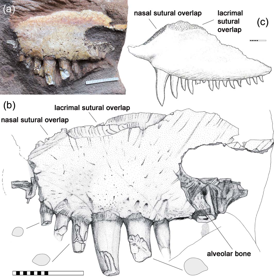
Holotype. Warwick County Museum Gz 1071, isolated maxilla (Fig. 2).
Gz 1071, holotypic maxilla of Hypselohaptodus grandis gen. nov., left lateral view. (a) Photograph of sandstone block. (b) Interpretative drawing, with cross sections of chosen tooth crowns. (c) Maxilla of Sphenacodon ferocior, lateral view, for comparison of similar sutural surfaces (mirrored from Eberth Reference Eberth1985, fig. 12). Scale bars = 2cm.

Locality. Kenilworth, Warwickshire, West Midlands, England.
Horizon. Kenilworth Sandstone Formation (Cisuralian), Enville Group, Pennine Basin (Old et al. Reference Old, Sumbler and Ambrose1987). This formation marks the lower-most Permian in its section (Powell et al. Reference Powell, Chisholm, McC Bridge, Rees, Glover and Besly2000), covering the upper-most Gzhelian to mid-Asselian (Warrington et al. Reference Warrington, Barclay, Leveridge and Waters2012). Gz 1071 is preserved in a red, coarse, arkosic sandstone with mud pebbles (Fig. 1).
Description. The high facial blade of the maxilla implies a tall skull. The ventral margin of the maxilla is moderately convex, with the strongest curvature in the precanine region. The presence or absence of a concave step is unknown, as is the entire anterior expansion of the maxilla. The canines are pronounced, almost twice the basal diameter of the surrounding teeth. The third postcanine is of moderate width compared to the first postcanine, indicating a high degree of variation in the postcanine tooth size, which clearly differs from the usually uniform row in other Sphenacodontia. There are three precanine teeth preserved. Median portions that reach more anteriorly than the broken labial area suggest a longer tooth row with at least a fourth precanine that has been lost due to crushing. In comparison, the maximum precanine count in sphenacodontids is three (only in Sphenacodon ferox Spielmann et al., Reference Spielmann, Rinehart, Lucas, Berman, Henrici, Harris, Lucas, Schneider and Spielmann2010, and juvenile S. ferocior Eberth, Reference Eberth1985) or four when including Neosaurus, whereas more basal haptodontiforms bear up to six or eight (Laurin Reference Laurin1993; Mazierski & Reisz Reference Mazierski and Reisz2010). The tooth type of Hypselohaptodus resembles that of basal sphenacodontians, e.g., Palaeohatteriidae, in its less derived morphology. Even close to their tips, the teeth are almost straight, by which Gz 1071 resembles Pantelosaurus. There is no indication of a distinct bulb-like thickening like in ‘Haptodus' garnettensis, Haptodus baylei, and sphenacodontids. The basal section is round and lacks plicidentine folding (except for a very shallow labial trough where the third postcanine is broken off), which is in strong contrast with Ianthodon and most sphenacodontids, but matches the condition in all therapsids, Tetraceratops, and a few Dimetrodon species (Brink et al. Reference Brink, LeBlanc and Reisz2014, Reference Brink, Maddin, Evans and Reisz2015). According to Spindler (Reference Spindler2015), there is indication for dentine folding in early haptodontiform material from Garnett, in Palaeohatteriidae, probably in Cutleria, and weakly developed as two cases of shallowly concave tooth sides in Tetraceratops. There are no striations or serrations on the tooth crowns of Hypselohaptodus, as far as can be revealed despite the broken-off tips. As mentioned by former workers (Paton Reference Paton1974; Currie Reference Currie1979), there is evidence for moderate carinae in Hypselohaptodus, at least on the posterior edge, where the subterminal region shows a blunt but distinct ridge. This might have not been surprising before the knowledge of well-preserved skulls assigned to Haptodus (Laurin Reference Laurin1993), but is in fact absent in less derived Haptodontiformes, while occurring in all sphenacodontids, the majority of therapsids, Cutleria, and a single juvenile specimen of Palaeohatteria (Spindler Reference Spindler2016). Tooth implantation in Gz 1071 is deeply thecodont. Alveolar bone is visible and bears a basal striation on several teeth.
The dorsal outline of the maxilla arches upwards posterior to the canine region, which is an unusual structure that occurs neither in the closest relatives of Sphenacodontia, namely Ophiacodontidae and Edaphosauridae, nor in any sphenacodontian except for Tetraceratops (Spindler Reference Spindler2015), Sphenacodon, and certain therapsids. Only in Tetraceratops and initially in Gz 1071, the maxillary ascending lamina is anteriorly bordered by a concave lacrimal suture, which is usually seen in more basal synapsids like certain varanopids and probably early caseasaurians, but also due to the enlarged nasal contact in therapsids like early dinocephalians and anomodontians (Langston & Reisz Reference Langston and eisz1981; Ivakhnenko Reference Ivakhnenko2003; Campione & Reisz Reference Campione and Reisz2010; Spindler et al. Reference Spindler, Falconnet and Fröbisch2016; respectively). In Gz 1071, this lobe is not an artefact of crushing, as the lacrimal contact is completely preserved. In the posterior portion, the maxillary lobe overlaps the lacrimal laterally. The posterior portion of the contact forms an acute-angled, V-shaped trough to envelop the lacrimal. Anteriorly, the maxillary lobe descends and leaves a simple lacrimal overlap above of and anterior to the canine region.
Paton (Reference Paton1974) classified the specimen as a ‘haptodontine' by the striking, plesiomorphic feature of a long lacrimal, presumably reaching the naris. In fact, the lacrimal sutural surface in Hypselohaptodus ends in a round groove dorsal to the precanines, not allowing any anterior expansion of the lacrimal beyond this point (Fig. 2b). Anterior to this, a down-sloping sutural surface is only bluntly preserved, apparently ignored as an artefact by Paton (Reference Paton1974) and Currie (Reference Currie1979, fig. 11). Since this pattern perfectly matches the morphology of Sphenacodon (Eberth Reference Eberth1985, fig. 12b), it is recognised as the long nasal–maxillary contact, revealing a short lacrimal that does not reach the naris. This is a diagnostic feature of Sphenacodontoidea (Reisz et al. Reference Reisz, Berman and Scott1992: Sphenacodontidae plus Therapsida).
2. Unnamed specimen
Diagnosis. No informative differential diagnosis possible.
Material. Warwick County Museum Gz 41, isolated dentary (Fig. 3), erroneously determined as Ophiacodon sp. by Paton (Reference Paton1974).
Gz 41, dentary of an undeterminable synapsid, originally ascribed to Ophiacodon by Paton (Reference Paton1974), in comparison with MCZ 1203 (below), hemimandible of O. retroversus, to demonstrate distinctness. Scale bars = 2cm.

Locality. One mile NW of Coventry, West Midlands county, England.
Horizon. Kenilworth Breccia of Kenilworth Sandstone Formation, late Gzhelian to early Asselian (Powell et al. Reference Powell, Chisholm, McC Bridge, Rees, Glover and Besly2000; Warrington et al. Reference Warrington, Barclay, Leveridge and Waters2012).
Description. The dentary is low anteriorly, although taller than in Ophiacodon. A striated impression of broken areas indicates a rather deep posterior mandible with a strong eminence in the coronoid region, very atypical for Ophiacodon and its closest relatives. As in early sphenacodontians, but again unlike ophiacodontids, the dorsal edge of the coronoid region is apparently formed by the dentary. The symphysis is slightly pronounced as in early sphenacodontians, less than in Stereophallodon or Stereorhachis, but distinct from the low tip in other ophiacodontids, or the gonys-like deep symphysis in Dimetrodon, Cryptovenator, and Sphenacodon. Paton (Reference Paton1974) counted 33 to 34 tooth positions, but also remarked the bad quality. From a revised observation, the tooth number is estimated lower than 30, probably as low as 27, which is in strong contrast to Ophiacodon, which has a minimum of 37 teeth (estimated from Ophiacodon uniformis, MCZ 1366).
The tooth shape is stout and maybe slightly bulbous, not as slender as usually seen in long-snouted ophiacodontids. That the teeth are almost uncurved does not allow for any taxonomic conclusions, since, e.g., Pantelosaurus exhibits straight teeth as well, whereas Ophiacodon can bear strongly curved teeth (Ophiacodon retroversus MCZ 1203; Fig. 3). By its stout teeth, Gz 41 resembles some sphenacodontid specimens in lateral outline. No indication of cutting edges is present in the admittedly broken condition of most tooth crowns. If carinae were indeed absent, this would mean distinctness from Stereorhachis and Baldwinonus. The presence of at least one enlarged fang near the dentary tip resembles the usual condition in sphenacodontians, but also Stereophallodon and Stereorhachis. In contrast, Ophiacodon lacks any initial lower caniniform (including the undescribed Garnett form, pers. obs.), except for the very slight condition in O. retroversus. Regarding the overall outline, Gz 41 shares some similarity with smaller sphenacodontids (Brink Reference Brink, Maddin, Evans and Reisz2015), with a short and tall snouted appearance unlike that of Ophiacodon and its closer relatives.
3. Preliminary phylogenetic implications
The original description counted Gz 1071 as the largest species of the basal sphenacodontian Haptodus, named as H. grandis (Paton, Reference Paton1974). Currie (Reference Currie1979) discussed the validity of a distinct species based on size and (erroneously) longer teeth. All subsequent authors accepted the generic assignment of Gz 1071, largely confirmed by the systematics of Romer & Price (Reference Romer and Price1940), who ranked Haptodus as the stem form of Sphenacodontidae (Sphenacodontinae in their sense), with no greater diversity expected but the designated species. The noticeable rareness of haptodontine-grade material is the reason for limited research on this former paradigm.
However, a specimen-level revision of Haptodus (Spindler Reference Spindler2015) could demonstrate that Gz 1071 does not group with the type species H. baylei from France or the similarly less derived specimens of ‘H.' garnettensis from Kansas. In general, the renamed holotype of Hypselohaptodus grandis shows plesiomorphic traits, so far considered to be haptodontine-grade characteristics, such as a high number of precanines and the absence of strongly recurved and laterally compressed postcanines. On the other hand, the shortened lacrimal is an unequivocal diagnostic character of Sphenacodontoidea.
It should be noted that systematics in this field are strongly directed towards distinguishing between haptodontine-grades versus sphenacodontids, while therapsids remained unconsidered as a possibility. Certain insights have changed this precondition. (1) Largely independent analyses have suggested that ‘Haptodus' garnettensis might be closer to Edaphosauridae than to Sphenacodontoidea (Laurin Reference Laurin1993; Spindler Reference Spindler2015; Spindler et al. Reference Spindler, Voigt and Fischer2019). In other words, either the position of Edaphosauridae within Haptodontiformes or the definition of Sphenacodontia is less certain than previously thought. (2) Cutleria, originally classified as a haptodontine (Lewis & Vaughn Reference Lewis and Vaughn1965; Laurin Reference Laurin1994; Brocklehurst & Brink Reference Brocklehurst and Brink2017), was found to be a less derived sphenacodontid by Benson (Reference Benson2012), for the first time introducing an intermediary concept besides of the classical genera like Sphenacodon and Dimetrodon. This perception gains further support from the sphenacodontid classification of Neosaurus (Watson Reference Watson1917; Romer & Price Reference Romer and Price1940), which was stated early, but interestingly not included in subsequent analyses. (3) Spindler (Reference Spindler2014, Reference Spindler2016) comprised arguments around the certain but frequently overlooked Carboniferous origin of therapsids, with the prediction of haptodontine-like missing links bridging the pelycosaur–therapsid morphological gap. In contrast, Tetraceratops could be shown to be a haptodontine-grade rather than a therapsid, and, independently from this, is an inappropriate representative of ancient therapsid morphology (Spindler Reference Spindler2015). As a so far unique hypothesis, Brocklehurst & Brink (Reference Brocklehurst and Brink2017, fig. 1) suggested a basal sphenacodontid position for Tetraceratops – after all, previous concepts are to be reconsidered.
A revised cladistic template to clarify the named issues is currently under construction. Possibly, the fragmentary nature of Gz 1071 will not allow a conclusive phylogenetic position for Hypselohaptodus. Preliminary tests (Spindler Reference Spindler2015) faced the issue that the less specialised tooth type of Hypselohaptodus conflicts with trends proposed for the sphenacodontid lineage. Distinctness from Haptodus clearly resulted from a specimen-level test. Basal to the node of Sphenacodontoidea, there were numerous hypotheses about the closer relationships of Hypselohaptodus, Cutleria, Palaeohatteriidae, and Tetraceratops.
Paton (Reference Paton1974) assigned the dentary Gz 41 to Ophiacodon for its elongated and curved morphology (along with several assumptions on the dentition). Concerning both characters, there is general parallelism between Sphenacodontia and some enigmatic representatives of the Ophiacodontidae, Stereophallodon, Stereorhachis, and Baldwinonus. This parallelism affects, for example, a pronounced mandible symphysis and lower fangs. Determination of Gz 41 as Ophiacodon is no longer convincing after comparison with certain specimens. It differs from the more robust Stereorhachis-type ophiacodontids only in minor aspects and, thus, could be a close relative of those. However, the strong mandibular eminence built up by the dentary differentiates Gz 41 from Ophiacodontidae. Despite sphenacodontian affiliations, it is impossible to assign it to any previously named genus. Interestingly, from its overall morphology and sizing, it is not generally precluded, though not testable, that Gz 41 may be close to Hypselohaptodus gen. nov. (certainly not conspecific), and, therefore, is not to be named. When including this specimen in the above phylotaxonomic considerations, the same conclusions can be made regarding Hypselohaptodus, stating an intermediary appearance between Haptodus-like and sphenacodontoid traits.
4. Comparisons and discussion of character histories
Until the origins of sphencodontoid lineages gain further resolution in general, the position of Hypselohaptodus remains questionable. Character histories regarding the jaw elements of early sphenacodontians (Spindler Reference Spindler2015) reveal a mosaic pattern that has been underestimated in previous analyses. Therefore, the characters observable in Hypselohaptodus deserve deeper comparisons.
Pantelosaurus is somewhat similar to Hypselohaptodus in that both show almost straight teeth. Those of Pantelosaurus are seemingly more slender. Both genera share a weak bending in the precanine region of the maxillary tooth margin, which at least in Hypselohaptodus lacks a concavity in the preserved portion of the ventral outline directly anterior to the canines. Pantelosaurus is smaller than Hypselosaurus and has a shallower maxillary dorsal lobe.
The maxillary tooth margin of Cutleria is evenly bent at a very low degree. Four or five precanine teeth are present. The best-preserved postcanines are stout, with rearward curved anterior but almost vertical posterior cutting edges. In this, Cutleria resembles Sphenacodontidae. As pointed out by Spindler (Reference Spindler2015), there is apparently a nasal contact on the anterior maxilla, restricting the lacrimal from the naris. This could underline Benson's (Reference Benson2012) hypothesis that Cutleria is a sphenacodontid, but the actual character combination is as intermediate as that of Hypselohaptodus.
Interestingly, the highly autapomorphic Tetraceratops is the only non-therapsid sphenacodontian that shares the postcanine ascending lobe in the maxillary blade to overlap a long lacrimal. Little can be said about the maxillary tooth shape. There is a very shallow precanine step, associated with a great diastema between the premaxillary overlap and a single, tiny precanine. This diastema and its entire surrounding are very untypical for therapsids, but resemble certain pelycosaur-grade synapsids (King Reference King1988; Sigogneau-Russell Reference Sigogneau-Russell1989; Ivakhnenko Reference Ivakhnenko2008; Spindler Reference Spindler2015; Lucas et al. Reference Lucas, Rinehart and Celeskey2018). Both Cutleria and Tetraceratops also demonstrate that the dentary Gz 41 is well within the spectrum of sphenacodontian jaws.
Another dentary from the Gzhelian of France, designated as ‘Stereorhachis blanzyancensis', could not be reliably assigned to a taxon by Falconnet (Reference Falconnet2013). It resembles Gz 41 in its unspecific tooth shape and similarly moderate curvature of the dentary ramus. The specimen is a little larger than Gz 41, but more robust in overall appearance and the mandible symphysis. Virtually none of the informative features can be tested in the broken fragment. The presence of cutting edges resembles the dentition of Stereorhachis and Baldwinonus, but reveals distinctness from Gz 41. From the spectrum of investigated jaws, ‘S. blanzyancensis' is either a Stereorhachis-like ophiacodontid or, less likely, another form around the sphenacodontoid node with deviation from the classical sphenacodontid tooth type.
‘Macromerion' schwarzenbergii, a supposed sphenacodontid known from an incomplete maxilla (Štamberg & Zajíc Reference Štamberg and Zajíc2008, fig. 281), has less curved tooth crowns. Evaluated from a cast (CM 11246), tooth crowns appear round in cross-section (Eberth Reference Eberth1985 mentioned a subquadrate basal section, apparently also from a cast), lacking the deeper folding of most sphenacodontids. The strong supracanine buttress on the anterior margin of the fragment resembles that of Sphenacodontidae, as well as of Stereophallodon or Baldwinonus (Brinkman & Eberth Reference Brinkman and Eberth1986). Again, a position close to Hypselohaptodus is possible, but cannot be answered until a detailed revision is undertaken. ‘Macromerion' differs from Hypselohaptodus in its even postcanine tooth lengths, tooth shape, and a taller maxilla.
Classical Sphenacodontidae were dominant terrestrial apex predators throughout the Early Permian and comprise the genera Sphenacodon, Ctenorhachis, Ctenospondylus, and Dimetrodon. Their cranial anatomy is largely uniform (Vaughn Reference Vaughn1969; Eberth Reference Eberth1985), but can be differentiated by details including enamel structure (Brink & Reisz Reference Brink and Reisz2014). The typical inventory includes a well-developed maxillary (precanine) step with a reduction of precanines (Eberth Reference Eberth1985, fig. 3; Reisz Reference Reisz1986, fig. 39). Opposing this rough picture, smaller skulls of Sphenacodon may lack a diastema and have only a shallowly concave step with fewer teeth, either due to taxic distinction (Spielmann et al. Reference Spielmann, Rinehart, Lucas, Berman, Henrici, Harris, Lucas, Schneider and Spielmann2010, fig. 20) or ontogenetic stages (Eberth Reference Eberth1985, fig. 36). It seems that during growth, replacement of precanine teeth ends, while the convexity of the maxillary tooth margin increases parallel to the concavity of the precanine step.
This pattern gains support from observations on Dimetrodon: the large skulls of Dimetrodon (Bathygnathus) borealis, D. limbatus, and D. grandis show one precanine tooth and a strong maxillary step (Brink et al. Reference Brink, Maddin, Evans and Reisz2015; pers. obs. MCZ 2779, OMNH 590). The small ‘D.' natalis (MCZ 5549, AMNH 4110; not juvenile according to Shelton et al. Reference Shelton, Sander, Stein and Winkelhorst2013) bears a row of precanines (high number according to Brink Reference Brink2015; three according to pers. obs.) on an inconspicuous maxillary step. The medium-sized skull of MCZ 2028 (originally labelled as D. milleri, new form according to Brink Reference Brink2015), has three precanines posterior to a distinct concave step with diastema. On the other hand, the large skull of Ctenospondylus (MCZ 3386) shows three precanines and a precanine step formed as a simple, purely convex bending. The medium-sized D. milleri (MCZ 1365) exhibits a diastema in a strong step with reduced precanine dentition. In conclusion, there is a phylogenetic signal intermixing with effects of ontogeny and specific size selection. For ‘D.' natalis, paedomorphosis could be discussed. Secodontosaurus is left out for its specialised nature that apparently caused reversals regarding the characters in question.
The typical marginal dentition of Sphenacodontidae, mostly seen in postcanines and dentary teeth, forms a low saw-like blade of densely placed, stout, teardrop-shaped tooth crowns with strongly curved anterior edges and nearly vertical rear edges. This type is rather diagnostic, though changeable, as the postcanines in D. grandis tend to be longer and less stout, somewhat similar to those in Hypselohaptodus. Interestingly, the mounted skull of D. grandis, observed from cast CM 29910, shows more teardrop-like teeth in the dentary, which makes minor differences between the teeth of Gz 41 and Gz 1071 less significant for taxic distinctness.
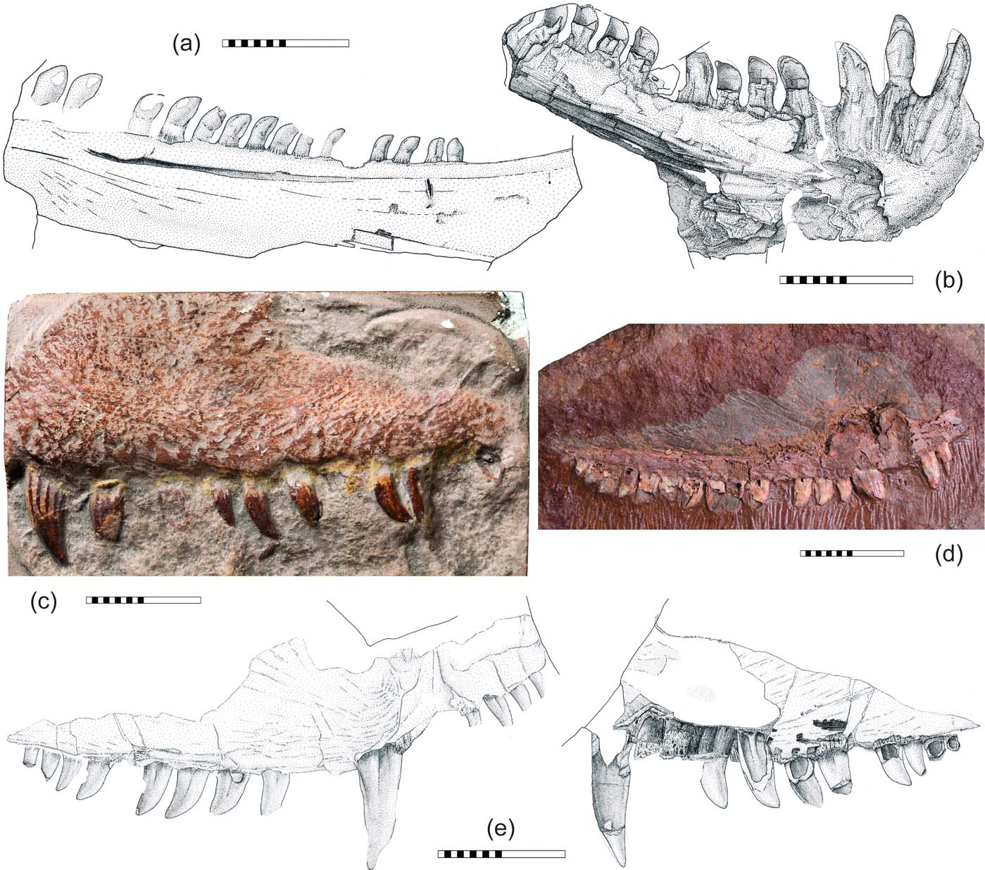
Sphenacodontidae are rare in Europe, but easily identified by the characteristics mentioned previously regarding tooth types and the precanine region. Since there is no general difference from the morphology of North American sphenacodontids, Hypselohaptodus appears all the more unique. The typical sphenacodontid dentition is seen in a few fragments from Southern France (Heyler Reference Heyler1969, pl. LII, 7; Falconnet Reference Falconnet2015, fig. 3; Fig. 4a). A strongly developed precanine step is present in new material from the Roadian of Sardinia (Romano et al. Reference Romano, Citton, Maganuco, Sacchi, Caratelli, Ronchi and Nicosia2018). Probably the oldest in this list is Cryptovenator from Germany, known from a dentary tip (Fröbisch et al. Reference Fröbisch, Schoch, Müller, Schindler and Schweiss2011; Fig. 4b). Its symphyseal area is tall, with three moderately enlarged fangs, which suggest that this genus is somewhat less derived than Sphenacodon.
Jaw fragments of various early Sphenacodontia from Europe. (a) MNHN.F.LOD 213, left dentary of indeterminate sphenacodontid (for photograph, see Falconnet Reference Falconnet2015, fig. 3a). (b) LFN—PW 2008/5599—LS, holotypic right mandible of Cryptovenator hirschbergeri. (c) NHMUK PV R 3729, cast of left labial surface of holotypic maxilla of Sphenacodon britannicus. (d) MNG 13433, referred right maxilla of Dimetrodon teutonis. (e) HN004 2009-00-1, holotypic maxilla of Neosaurus cynodus, two counterslabs. All specimens documented via original fossil work, except for (c): courtesy of S. Chapman. Scale bars=2cm.

The maxilla of Sphenacodon (Oxyodon) britannicus GSM 22893/22894 (catalogue number after Paton Reference Paton1974; available as cast NHMUK PV R 3729) from the same locality as the type of Hypselohaptodus is badly preserved (Paton Reference Paton1974; Fig. 4c). Its postcanine teeth appear long and slender, while their curvature is in sphenacodontid fashion. The lingual part (Paton Reference Paton1974, fig. 1b) reveals more details, such as two precanines and the absence of a distinct step and diastema. Generic assignment to Sphenacodon is tentatively accepted according to dental morphology (Paton Reference Paton1974 mentions unserrated cutting edges; for cross-section, see von Huene Reference von Huene1908, fig. 1). Moreover, the outline of the dorsal blade with the highest point of the maxilla situated posterior to the canine position resembles Sphenacodon more than Dimetrodon. However, species validity is not supported.
The only evidence for Dimetrodon in Europe is a set of small specimens from the Bromacker locality in Germany, designated as D. teutonis (Berman et al. Reference Berman, Reisz, Martens and Henrici2001; Fig. 4d). An isolated maxilla, MNG 13433, is determined on the base of faunal association, not a diagnostic character combination. Because it lacks tooth serrations (Berman et al. Reference Berman, Henrici, Sumida and Martens2004) it might represent a less derived lineage (Brink & Reisz Reference Brink and Reisz2014, fig. 6), if genus determination is correct at all. The steeper anterior outline of the dorsal blade supports its classification as Dimetrodon in contrast with Sphenacodon. There are at least two precanines, typical sphenacodontid postcanines, and no maxillary step.
Neosaurus from France, recently described in detail by Falconnet (Reference Falconnet2015), is of crucial importance for the understanding of early sphenacodontid character histories (Fig. 4e). Its postcanines have the diagnostic morphology of classical sphenacodontids. There is a long precanine zone with a strongly upward bending ventral margin, but no concave step in the preserved region. In clear contrast with Hypselohaptodus, the outer surface texture dorsal to the canine bears curved striae that resemble the pattern of Dimetrodon incisivus (Brink Reference Brink2015, fig. 5-2a, c) and probably yield information about uneven growth directions in this region. Four precanine teeth of similar size are preserved, representing the highest number in any doubtless sphenacodontid and supporting a valid genus diagnosis. Eberth (Reference Eberth1985) suggested a juvenile status for this specimen, but could not document a juvenile Sphenacodon with more than three precanines. As in other sphenacodontids and Hypselohaptodus, but in contrast with ‘Haptodus' garnettensis, tooth implantation is deeply thecodont. Neosaurus might represent a basal sphenacodontid, distinguishable from Cutleria by its longer canines. Distinctness from Cryptovenator is not conclusively testable, but is suggested by a correlation of the precanine step with mandibular symphysis and caniniform dentition.
In the majority of less derived therapsids, both a maxillary step and a distinct precanine diastema are only slightly developed, with the first pronounced occurrence in Rubidgina, Lemurosaurus, or certain gorgonopsians (Sigogneau-Russell Reference Sigogneau-Russell1989). Precanine marginal teeth are absent except for rare forms like Niaftasuchus, Microsyodon, or the specialised Nikkasaurus (Ivakhnenko Reference Ivakhnenko2003). The characteristic postcanine and dentary tooth type of all certain sphenacodontids does not occur in any of the therapsids, of which Ivakhnenko (Reference Ivakhnenko2008) documented many in detail. This supports the sphencodontid tooth type as a strongly diagnostic trait of this branch.
In conclusion, sphenacodontian maxillary characters show a mosaic distribution and sometimes gradual development among taxa (Fig. 5). Cautious trend reconstructions and the diagnostic tooth type of Sphenacodontidae allow for an exclusion of Hypselohaptodus from this lineage. Although it could be a sphenacodontid as specialised as Secodontosaurus, this remains very speculative. Less curved postcanines in Sphenacodontidae appear to be restricted to large specimens. Furthermore, the reduction of precanine dentition occurred as a homoplasy in Sphenacodontidae and Therapsida. Thus, if Hypselohaptodus is precluded from Sphenacodontidae, its initial stage of sphenacodontoid trends implies either another side branch, or the hypothesis that Hypselohaptodus is a potential early therapsid. More sphenacodontian characters need to be revisited carefully before a comprehensive phylogenetic analysis is completed.
Comparison of various early synapsid skulls (Ophiacodontidae and Sphenacodontia), focusing on maxillae and dentaries. Dimetrodon limbatus from Romer & Price (Reference Romer and Price1940) and pers. obs. MCZ 2779; D. teutonis from MNG 13433; D. grandis from Romer & Price (Reference Romer and Price1940) and pers. obs. OMNH 590; D. milleri from Romer & Price (Reference Romer and Price1940) and pers. obs. MCZ 1365; ‘D.' natalis AMNH 4110 and indeterminate skull MCZ 2028 courtesy of K. Brink, and pers. obs.; Ctenospondylus spp. based on pers. obs. of type of C. ninevehensis MCZ 3386 plus reconstruction of C. casei from Vaughn (Reference Vaughn1969, fig. 3c); Neosaurus cynodus from HN004 2009-00-1; Cryptovenator hirschbergeri from LFN—PW 2008/5599—LS; Sphenacodon britannicus from Paton (Reference Paton1974) and photographs of NHMUK PV R 3729; S. ferocior including juvenile from Eberth (Reference Eberth1985), scaled from various specimens; S. ferox from Spielmann et al. (Reference Spielmann, Rinehart, Lucas, Berman, Henrici, Harris, Lucas, Schneider and Spielmann2010); Cutleria wilmarthi and Tetraceratops insignis from Spindler (Reference Spindler2015); Pantelosaurus saxonicus and Palaeohatteria longicaudata from Spindler (Reference Spindler2016); ‘Macromerion' schwarzenbergii from Fritsch (Reference Fritsch1889), Štamberg & Zajíc (Reference Štamberg and Zajíc2008), and pers. obs. of cast CM 11246; Hypselohaptodus grandis from Gz 1071, putative dentary Gz 41, this study; Stereorhachis dominans prepared in cooperation with J. Falconnet; Baldwinonus trux and Stereophallodon ciscoensis from Brinkman & Eberth (Reference Brinkman and Eberth1986); Archaeosyodon praeventor from Kammerer (Reference Kammerer2011); Eotitanosuchus olsoni from Ivakhnenko (Reference Ivakhnenko2008). All drawn to same scale.

5. Implications for evolutionary history
The monophyly of Sphenacodontidae plus Therapsida forming the Sphenacodontoidea is based on an inclusion of classical sphenacodontids (Sphenacodon, Ctenorhachis, Ctenospondylus, Dimetrodon) and generalised therapsids (Reisz et al. Reference Reisz, Berman and Scott1992). No subsequent analysis has raised doubt on this, plotting less derived taxa outside Sphenacodontoidea. Possible parallelism within Sphenacodontoidea is discussed by Spindler (Reference Spindler2016). However, no haptodontine-like taxon can be stated as a potential ancestral representative within one of the two sphenacodontoidean lineages. This underlines how quickly diversification of early sphenacodontians must have taken place in the latest Carboniferous.
The rarity of haptodontine-grade sphenacodontians might be interpreted as reflecting an intermediate ecological pattern between the abundantly recorded Sphenacodontidae and missing Early Permian therapsids. The palaeoecological model explains how early Permian Sphenacodontians diverged into members of moist lowland assemblages and dry uplands that are rarely preserved (Fig. 6; Spindler Reference Spindler2014). Increasing overlap of dry habitats with the fossil-preserving basins is in accordance with specific environmental information on aridification and wet phases (Roscher & Schneider 2006; Roscher et al. 2008). Interestingly, the above comparisons with less derived sphenacodontians comprise mostly European finds from intramontaneous settings, whereas North American ‘haptodontines' are either more isolated than European finds or come from Carboniferous lowlands (Peabody Reference Peabody1957; Reisz et al. Reference Reisz, Heaton and Pynn1982; Sumida & Berman 1993). In this ecological regard, certain forms like Hypselohaptodus do indeed offer a potential model for the initial therapsid appearance. However, the rare and phylogenetically challenging ‘haptodontines' do not simply correlate with arid phases, except for Tetraceratops (Pantelosaurus from a wet phase deposition is likely allochthonous, see Spindler Reference Spindler2016; Hypselohaptodus comes from a proximal system, likely washed in from upland habitats; no exact age is known for Cutleria).
Principal eco-evolutionary model for early Sphenacodontia. The fossil record is reflected such that therapsids appear in the Middle Permian after the transition from a pelycosaur-like ancestor. The ancient radiation of known therapsid subclades is not reported from the preserved basins. Early Permian therapsids inhabited arid, extra-basinal environments. Sphenacodontids were adapted for a range of habitats in the equatorial basins. Climatic changes including a superior aridification and wet phases (Roscher & Schneider 2006; Roscher et al. 2008) caused a biome shift, which certain taxa may have followed in their distribution. These fluctuations crossed the preserved basins that represent the palaeontological window of fossil record. The decline and extinction of both the Sphenacodontidae and wetter refuge habitats correlates with the invasion of therapsids. Biome shift is the main trigger for this faunal turnover, instead of direct competition. The sudden occurrence of Therapsida in the fossil record is not explained by a stratigraphical hiatus (formerly known as Olson's gap to cover a presumed ‘pelycosaur' – therapsid transition). The predicted earliest therapsids are pelycosaur-like in appearance and habitat, then diverging from Sphenacodontidae.

Ophiacodontids from Europe are extremely rare. Although they include a basal form (cf. Archaeothyris, Reisz Reference Reisz1975) and a derived form (Stereorhachis, Falconnet Reference Falconnet2013), the only common and well-documented genus from this family, Ophiacodon, is not known from Europe, after the only evidence is herein rejected. It has been previously discussed that Ophiacodon performed piscivory (Case Reference Case1907; Romer & Price Reference Romer and Price1940), based on snout elongation associated with atypically high tooth numbers, and heron-like eye orientation capable of three-dimensional vision (Berman et al. Reference Berman, Reisz, Bolt and Scott1995). Felice & Angielczyk (Reference Felice, Angielczyk, Kammerer, Angielczyk and Fröbisch2014) demonstrated that Ophiacodon species lacked distinct swimming adaptations. Piscivory, however, can also be performed as shore ambush predation. As the Late Palaeozoic basins of Europe are mainly restricted to intramontaneous basins, palaeobiogeographical distribution had to negotiate barrages and possibly watersheds. Although the European basins were usually rich in fish, except for the fish-free assemblage of Niederhäslich, Germany (Schneider Reference Schneider1993), they were partially cut from extended coastal lowlands. This is a general difference from most North American ‘pelycosaur' assemblages. Basin constraints should be considered additional to assemblage analyses, like those recently carried out by Pardo et al. (Reference Pardo, Small, Milner and Huttenlocker2019). Although the authors detected a W–E lag in the Permian increase of dryland assemblages, in which the Palaeo-Tethys might have functioned as a buffer, there was apparently no refuge for Ophiacodon–Varanosaurus-type ophiacodontids in the eastern equatorial zone. Since these ophiacodontids represent the only Late Pennsylvanian to Early Permian synapsids from North America that lack a European counterpart (in terms of disparity, not genus diversity), it can be tentatively confirmed that this morphotype was strictly bound to certain constraints of limnic environments, possibly some type of prey, cryptic habitats for offspring, or unknown narrow tolerance ranges.
6. Acknowledgements
This study was derived from a dissertation project kindly supervised by Jörg W. Schneider (Freiberg) and P. Martin Sander (Bonn). Great thanks for collection assistance and specimen access are owed to Jon Radley (Warwick), Jessica Cundiff (Cambridge, MA), Kyle Davies, Jennifer Larson, and Richard Cifelli (Norman, OKL), and Thomas Bach (Thallichtenberg). Invaluable exchange was provided by Robert R. Reisz (Mississauga), Kirsten S. Brink (Vancouver), and Jocelyn Falconnet (Paris). Photographs of Sphenacodon britannicus were used by courtesy of Sandra Chapman (London), with additional collection aid by Mike Day (London). Funding received in 2012 to 2015: Deutsche Forschungsgemeinschaft SCHN 408/20-1. Neil Brocklehurst (Oxford) deserves deep gratitude for language improvement and palaeoecological discussion prior to submission, as well as during the review. A second referee also greatly helped to improve the quality of this study.