Introduction
Genetic selection of dairy cows has focussed primarily on increased milk production over the past three decades, and this has been associated with declining fertility in many countries (Butler, Reference Butler2000; Royal et al., Reference Royal, Darwash, Flint, Webb, Woolliams and Lamming2000; Evans et al., Reference Evans, Wallace, Shalloo, Garrick and Dillon2006; Petersson et al., Reference Petersson, Gustafsson, Strandberg and Berglund2006; Garcia-Ispierto et al., Reference Garcia-Ispierto, Lopez-Gatius, Santolaria, Yaniz, Nogareda and Lopez-Bejar2007). Components of reduced fertility include delayed resumption of oestrous cycles post partum, greater incidence of abnormal oestrous cycles and poorer conception rates to first and subsequent inseminations (Royal et al., Reference Royal, Woolliams and Flint2002). The combined effects of these components are longer calving intervals and higher replacement rates, leading to reduced lifetime performance. High replacement rates are of particular concern because when average herd life of cows drops below three lactations (replacement rate 33%), insufficient female replacements can be produced to maintain herd size (Garnsworthy, Reference Garnsworthy2005).
Several management factors that affect fertility have changed in parallel with increasing genetic merit for milk production. For example, average herd size has increased and labour input per cow has decreased (Defra, 2007), both of which can result in less attention to detail, increased disease incidence and less time for oestrous detection. For the purposes of this review, however, we will concentrate on biological factors related to fertility.
Poor fertility has negative consequences for the economics of milk production; Stott et al. (Reference Stott, Veerkamp and Wassell1999) calculated that poor fertility could reduce gross margins by approximately 20%; Evans et al. (Reference Evans, Wallace, Shalloo, Garrick and Dillon2006) calculated that, of the potential increase in farm profitability resulting from improved genetic merit between 1990 and 2003, 50% had been lost due to poor fertility. Poor fertility also increases the environmental impact of dairy systems; involuntary culling for failure to conceive means that more cows are required per unit of milk produced and more non-productive replacements are required per milking cow in the herd. These extra animals increase resource use (feed, fertiliser, fossil fuel) and emissions of pollutants (methane, ammonia, nitrate, nitrous oxide). It has been estimated that if fertility could be restored to levels achieved in the 1980s, total emissions of methane by dairy herds would be reduced by 24% and emissions of ammonia by 17% (Garnsworthy, Reference Garnsworthy2004).
Reproductive events
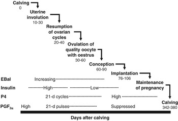
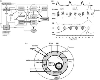
In dairy cows, successful reproduction is the result of a chain of events including resumption of oestrous cycles post partum, development and ovulation of a healthy oocyte, fertilisation, embryo development, implantation in the uterus, maintenance of pregnancy and parturition (Figure 1). Failure at any stage results in failure of the whole process. The ovaries play a central role in the reproduction chain, so compromised ovarian function, manifested as anoestrus or failure to conceive and maintain pregnancy, is often a major cause of poor fertility. Ovarian function is regulated by a complex interaction of factors and feedback mechanisms at the cell (intra-follicle), organ (ovary) and animal (e.g. pituitary, pancreas, liver, adipose tissue) levels (Figure 2). These ovarian regulatory mechanisms must operate in concert with the animal’s overall homoeostatic and homeorhetic regulatory mechanisms, with the systems being linked by signals from metabolic hormones and metabolites (see Garnsworthy and Webb, Reference Garnsworthy and Webb1999; Webb et al., Reference Webb, Garnsworthy, Gong and Armstrong2004; Webb et al., Reference Webb, Garnsworthy, Campbell and Hunter2007).
Sequence of reproductive events in the dairy cow. Each event depends on the success of preceding events. Numbers indicate optimum range (days after calving) to achieve an average calving interval of 365 days. Major temporal factors that influence success are: energy balance (EBal), which should start to increase early in lactation; insulin, which stimulates resumption of oestrous cycles, but may reduce oocyte quality; progesterone (P4), which is low during anoestrus, high during luteal phases of cycles, and low during follicular phases of cycles; and prostaglandin-F2α (PGF2α), which stimulates uterine involution and corpus luteum regression, but has to be suppressed for successful implantation and maintenance of pregnancy. For detailed explanation, see text.

Interactions among physiological regulatory mechanisms at (a) animal, (b) ovarian and (c) follicular levels. At the animal level (a), plasma concentrations of glucose, amino acids and fatty acids are determined primarily by dietary intake and mammary uptake. When plasma glucose is high, insulin stimulates the uptake of glucose by the liver, muscles and adipose tissue, and stimulates lipogenesis. When plasma glucose is low, glucagon stimulates gluconeogeneisis from amino acids, release of glycogen from liver and muscle, and lipolysis. Growth hormone regulates muscle deposition and milk yield, and also regulates IGF and IGF binding protein (IGFBP) synthesis in the liver. Leptin provides feedback from adipose tissue to regulate feed intake. All of these metabolic hormones and metabolites interact with the hypothalamus–pituitary–ovarian axis. The hypothalamus releases GnRH to regulate FSH and LH release from the pituitary gland, with feedback from ovarian oestradiol (E2) and inhibin (In). At the ovarian level (b), FSH stimulates follicle recruitment and early growth in two or three waves per cycle. Pulsatile LH is then required for continued growth and development of the dominant ovulatory follicle, which secretes E2. The corpus luteum (CL), which develops after ovulation, secretes progesterone (P4) until prostaglandin-F2α is released by the non-pregnant uterus to cause CL regression. At the follicular level (c), gonadotrophins and metabolic hormones work in conjunction with locally produced growth factors (e.g. IGFBP, bone morphogenic proteins (BMP) and growth differentiation factor-9 (GDF-9)) released by granulosa and theca cells, and the oocyte, to regulate development of the follicle and cumulus-oocyte complex (COC). For detailed explanation, see text and also: Garnsworthy and Webb (Reference Garnsworthy and Webb1999); Webb et al. (Reference Webb, Garnsworthy, Gong and Armstrong2004); Webb and Campbell (Reference Webb and Campbell2007); Webb et al. (Reference Webb, Garnsworthy, Campbell and Hunter2007).

Regulation of blood glucose and body fatness
Metabolic regulation of nutrient partitioning (Figure 2a) is important for successful lactation and represents the physiological basis for improvements in productive efficiency of high-merit dairy cows (Bauman, Reference Bauman2000). Homoeostatic controls of blood glucose are of primary importance in dairy cows because up to 80% of glucose turnover is used for mammary synthesis of lactose, which is the major osmotic component of milk that determines milk yield (Bauman and Currie, Reference Bauman and Currie1980). Insulin and glucagon play major complementary roles in glucose homoeostasis: insulin stimulates glucose uptake by tissues, promotes protein synthesis and increases lipogenesis; glucagon stimulates gluconeogenesis and glycogenolysis (Vernon, Reference Vernon1988). Both insulin and glucagon respond rapidly to changes in nutrient absorption and utilisation throughout the day. Long-term regulation of nutrient partitioning is by homeorhetic mechanisms, which attenuate responses of specific tissues to homoeostatic signals (Bauman, Reference Bauman2000). Thus, for example, when growth hormone is increased (at the onset of lactation, by bovine somatotrophin (BST) administration, or by genetic selection for high milk yield), less nutrients are directed to body fat reserves following a meal because of altered responses to insulin, and more nutrients are taken up by the mammary gland to support milk synthesis (Bauman, Reference Bauman2000).
Some of the effects of insulin and growth hormone are mediated through the IGF system. IGF-I is synthesised primarily in the liver, in response to growth hormone, and IGF receptors are found in most tissues (Baumrucker, Reference Baumrucker2000). IGF is transported in the bloodstream by six high-affinity binding proteins (IGFBP), whose specific characteristics allow local regulation of IGF availability in target tissues (Jones and Clemmons, Reference Jones and Clemmons1995). During early lactation, growth hormone receptors in the liver are down-regulated and circulating concentrations of IGF-I are dramatically reduced despite elevated growth hormone concentrations (Butler et al., Reference Butler, Marr, Pelton, Radcliff, Lucy and Butler2003). This down-regulation can be overcome, however, and circulating IGF-I concentrations restored, by elevated plasma insulin concentrations during early lactation (Butler et al., Reference Butler, Marr, Pelton, Radcliff, Lucy and Butler2003).
Leptin also has a homeorhetic role by acting as an indicator of body fatness and by providing a feedback mechanism to control feed intake, although the overall regulatory system is complex (Vernon et al., Reference Vernon, Denis and Sørensen2001). In addition to its effects on feed intake, leptin has been found to modulate nutrient transfer and partitioning by interaction with other hormones including insulin, glucagon, glucocorticoids, growth hormone, IGF-I, cytokines and thyroid hormones (Hill, Reference Hill2004). It appears that both insulin and IGF-I are involved in regulating leptin responses to nutrition (Armstrong et al., Reference Armstrong, Gong and Webb2003). Other factors secreted by adipose tissue (tumour necrosis factor α and resistin) have also been shown to interact with leptin in the regulation of body fatness (Vernon et al., Reference Vernon, Denis and Sørensen2001).
Regulation of body fatness has important implications for energy balance in early lactation. During the first few weeks of lactation, high-yielding dairy cows are usually in negative energy balance because energy output in milk exceeds energy intake from the diet, implying that body fat reserves are being mobilised to make up the energy deficit. This mobilisation can be energetically equivalent to one-third of the milk produced (Bauman and Currie, Reference Bauman and Currie1980). Body fat mobilisation is not, however, a passive response to high milk yield and low feed intake. As reviewed by Garnsworthy (Reference Garnsworthy2007), there is much evidence to indicate that dairy cows adjust their feed intake to regulate body fatness over the first 3 months of lactation.
The concept that dairy cows regulate their feed intake to achieve a target level of body fatness in early lactation was proposed by Garnsworthy and Topps (Reference Garnsworthy and Topps1982), based on observations of cows fed to achieve different levels of body condition score (BCS) at calving. Cows with a BCS above their target had lower feed intakes and lost BCS throughout the study; cows with a BCS below their target had higher feed intakes and gained BCS throughout the study. Consequently, BCS of all cows converged to an average of 2.5 (1 to 4 scale) by week 12 of lactation. Importantly, there was no difference in milk yield among treatment groups, indicating that homeorhetic mechanisms were actively regulating body fatness through the negative feedback effect of body fat on feed intake.
Numerous studies in the 1980s and 1990s (reviewed by Broster and Broster, Reference Broster and Broster1998; Garnsworthy, Reference Garnsworthy1988; Stockdale, Reference Stockdale2001) confirmed the strong negative relationship between BCS at calving and change in BCS during early lactation. More recently, Garnsworthy (Reference Garnsworthy2007) compared responses published between 1980 and 1993 with responses published between 2000 and 2006. The BCS at calving resulting in no net change in BCS over the first 12 weeks of lactation has decreased from 2.5 (1 to 5 scale) in older studies to 2.1 in recent studies. This suggests that the average biological target BCS has decreased with increasing genetic merit for milk production, so modern dairy cows are genetically ‘thinner’.
It is important to distinguish between phenotypic and genetic body fatness. Dechow et al. (Reference Dechow, Rogers and Clay2002) examined correlations between BCS and BCS loss in 310 000 lactation records. Phenotypically, an increase in BCS at calving was associated with greater BCS loss in early lactation, as expected if cows are attempting to reach biological targets for BCS. Genetically, however, higher BCS at calving was correlated with less BCS loss during early lactation. In other words, management conditions that increase BCS at calving result in greater BCS loss, but genetically fat cows maintain BCS in early lactation.
Negative energy balance and fertility
Several studies have shown that high genetic merit, negative energy balance, body fat mobilisation and low plasma insulin are all associated with delayed first ovulation post partum and reduced pregnancy rates (see reviews by Garnsworthy and Webb, Reference Garnsworthy and Webb1999; Butler, Reference Butler2003; Pryce et al., Reference Pryce, Royal, Garnsworthy and Mao2004; Butler, Reference Butler2005; Garnsworthy, Reference Garnsworthy2007). Lopez-Gatius et al. (Reference Lopez-Gatius, Yaniz and Madriles-Helm2003) performed a meta-analysis of 15 papers corresponding to nearly 8000 cows to examine the relationships between BCS and reproductive performance. Compared with cows losing 0 to 0.5 BCS, cows losing 0.5 to 1.0 BCS took 3.5 days longer to conceive, and cows losing >1.0 BCS took 10.6 days longer to conceive; cows gaining BCS took 3.7 days less to conceive. Similarly, Butler (Reference Butler2005) reported that cows losing less than 0.5 BCS over the first 30 days post partum took an average of 30 days from calving to first ovulation; cows losing 0.5 to 1.0 BCS took 36 days; cows losing more than 1.0 BCS took 50 days. In addition, Butler (Reference Butler2005) concluded from several studies that conception rate decreases by 10% per 0.5 unit BCS loss. It is clear, therefore, that reducing the extent and duration of negative energy balance and BCS loss should be beneficial for fertility.
Three basic strategies are available to reduce the extent and duration of negative energy balance and BCS loss in early lactation. The first strategy is to reduce BCS at calving so that energy intake is not limited by the negative feedback effect of BCS, as discussed above; a recent study showed that high-genetic merit cows can maintain a BCS of 2.5 throughout lactation (Yan et al., Reference Yan, Mayne, Keady and Agnew2006). The second strategy is to feed low-protein diets that reduce body fat mobilisation (Garnsworthy and Jones Reference Garnsworthy and Jones1987; Westwood et al., Reference Westwood, Lean, Garvin and Wynn2000; Schei et al., Reference Schei, Volden and Baevre2005). The third and the commonest strategy is to increase dietary energy concentration by increasing the starch or fat components of the diet at the expense of forage components. Such changes in carbohydrate source have implications for rumen function, milk composition, nutrient partitioning and metabolic hormones, particularly insulin (Sutton, Reference Sutton1989; Sutton et al., Reference Sutton, Dhanoa, Morant, France, Napper and Schuller2003; Reynolds, Reference Reynolds2006). These implications are discussed in subsequent sections.
Resumption of oestrous cycles
Metabolic hormones and reproductive function were measured in dairy cows selected for high and low genetic merit for milk yield (Gutierrez et al., Reference Gutierrez, Gong, Bramley and Webb2006). Resumption of normal oestrous cycles post partum occurred approximately 8 days later in the high-merit cows, and this was associated with lower plasma insulin concentrations. Interestingly, these differences occurred despite no difference in milk yield between the two selection lines during this period, suggesting other underlying mechanisms linked to selection for total milk yield. A further study investigated whether feeding diets designed to increase circulating insulin concentrations during the early post partum period can overcome the delay in first ovulation post partum shown by cows selected for increased milk yield (Gong et al., Reference Gong, Armstrong, Baxter, Hogg, Garnsworthy and Webb2002a). Two isoenergetic diets were formulated to either stimulate or depress plasma insulin concentrations, and these were fed to cows of high or low genetic merit. Again, first ovulation and resumption of normal oestrous cycles post partum were delayed in cows of high genetic merit. This was associated with lower circulating insulin concentrations, but did not involve an alteration in basal plasma gonadotrophin concentrations or patterns of ovarian follicular development during the early post partum period. Feeding the diet designed to increase circulating insulin concentrations advanced first ovulation post partum so that 100% of low-merit cows and 80% of high-merit cows ovulated within the first 50 days of lactation when fed on the high-insulin diet, compared with 60% of low-merit cows and 50% of high-merit cows fed on the control diet.
These data suggest that genetic influences on reproductive performance can be partially overcome through dietary manipulation because the responses observed by Gong et al. (Reference Gong, Armstrong, Baxter, Hogg, Garnsworthy and Webb2002a) were independent of milk yield and energy balance, as were the differences between cows of high and low genetic merit in the study of Gutierrez, et al. (Reference Gutierrez, Gong, Bramley and Webb2006). It appears that insulin acts as a metabolic signal to the reproductive system, signalling that nutritional status is adequate and conception can proceed. The exact mechanism for this signalling is still unclear, but it probably involves interactions between insulin, IGFs and gonadotrophins (Figure 2a).
Regulation of follicle development
Ovarian follicular growth is a developmental process during which follicles leave the primordial pool, increase in diameter and complexity, and differentiate into the three cell types (oocyte, granulosa and theca) that ultimately constitute the preovulatory follicle (Webb et al., Reference Webb, Garnsworthy, Campbell and Hunter2007). Functional changes include changes in the steroidogenic capability of follicles, particularly during the antral stages of development, the acquisition of granulosa cell FSH receptors during the preantral stage of development and the acquisition of granulosa cell LH receptors around the time of selection of the dominant follicle (see Webb et al., Reference Webb, Nicholas, Gong, Campbell, Gutierrez, Garverick and Armstrong2003 and Reference Webb, Garnsworthy, Gong and Armstrong2004). These later stages of antral follicle development are characterised by two or three waves of follicular growth (Figure 2b) during each oestrous cycle (Adams, Reference Adams1999). Each wave of follicle growth involves recruitment of a group of follicles that grow to approximately 6 to 8 mm in diameter. One follicle is then selected for continued growth and becomes dominant. The remaining follicles become atretic and regress. The precise mechanism for selection of dominant follicles remains to be fully elucidated, but does involve the action of gonadotrophins as well as locally produced factors (Webb et al., Reference Webb, Garnsworthy, Gong and Armstrong2004). The very early stages of folliculogenesis can occur without gonadotrophins but FSH, along with other extra-ovarian and locally produced growth factors, may affect the rate of preantral follicle growth (Hulshof et al., Reference Hulshof, Figueiredo, Beckers, Bevers, van der Donk and van den Hurk1995; Gutierrez et al., Reference Gutierrez, Ralph, Telfer, Wilmut and Webb2000; Webb et al., Reference Webb, Garnsworthy, Gong and Armstrong2004; Webb and Campbell, Reference Webb and Campbell2007). Both FSH and LH exert their effects on follicular somatic cells via specific membrane-bound receptors that exhibit alternate patterns of expression. Granulosa cells of large oestrogenic (dominant) antral follicles develop LH receptors (Bao and Garverick, Reference Bao and Garverick1998; Webb et al., Reference Webb, Campbell, Garverick, Gong, Gutierrez and Armstrong1999a), and this event is critical to the process of follicle selection. In addition, changes in the expression of mRNAs for both gonadotrophin receptors and steroidogenic enzymes (e.g. P450arom) (Bao and Garverick, Reference Bao and Garverick1998; Webb et al., Reference Webb, Campbell, Garverick, Gong, Gutierrez and Armstrong1999a; Webb and Campbell, Reference Webb and Campbell2007) occur at this stage of development. Thus the presence of LH receptors on granulosa cells enables the dominant follicle to switch its gonadotrophic dependence from FSH to LH and attain dominance over other antral follicles, which remain FSH-dependent (Webb et al., Reference Webb, Garnsworthy, Campbell and Hunter2007).
Development of follicles depends also on interactions with a range of local and circulating growth factors. For example, treatment with (BST), through increasing circulating insulin and IGF concentrations, can increase both the number of gonadotrophin-responsive follicles and their quality (Gong et al., Reference Gong, Bramley and Webb1991 and Reference Gong, Bramley and Webb1993). Nutrition also influences follicle development (Garnsworthy and Webb, Reference Garnsworthy and Webb1999), with short-term changes in plane of nutrition affecting small antral follicle (1 to 4 mm) recruitment, without affecting circulating concentrations of FSH (Gutierrez et al., Reference Gutierrez, Oldham, Bramley, Gong, Campbell and Webb1997b; Gong et al., Reference Gong, Armstrong, Baxter, Hogg, Garnsworthy and Webb2002a), resulting in a larger number of ovulations after a superovulatory gonadotrophin challenge (Gong et al., Reference Gong, Lee, Garnsworthy and Webb2002b). Increased dietary energy decreases steady-state concentrations of mRNAs encoding IGFBP-2 and -4 in small antral follicles, which in turn increase the bioavailability of locally produced IGF-II and systemically derived IGF-I in these follicles (Armstrong et al., Reference Armstrong, Gong and Webb2003; Webb et al., Reference Webb, Nicholas, Gong, Campbell, Gutierrez, Garverick and Armstrong2003). Hence this work provides important evidence that diet can have direct effects at the ovarian level, as well as the whole animal level, to influence follicular development and oocyte quality.
Regulation of oestrous cycles
Oestrous cycles have a follicular phase, at the end of which there is a surge release of gonadotrophins (LH and FSH) from the anterior pituitary gland accompanied by ovulation, and a luteal phase with the secretion of progesterone from the newly formed corpus luteum (Figure 2a and b). The oestrous cycle is driven primarily by GnRH, which is secreted into the hypophyseal portal system in a pulsatile fashion from the terminals of neurons originating around the preoptic area and mediobasal hypothalamus at the base of the brain. GnRH neurons are regulated by feedback from oestradiol and progesterone (Clarke and Pompolo, Reference Clarke and Pompolo2005). Both FSH and LH are released from the anterior pituitary in response to GnRH (Webb et al., Reference Webb, Gong, Law and Rusbridge1992; Wolfenson et al., Reference Wolfenson, Inbar, Roth, Kaim, Bloch and Braw-Tal2004). Each pulse of GnRH is followed by a pulse of LH that is released into the peripheral circulation. In contrast, there is a less well-defined association between GnRH and FSH release, which is thought to be dependent on the rate of FSH synthesis. Both ovarian-derived oestradiol and inhibin feedback to inhibit FSH release by suppressing FSH gene expression (Pawson and McNeilly, Reference Pawson and McNeilly2005).
A number of growth factors (e.g. insulin, IGF-I and leptin) and metabolites (e.g. glucose, free fatty acids and ammonium) influence GnRH release from hypothalamic neurons (Sinclair and Webb, Reference Sinclair and Webb2005). Leptin and IGF-I receptors are expressed within the anterior pituitary, and both leptin and IGF-I increase LH secretion in primary cultures of anterior pituitary cells (Daftary and Gore, Reference Daftary and Gore2005; Zieba et al., Reference Zieba, Amstalden and Williams2005).
Extra-ovarian factors, such as metabolic hormones and nutrients, interact with growth factors produced locally within the ovary (Figure 2c) to induce changes in follicle dynamics and oocyte quality (Webb et al., Reference Webb, Garnsworthy, Gong and Armstrong2004). A large number of in vitro studies have demonstrated the direct action of metabolic factors on granulosa and theca cells (Webb et al., Reference Webb, Campbell, Garverick, Gong, Gutierrez and Armstrong1999a and Reference Webb, Gosden, Telfer and Moor1999b; Lucy, Reference Lucy2000; Spicer et al., Reference Spicer, Chamberlain and Maciel2002; Armstrong et al., Reference Armstrong, Gong and Webb2003). Some cell culture studies have shown bovine granulosa cells to be critically dependent on the presence of physiological concentrations of insulin (Gutierrez et al., Reference Gutierrez, Campbell and Webb1997a). Moreover, diet-induced increases in circulating concentrations of insulin are correlated with increased oestradiol production in cultured granulosa cells from small antral (1 to 4 mm) follicles (Armstrong et al., Reference Armstrong, Gong, Gardner, Baxter, Hogg and Webb2002), demonstrating a direct action of metabolic hormones on follicle function.
Oocyte quality
At the follicle level, cell proliferation and development are controlled largely by endocrine and paracrine growth factors and nutrient supply (Figure 2c). Collectively, these determine maturation and developmental competence of the oocyte. Studies have highlighted the link between nutrient intake and oocyte developmental competence in cattle (reviewed by O’Callaghan and Boland, Reference O’Callaghan and Boland1999; Boland et al., Reference Boland, Lonergan and O’Callaghan2001). From our own studies in cattle we know that diets containing high levels of rumen-degradable nitrogen can increase concentrations of ammonium and urea in plasma and follicular fluid, which can lead to significantly reduced cleavage rates after in vitro maturation (IVM) and fertilisation (IVF), and reduced blastocyst yields during in vitro culture (IVC) (Sinclair et al., Reference Sinclair, Kuran, Gebbie, Webb and McEvoy2000a). Such diets also depress plasma insulin concentrations significantly (Sinclair et al., Reference Sinclair, Sinclair and Robinson2000b), which may contribute to this phenomenon. In heifers, high-energy (1.6 times maintenance ME requirements) and high-protein (27 g/MJ ME) diets, although enhancing ovarian follicle development, led to a significant reduction in oocyte quality, determined by the ability of oocytes to mature, fertilise and develop to the blastocyst stage during IVC (Armstrong et al., Reference Armstrong, McEvoy, Baxter, Robinson, Hogg, Woad, Webb and Sinclair2001). The ovarian IGF system has the potential to interact directly with the oocyte through the Type-I IGF receptor, so that the reduced levels of IGFBP-2 and -4, together with increased concentrations of both IGF-I and insulin reported by Armstrong et al. (Reference Armstrong, McEvoy, Baxter, Robinson, Hogg, Woad, Webb and Sinclair2001), may have led to the oocyte being ‘overstimulated.’ Indeed, we (Adamiak et al., Reference Adamiak, Mackie, Watt, Webb and Sinclair2005) have subsequently demonstrated that the effects of high levels of feeding on oocyte quality in cattle are cumulative, are linked to insulin metabolism and are dependent on the initial BCS of the animal. Although feeding a high-energy (2.0 times maintenance ME requirements) diet to thin heifers (BCS 2.0) improved early embryo development following IVM, IVF and IVC, the same diet offered to fat heifers (BCS 3.75) reduced embryo development significantly. Furthermore, the effects were cumulative, with embryo development for the fat heifers on twice maintenance deteriorating over the 9-week experimental period. A significant proportion of these animals were hyperinsulinaemic (plasma insulin >1.66 ng/ml), and this condition was associated directly with impaired oocyte quality. As observed in the study of Armstrong et al. (Reference Armstrong, McEvoy, Baxter, Robinson, Hogg, Woad, Webb and Sinclair2001), high-energy diets enhanced ovarian follicular development. Therefore, from studies with heifers it appears that diets that promote ovarian follicular development may be detrimental for oocyte quality.
Results of our subsequent studies with lactating dairy cows are consistent with results from heifer studies, although insulin concentrations are lower in lactating dairy cows (e.g. 0.32 ng/ml for a high-insulin diet v. 0.21 ng/ml for a low-insulin diet in the study of Gong et al., Reference Gong, Lee, Garnsworthy and Webb2002b). These dairy cow studies indicate that high-insulin status may encourage early resumption of oestrous cycles, but high insulin might not be beneficial for oocyte quality. In one experiment (Fouladi-Nashta et al., Reference Fouladi-Nashta, Gutierrez, Garnsworthy and Webb2005), cows were fed diets with either low or high starch content in order to induce differences in plasma insulin. Oocytes were collected for IVF and cultured to examine developmental potential to the blastocyst stage. For the high-insulin diet, 26% of cleaved embryos developed to blastocysts, compared with 41% for the low-insulin diet. Furthermore, the high-insulin diet produced a significantly higher number of grade 4 (poor quality) oocytes (high insulin 28%, low insulin 13%). In another experiment, addition of fat to a high-starch diet improved blastocyst yield from 29% to 38% (Fouladi-Nashta et al., Reference Fouladi-Nashta, Gutierrez, Gong, Garnsworthy and Webb2007).
Embryo development and implantation
Progesterone secreted by the developing corpus luteum during the post-ovulatory period plays a critical role in supporting uterine function and numerous studies have linked poor progesterone secretion at this time to poor embryo development and early embryo loss (e.g. Mann and Lamming, Reference Mann and Lamming1999 and Reference Mann and Lamming2001). Progesterone secretion is related directly to the size of the corpus luteum, which can be increased by feeding supplemental dietary fat (Staples and Thatcher, Reference Staples and Thatcher2005). Metabolic hormones might also play a role in embryo development; for example, we found a lower proportion of elongated embryos (>10 cm at 16 days after insemination) in heifers with a low insulin to glucagon ratio, although progesterone concentrations were not affected by treatment (Mann et al., Reference Mann, Green, Sinclair, Demmers, Fray, Gutierrez, Garnsworthy and Webb2003).
Embryo mortality, during the pre-implantation period, is a key contributor to reduced fertility in cattle, with up to 40% of total embryo losses occurring between days 8 and 17 of pregnancy (Thatcher et al., Reference Thatcher, Guzeloglu, Mattos, Binelli, Hansen and Pru2001). This stage of embryonic loss coincides with the inhibitory effects of interferon-τ, produced by trophectoderm cells of embryos, on prostaglandin release from the uterus (Flint, Reference Flint1995). This suggests that a proportion of embryos are unable to inhibit prostaglandin-F2α release, leading to regression of the corpus luteum, reduction in progesterone production and termination of pregnancy (Flint, Reference Flint1995; Thatcher et al., Reference Thatcher, Binelli, Burke, Staples, Ambrose and Coelho1997). Hence the quality of the pre-implantation embryo and its ability to communicate with the cells of the uterus are critical for the establishment and continuation of pregnancy.
Integration of physiological mechanisms
From the previous sections, it is clear that several physiological mechanisms influence fertility in dairy cows. As illustrated in Figures 1 and 2, these mechanisms operate at different levels (cell, ovary, animal) and on different timescales (minutes, hours, days, weeks). The interactions between regulatory systems require an integrated approach when addressing fertility issues because changes in one system can have consequences for other systems. It is necessary to consider general homoeostatic and homeorhetic mechanisms simultaneously with regulation of ovarian cycles, folliculogenesis and oocyte development.
In reviewing reproductive mechanisms affected by energy balance, Butler (Reference Butler2003) found that negative energy balance is strongly associated with attenuation of LH pulse frequency and low levels of blood glucose, insulin and IGF-I that collectively limit oestrogen production by dominant follicles. Negative energy balance was also associated with a diminished quality of oocytes, reduced embryo development and lower serum progesterone concentrations. Thus, it would seem that the primary goal when addressing poor fertility is to reduce the extent and duration of negative energy balance. As discussed previously, however, strategies designed to reduce negative energy balance will alter circulating concentrations of metabolites and hormones.
Circulating concentrations of metabolites and hormones are determined by the balance between nutrient intake, milk production level and body tissue reserves, relative to the animal’s genetic potential. As outlined above, these factors are regulated by homoeostatic and homeorhetic mechanisms, but they can be manipulated by altering diet composition. Insulin appears to be the most important hormone that links metabolic status to the regulation of ovarian function. After addressing the issue of negative energy balance, the next most promising strategy for improving fertility is to promote resumption of oestrous cycles by ensuring that insulin status is adequate.
There is a potential conflict between strategies designed to start cows cycling and production of good-quality oocytes. High plasma insulin concentrations stimulate resumption of oestrous cycles, but have detrimental effects on oocyte developmental competence. This presents an interesting challenge for researchers and producers, and might explain why the fertility problem is so widespread in dairy cows. Our recent studies addressed these differential responses by nutritional manipulation of insulin at different stages of the reproductive cycle. In an initial experiment (Garnsworthy, Sinclair, Webb et al.: unpublished), increasing insulin status immediately post partum, and then reducing insulin status during the mating period, doubled the pregnancy rate at 120 days in milk. This suggests that progress towards restoring fertility can be made by an integrated approach that allows for interactions between physiological mechanisms that regulate metabolism and reproduction.
Acknowledgements
Fertility research at Nottingham has been sponsored by defra, LINK (SEERAD, ABNA Ltd, BOCM PAULS Ltd and Provimi Ltd), BBSRC and NIH. We acknowledge the following for significant contributions to the work discussed in this paper: APF Flint, AA Fouladi-Nashta, CG Gutierrez, JG Gong, DG Armstrong, GE Mann, N Saunders, H Russell and M Mitchell.